|
|
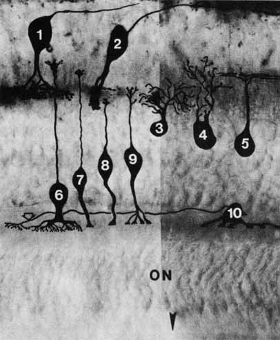
| Fig. 27 Drawing of Golgi-stained neural cells between the nerve fiber and the outer plexiform layers: (1) stratified diffuse ganglion cell; (2) midget ganglion cell; (3) stratified diffuse amacrine cell; (4) diffuse amacrine cell; (5) bistratified amacrine cell; (6) rod bipolar cell; (7) invaginating midget bipolar cell (to a single-cone pedicle); (8) flat midget bipolar cell (to a single-cone pedicle); (9) flat bipolar cell (diffuse cone bipolar to 6 or 7 cones); (10) horizontal cell. Arrowhead indicates external limiting membrane. Open arrow points to horizontal cell axon (going to rod spherules only). ON, outer nuclear layer. |