|
|
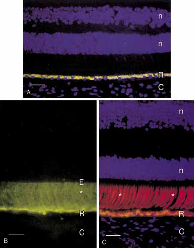
| Fig. 42 A. Immunofluorescence micrograph of adult human retina demonstrating autofluorescence from lipofuscin granulates in the retinal pigment epithelium (R). Cell nuclei in the outer and inner nuclear layers (n) are counterstained blue with DAPI. C, choroid. Bar = 30 microns. B. Immunofluorescence micrograph demonstrating interphotoreceptor retinoid binding protein (* green label) in the subretinal space of adult human retina. The internal limit of the subretinal space is the external limiting membrane (E). Bar = 0 microns. C. Immunofluorescence micrograph demonstrating cone matrix sheaths (* labeled red with peanut agglutinin lectin) that surround individual cone outer segments in the subretinal space. Bar + 20 microns. (Milan A, Smithe JE, John SK: Anatomy and cell biology of the human retina. In Tasmam W, Jaegar EA (eds): Duane's Clinical Ophthalmology, Vol. 3. Philadelphia: JB Lippincott Co, 2001:1–22) |