|
|
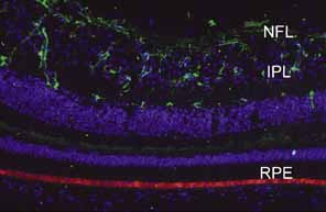
| Fig. 41 Immunofluorescence photomicrograph of a normal human retina with astrocytes (green) confined to the nerve fiber (NFL) and inner plexiform layers (IPL). The astrocytes are stained with anti-glial fibrillary acidic protein (GFAP). The retinal pigment epithelium (RPE) contains lipofuscin (orange). Cell nuclei are stained with DAPI (blue). 20× objective. (Courtesy of Scheie Eye Institute) |