|
|
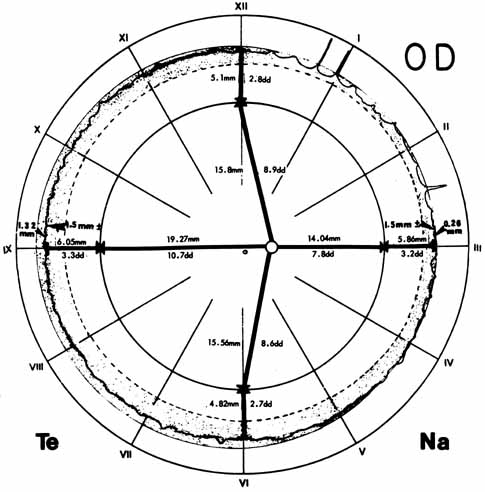
| Fig. 14 Diagram used for drawing retinal detachments, tumors, and vascular lesions shows the equator as the innermost of the large circles and the ora as the middle circle. Beyond the ora is the pars plana so that the outermost circle represents the posterior edge of the pars plicata. The dimensions in millimeters and in disc diameters refer to the distances from the disc. The fovea is in the geometric center of the diagram. The vitreous base (shaded band) is narrower nasally than temporally. The normal vitreous base extends from posterior to the ora serrata to anterior to all but the longest dentate processes. In senile anterior retraction of the vitreous base, retinal tears may form in the zone between the posterior edge of the vitreous base and the anterior edge of the ora. Te, temporal; Na, nasal; OD, right eye. (Straatsma BR, Foos RY, Spencer LM: The retina—Topography and clinical correlations. In New Orleans Academy of Ophthalmology Symposium on Retina and Retinal Surgery. St. Louis: CV Mosby, 1969) |