|
|
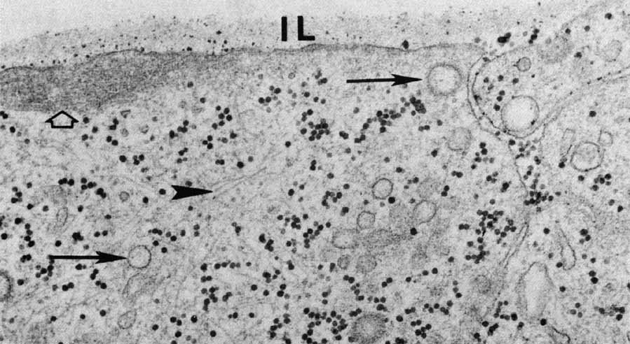
| Fig. 43 Internal limiting membrane (IL) of the retina. Portions of two Müller cells with dense fibrillar patches adjacent to the cytoplasmic face of the plasmalemma are seen (open arrow). The cytoplasm contains ribosomes and glycogen granules (the larger, somewhat irregular, darker figures), microfibrils, microtubules (arrowhead), and various membrane-bound structures, including some cored vesicles (arrow). There is a slightly less electron-dense strip adjacent to the vitreal surface of the plasmalemma, followed by the more osmiophilic basal lamina (original magnification ×48,000). |