|
|
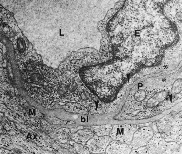
| Fig. 51 Capillary wall shows the separation of the lumen (L) from the surrounding Müller cell (M) and bipolar axons (Ax). The endothelial cell's (E) basal lamina (bl) encases the intramural pericyte (P), whose inner surface shows microfibrils (asterisks) similar to those of the endothelial cell. Pinocytotic vesicles are along the outer walls of the endothelial cell and pericyte (arrowheads) (original magnification ×24,000). |