|
|
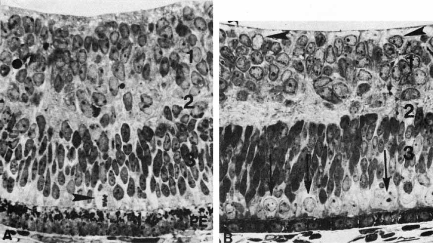
| Fig. 18 A. In the 24-mm human embryo (approximately 7 weeks' gestation), the developing retina is divided into two layers. The nuclei of the cells of the inner neuroblastic layer (1) are oriented in various directions without any layering. The layer of Chievitz (2) is composed of interconnecting processes and also contains nuclei that are migrating from the outer toward the inner retina. The closely packed cells of the outer neuroblastic layer (3) have nuclei of variable densities. Mitoses (arrowhead) occur in the outer layer. The underlying pigmented epithelium (PE) cells show clumping of their granules at their apical ends (original magnification ×720). B. In the retina of the 50-mm fetus (10 weeks' gestation), cells of the inner neuroblastic layer (1) have started to differentiate. Immature ganglion cells (arrowheads) are near the inner limiting membrane. The layer of Chievitz (2) contains some cells presumably in transit between the two nuclear layers. (3) Outer neuroblastic layer. The future cone nuclei (arrows) are positioned near the monolayered pigmented epithelium (original magnification ×720). |