|
|
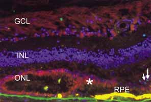
| Fig. 22 Immunofluorescence photomicrograph of a retina section from a post mortem donor with geographic atrophy from age-related macular degeneration (AMD). Nuclei are labeled with DAPI (blue). Retinal pigment epithelium (RPE) is yellow as a result of autofluorescence. At the edge of atrophy, RPE disappears first and photoreceptors form a psuedorosette. Photoreceptors label with anti_Fas receptor (red, asterisk), suggesting that the Fas pathway may participate in photoreceptor death in AMD. Dying photoreceptors whose nuclei are TUNEL positive (green-arrow) are present consistent with apoptosis. Outer nuclear layer (ONL), inner nuclear layer (INL), ganglion cell layer (GCL). Scale bar 50 microns. (Courtesy of Joshua L. Dunaief, MD PhD, Scheie Eye Institute, University of Pennsylvania) |