|
|
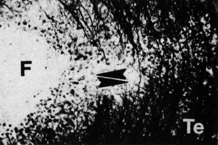
| Fig. 35 Golgi preparation shows the devision of the nerve fiber layer temporal to the fovea (F) into perpendicular fascicles above and below the horizontal line. Temporal (Te) fasicles cross the midline raphe. Upper arrowhead represents the porition of the midline raphe. Lower arrowhead indicates a perpendicular fascicle crossing the midline. (Courtesy of Professor Vrabec) |