Chapter 9
Pathology of the Cornea-Sclera
HENRY D. PERRY and J. DOUGLAS CAMERON
Main Menu   Table Of Contents

Search

CONGENITAL ABNORMALITIES OF THE CORNEA
CONGENITAL OPACITIES OF THE CORNEA
DYSGENESIS OF THE CORNEA
CHORISTOMAS OF THE CORNEA
INFLAMMATORY CONDITIONS OF THE CORNEA
CORNEAL DYSTROPHIES
CORNEAL DEGENERATION
PATHOLOGY OF THE SCLERA
REFERENCES

CONGENITAL ABNORMALITIES OF THE CORNEA
Developmental abnormalities of the cornea are usually associated with other ocular and nonocular abnormalities because the cornea develops in conjunction with several germinal layers. Surface ectoderm develops into corneal epithelium, Bowman's layer, and crystalline lens. Anomalies arising from defects of surface ectoderm range from cryptophthalmos to cataracts.1 Neural crest mesenchyme develops into the corneal stroma, the anterior chamber angle filtering apparatus, and the iris stroma. Associated anomalies of neural crest mesenchyme range from a localized faint corneal stromal opacity to major alterations of the anterior chamber filtering mechanism (Axenfeld's anomaly) and congenital glaucoma. Neuroectoderm develops into the iris pigment epithelium. Abnormalities of the optic cup may lead to such anomalies as microcornea and megalocornea and possibly macular hypoplasia.2,3 Because the cornea develops at an early embryonic stage, coexisting abnormalities in other organ systems are to be expected. Facial, dental, genitourinary, cardiac, and skeletal abnormalities commonly are associated with corneal defects.4–7 Many types of pathologic processes may lead to developmental abnormalities of the cornea that are indistinguishable clinically from one another. Congenital corneal opacities have been associated with the connective tissue disorder Ehlers-Danlos syndrome,8 with congenital infection,9 or as a manifestation of a recognized teratology.10,11 Some types of processes, such as corneal keloid, may be expressed long after birth.12

No all-encompassing system of classification exists for developmental abnormalities of the cornea. Classifications of appearance and shape are not mutually exclusive (e.g., sclerocornea often is associated with cornea plana and vice-versa). Classification by neural crest origin is appropriate for some elements of the cornea (e.g., the endothelium) but not for all components of the cornea.13 Classification by direct cause on a biochemical level is becoming known for some of the genetically determined corneal diseases (formerly known as corneal dystrophies). The ultimate classification will be based on gene locus abnormalities responsible either for molecules of abnormal tissue induction or for abnormal tissue function.

AGENESIS OF THE CORNEA

Total absence of the cornea as an isolated finding without major abnormalities of the surrounding structures has not been reported.

Cryptophthalmos is a rare condition in which the eyelids fail to form. The surface ectoderm component of the cornea is absent in cryptophthalmos; however, the neural crest components are present and make up the external surface of the globe and the anterior segment, even though these tissues may not be normal.14

Cryptophthalmos may present as an isolated abnormality or may be one of a spectrum of congenital abnormalities. Fraser syndrome is an autosomal recessive multiple malformation syndrome whose major manifestations are cryptophthalmos, syndactyly, laryngeal atresia, and urogenital defects.15–17 Cryptophthalmos is found in 93% of cases. In Fraser syndrome there is a stillborn rate of 26% and a first year mortality of 19%.18

ABNORMALITIES OF SIZE

Microcornea

Microcornea exists when the largest corneal diameter is less than 10 mm.19 The globe in microcornea is normal in size.5 This is in contrast to microphthalmia, in which the cornea and the globe are both small. Microcornea may be entirely normal in clinical appearance or may be associated with sclerocornea.20 Associated ocular findings include cataract, corectopia, high myopia, macular hypoplasia,3 and nystagmus. Although microcornea rarely is associated with congenital glaucoma, approximately 20% of individuals who have microcornea will develop complex mechanism glaucoma later in life.21 Associated systemic conditions include skeletal abnormalities,22 Weill-Marchesani syndrome, Ehrlos-Danlos syndrome,23 and Norrie's disease. The inheritance pattern of microcornea is autosomal dominant or X-linked.1,20,24 Histologically, the cornea is normal.

Megalocornea

Megalocornea exists when the largest corneal diameter is greater than 13 mm (Fig. 1).19,25 Megalocornea is a primary overgrowth rather than a secondary distention of the cornea.

Fig. 1. Megalocornea. A. The corneal diameter measures 15 mm. The remainder of the eye is normal. B. Megaloglobus consists of an enlarged cornea and globe. C. Megalocornea in the brother of the patient shown in A (a third brother also had megalocornea). The corneal diameter measures 16 mm. D. The crystalline lens is dislocated, and chronic open angle glaucoma is present. (Courtesy of SEI Photoarchives.)

The condition is not progressive. The cornea clinically is normal, although associated prominent iris processes and a heavily pigmented trabecular meshwork may occur. Myopia, high astigmatism, anterior embryotoxon, Krukenberg's spindles, a pigmentary-like glaucoma, and cataract also have been found in association with megalocornea. The lens may dislocate later in life and cause secondary glaucoma. Associated systemic conditions include osteogenesis imperfecta, Marfan syndrome, and Alport's syndrome. The inheritance pattern most commonly found is sex-linked recessive (Xq21.3-q22 region)26 although autosomal dominant and autosomal recessive cases have been identified.

Histologically, megalocornea shows a normal-sized cornea but an increased length and thickness from the end of Bowman's membrane to sclera (limbal region). Endothelial cell density by specular microscopy is normal. An increased endothelial cell population suggests a process of total corneal hyperplasia.27

ABNORMALITIES OF CURVATURE

Cornea Plana

Cornea plana is an abnormal flattening of the curvature of the cornea that decreases the refractive power or the cornea. This is a rare condition occurring worldwide with a high prevalence in Finland.28,29 The genetic defect has been mapped to the long arm of chromosome 12 for both the mild dominantly inherited form and the more severe autosomal recessive form.30,31 Clinical findings among those more severely affected include a greatly reduced corneal refraction (25–35 D ) causing high hyperopia, slight microcornea, an extended limbus zone, a central deep corneal opacity, and marked arcus senilis, even before the age of 20 years.29 Mutations in keratocan (KERA), a small leucine-rich proteoglycan, have been shown to be responsible for cases of autosomal recessive cornea plana.32–34

The cornea generally is normal in diameter. Cornea plana often is associated with sclerocornea and may be associated with microcornea, posterior embryotoxon, congenital cataract, iris and ciliary body coloboma, and macular aplasia. Diffuse, deep stromal opacities may be present. The anterior chamber may be shallow, and the upper lid may appear ptotic. Astigmatism usually is present, although the eyes may be either myopic or hyperopic. The corneal tissue histologically is normal. The axial length of eyes that contain cornea plana is normal.35 By in vivo confocal microscopy the thickness of the epithelium is observed to be reduced and Bowman's membrane is absent. The overall corneal thickness was normal or slightly reduced; however, the cytology of the keratocytes is abnormal, as is the appearance of the subbasal nerve plexus. Backscattering of light is noted. The epithelial and endothelial cell morphology is normal.36

Keratoglobus

Keratoglobus is a bilateral abnormal steepening of a uniformly thin cornea that is generally normal in diameter. Clinically the cornea has a protruding, globular appearance. The condition usually is stable and asymptomatic except for isolated instances of spontaneous ruptures of Descemet's membrane.37 Glaucoma, cataracts, and lens dislocations are not associated with keratoglobus as they are with megalocornea. No definite inheritance pattern has been noted for keratoglobus.

The corneal stroma is approximately one-third normal thickness except at the periphery, where it approaches normal thickness. The corneal epithelium is diffusely thin. Defects may occur in Bowman's membrane either centrally or peripherally, and focal areas of Descemet's membrane may be ruptured, similar to that observed in keratoconus.38,39 The areas of Descemet's membrane rupture may show endothelial repair and scarring of the overlying stroma.40

Keratoglobus may be the result of arrested buphthalmos, a variant of megalocornea, or most likely an extreme form of keratoconus.41

Keratoconus

See the the section on corneal dystrophies.

Back to Top
CONGENITAL OPACITIES OF THE CORNEA

NONSPECIFIC OPACITIES

There are multiple nonspecific types of congenital corneal opacities. Although arrested embryogenesis or intrauterine inflammation may cause the entity, the opacities have the same clinical characteristics as acquired changes following trauma. In order of progressive severity, the degree of opacification is called a facet when only Bowman's membrane is involved; a nebula when the area of opacification is diffuse, cloudlike, and has indistinct borders; a macula when the area is dense and has a circumscribed border; and a leukoma when the cornea is opaque. Adherent leukoma is a subgroup in which a portion of iris is fused to the posterior surface of the opaque corneal tissue, similar to the findings of some healed acquired corneal perforations (Fig. 2).

Fig. 2. Adherent leukoma. A. Proliferated fibrous tissue attaches the iris to the cornea through a gap in Descemet's membrane of a 3-week-old wound. An overlying scar(s) is present through the full thickness of the cornea. After organization and shrinkage of the fibrous membrane, the scar will look much like the scar of adherent leukoma seen in B. Peripheral adherent leukoma (arrow) in a 12-year-old girl who had accidental perforation of the globe by a pair of scissors 5 weeks previously. The perforation of the cornea was repaired on the day of the injury. Sympathetic uveitis developed 2 days before the photograph was taken. C. The peripheral iris is adherent to the corneal stroma through a gap in Descemet's membrane. The overlying stroma is scarred. (Courtesy of SEI Photoarchives.)

ANTERIOR EMBRYOTOXON

Anterior embryotoxin, synonymous with arcus juvenilis, consists of a yellow-white linear deposit central and distinct from the limbus and separated by clear cornea (Fig. 3). The condition, seen most often in men, becomes more intense with age. The adult acquired type of deposit, arcus senilis, has been found to be a clinical characteristic of type II hyperlipidemia and is considered to be a risk factor for coronary heart disease and cardiovascular disease.42 The corneal finding is a manifestation of a systemic disease and is not congenital.

Fig. 3. Anterior embryotoxin (arcus juvenilis) in a 28-year-old woman with familial hypercholesterolemia.

Lipid can be shown by special staining methods to be located at the junction of the peripheral cornea and sclera, most densely deposited adjacent to Bowman's membrane, and next to Descemet's membrane.

The deposit is more likely due to defective clearing of lipid than excessive deposition of lipid.43

CORNEAL KELOIDS

Corneal keloids are hypertrophic scars of the cornea that may be present at birth following intra-uterine trauma. but more often they appear spontaneously or after minor trauma in early childhood. The opacity appears to be an inappropriate repair response of the corneal tissue to trauma. A sector of the cornea or the entire cornea may be involved. The inappropriate repair response usually coexists in the skin. Black persons more commonly are affected than others. Recurrence is the rule, particularly following attempts at surgical excision.44 No inheritance pattern has been recognized.

The stromal nodules are composed of proliferating myofibroblasts, activated fibroblasts, and haphazardly arranged fascicles of collagen. Immunohistochemical stains show spindle cells that express immunoreactivity for vimentin and alpha smooth muscle actin.45

Keloid formation may be the result of excessive local delivery of amino acids and unknown noxious substances through leaking corneal vessels.46 Occasional cases show progression with severe visual loss.47

CENTRAL DYSGENESIS OF THE CORNEA

Central dysgenesis of the cornea involves abnormalities of the neural crest mesenchymal derivatives that make up the central cornea posterior to Bowman's membrane. Bowman's membrane may be involved secondarily.

PETERS' ANOMALY

Peters' anomaly includes absence of central corneal endothelium, Descemet's membrane, and variable amounts of corneal stroma (Fig. 4). In most cases Bowman's membrane also is absent. Peters' anomaly may be caused by primary dysgenesis of the corneal endothelial mesoderm, primary dysgenesis of keratocyte and endothelial neural crest mesoderm, or secondary endothelial degeneration due to late anterior displacement of a normally developed crystalline lens.48 In addition, it has been suggested that abnormal apposition of an ectopic lens to the developing cornea during the second or third month of gestation may be the cause of exceptional cases of peripheral Peters' anomaly.49

Fig. 4. Peters' anomaly. A. Note the central corneal scar in the right and left eyes. The lens was adherent to the back of the corneal scar. Iris abnormalities also were present. B. The anterior segment shows a posterior corneal defect, a “top hat” appearance of the lens, and total adherence of the anterior surface of the iris to the cornea. C. High magnification shows termination of the endothelium and Descemet's membrane (arrow), corneal thinning, and localized absence of Bowman's membrane. The lens (lower left) is artifactually separated from the cornea. D. A PAS-positive membrane (lens capsule) is shown (top) adherent to the posterior corneal surface (arrow). The lens cortex (c) is artifactually separated from the rest of the lens (bottom). (Courtesy of SEI Photoarchives.) (B–D modified from Scheie HG, Yanoff M: Peter's anomaly and total posterior coloboma of retinal pigment epithelium and choroid. Arch Ophthalmol 87:525, 1972.)

Associated anterior segment anomalies include corectopia, iris hypoplasia, anterior polar cataract or other lens abnormalities, and iridocorneal adhesion. (Fig. 5) Corneal perforations secondary to Peters' anomaly have been reported at birth.50,51 Systemic anomalies include Potter's syndrome (agenesis of the urinary tract) and intestinal malrotation.52–54 Generally, no specific inheritance pattern has been noted, although a family that had an autosomal dominant inheritance pattern has been reported.55

Fig. 5. Peters' syndrome complicated by buphthalmos. The corneal anterior segment of the right eye has expanded because of the influence of increased intraocular pressure on scleral tissue that is still elastic in young people.

Histopathologic findings include absence of Descemet's membrane, corneal endothelium, and usually Bowman's membrane, as well as thinning of corneal stroma. The defects in Descemet's membrane, although usually single and central, may be multiple and isolated to the periphery or may be limited to an area of adhesion of iris.56 Descemet's membrane has been found to have embryonal ultrastructural characteristics combined with attenuated endothelium.57 The corneal stromal lamella are more irregular and closely packed when compared with normal. Immunohistochemical markers indicate that a normal complement of collagens type I, III, IV, V, and VI occurs in Peters' anomaly; however, an increased concentration may occur of the adhesive protein fibronectin, which is known to play a role in the embryologic development of the cornea.58–60

LOCALIZED POSTERIOR KERATOCONUS

Localized posterior keratoconus, which usually presents as an isolated anomaly, consists of central or paracentral depressions of the posterior contour of the cornea. Descemet's membrane and the corneal endothelium are present. The mechanism of origin is unknown but may be a mild form of Peters' anomaly. The condition tends to be unilateral, relatively central in the cornea, and sporadic. No relationship exists with anterior keratoconus. Vision is not affected except in extreme cases in which the anterior corneal curvature is secondarily altered. Associated systemic defects include median facial clefting and severe genitourinary abnormalities, which suggests that the defect occurs early in the gestational period.

Histopathologic changes include disarray of corneal stromal collagen in the area of abnormal posterior curvature. Bowman's membrane has been absent in some cases. In the area of stromal thinning, an abnormal anterior banding and a multilaminar configuration of Descemet's membrane occurs. Knoblike excrescences of Descemet's membrane around the periphery of the corneal defect have been observed, suggesting early embryonic iridocorneal adhesion.61–63

PERIPHERAL DYSGENESIS OF THE CORNEA AND IRIS (MUTATIONS IN PAX 6 GENE)

The PAX family of genes is highly conserved throughout species indicating the fundamental nature of function in development. The gene products from the PAX genes are necessary for initiating development of certain tissues and organs. The PAX 6 gene is thought to initiate development of the eye. The following group of disparate entities are part of the anterior segment dysgenesis group as illustrated by the Step ladder classification and linked as they involve mutations in at least three genetic loci. The most important reason for segregating these conditions is that they confer a 50% or greater risk of developing glaucoma.64,65

AXENFELD'S ANOMALY

Isolated Axenfeld's anomaly (posterior embryotoxon) consists of a clinically prominent Schwalbe's line (terminal end of Descemet's membrane) plus a variable number of iris processes extending from the peripheral iris to Schwalbe's line (Fig. 6). The condition is most likely a developmental arrest, late in gestation, of tissues derived from neural crest cells.66 The line appears as a deep linear opacity of the peripheral cornea of variable prominence and extent and is most often found temporally. The prevalence rate is approximately 15% to 25%.67,68 No race or sex predilection exists. Although the majority of the eyes are normal, an associated partial iris coloboma and other anomalies may occur.68 Axenfeld's anomaly may be associated with non-ocular abnormalities as part of Axenfeld-Rieger's syndrome (see later).

Fig. 6. Axenfeld's anomaly (posterior embryotoxin). A. The only abnormality visible from the 2-o'clock to 4-o'clock positions adjacent to the limbus is a “ropy” corneal opacity. The other eye is normal. B. A corneal opacity is present over 360 degrees near the limbus at the level of Descemet's membrane. C. Scanning electron micrograph shows the iris processes spanning the angle and attaching to the anteriorly displaced Schwalbe's ring. Artifactually broken ends of the iris processes are indicated by the arrows. D. Macroscopic appearance of the iris processes attaching to Schwalbe's ring. E. Iris processes attach to the anteriorly displaced Schwalbe's ring. (Courtesy of SEI Photoarchives.)

Histologically, Axenfeld's anomaly consists of dense collagen and ground substance covered by a monolayer of flattened endothelial or spindle-shaped cells at the terminal end of Descemet's membrane. The endothelium is contiguous with the endothelium covering the trabecular beams.68 Associated iris processes are composed of normal-appearing iris stroma.

RIEGER'S SYNDROME

Peripheral dysgenesis (isolated Rieger's syndrome) encompasses a wide spectrum of developmental abnormalities of anterior chamber angle tissues of neural crest origin associated with systemic anomalies.70 This group of abnormalities is important clinically because it is associated with an increased prevalence of glaucoma. The defects are thought to result from a developmental arrest in the third month of gestation. Rieger's syndrome probably includes those entities sometimes described as mesodermal dysgenesis and anterior chamber cleavage syndrome and most accurately called anterior segment dysgenesis.

Histopathologic changes are characterized by retention of primordial endothelial tissue on the iris and by anterior chamber angle and peripheral iris strands (Fig. 7). Continued contraction of component membranes causes progressive changes of the iris architecture. Glaucoma results from arrested development of the anterior chamber angle structures, characterized by incomplete maturation of the trabecular meshwork and Schlemm's canal and a high insertion of the peripheral iris.70

Fig. 7. Rieger's syndrome. A. Posterior embryotoxon, marked iridocorneal processes and iris hypoplasia are present. B and C. Arrows show the central location of Schwalbe's ring. (Courtesy of SEI Photoarchives.)

AXENFELD-REIGER SYNDROME

Because Rieger's syndrome is often associated with Axenfeld's anomaly as one of its ocular findings, along with marked anomalous development of the iris and systemic anomalies mainly consisting of facial and dental abnormalities, the clinical term Axenfeld-Rieger syndrome has been suggested.66 Corectopia (displacement in the direction of prominent peripheral tissue strands), dyscoria, slit pupil, iris hypoplasia, and prominent iris strands have been reported. Glaucoma eventually may be found in 50% of affected people; however, the glaucoma may not be manifested until childhood or early adulthood. Extraocular abnormalities include enamel hypoplasia, conical and misshapen teeth, hypodontia; impactions, underdevelopment of the maxilla, mandible, and cranial base (anterior and posterior), low-set ears, wide nasal bridge, bilateral microcondyles, and bilateral choanal atresia. Autosomal dominant inheritance patterns in families have been identified.72

Three chromosomal loci have recently been demonstrated to link Axenfeld-Rieger syndrome and related phenotypes. These loci are on chromosomes 4q25, 6p25, and 13q14. The genes at chromosomes 4q25 and 6p25 have been identified as PITX2 and FKHL7, respectively. Mutations in these genes can cause a wide variety of phenotypes that share features with Axenfeld-Rieger syndrome. Axenfeld anomaly, Rieger anomaly, Rieger syndrome, iridogoniodysgenesis anomaly, iridogoniodysgenesis syndrome, iris hypoplasia, and familial glaucoma iridogoniodysplasia all have sufficient genotypic and phenotypic overlap such that they should be considered one condition.64

Back to Top
DYSGENESIS OF THE CORNEA

CONGENITAL CORNEAL ECTASIA

Congenital corneal ectasia is an opaque, ectatic cornea extending between the lids. If the ectactic cornea is lined by adherent iris, the condition is called corneal staphyloma. The cornea then has a blue hue caused by posterior approximation of atrophic iris tissue. Lens opacities are common. The posterior segment is normal. No inheritance pattern is evident. Congenital corneal ectasia is thought to be due to failure of migration of embryonic mesoderm to form corneal endothelium and iris stroma at approximately 7 weeks' gestation.73,74

Histologically, the corneal epithelium is normal in thickness but may be keratinized secondary to exposure. Often, local attenuation of Bowman's membrane occurs. The stroma is thickened, disorganized, hypercellular, and vascularized. A double layer of pigment-containing cells lines the posterior corneal stroma. Usually no sign of an inflammatory infiltrate is present. Descemet's membrane and corneal endothelium are absent.

Corneal ectasia has become a major concern related to laser-assisted in-situ keratomileusis (LASIK) surgery. Increasing numbers of cases of progressive corneal ectasias have been reported, with many leading to keratoconus.75

SCLEROCORNEA

Sclerocornea is a totally opacified cornea that shows clinical and histologic features of sclera. The condition often is bilateral. Superficial or deep vascularization of the tissue may occur. Associated clinical conditions include nystagmus, strabismus, aniridia, cornea plana, macular hypoplasia, horizontally oval cornea, and glaucoma.76 As described in Mieten's syndrome, sclerocornea may present with congenital cerebral dysfunction, deafness, cryptorchidism, pulmonary disease, brachycephaly, and defects of the face, ears, and skin. Sclerocornea may be inherited in an autosomal dominant pattern. It may result from intrauterine inflammation and other nonspecific causes.

Histologically, increased numbers of collagen fibrils with a variable collagen diameter occur in the normal corneal stroma. Descemet's membrane appears thin.77,78 Bowman's membrane may be absent.79

Sclerocornea has been associated with an interstitial deletion of the short arm of chromosome 6(46XY del[6 [p22 p24])80 and a microdeletion of Xp22.3.81,82 Recent evidence supports linkage to mutations in the distal arm of chromosome 6.80

Back to Top
CHORISTOMAS OF THE CORNEA

CORNEAL DERMOID

Corneal dermoids are choristomas of mesenchymal elements covered by epithelium. The location and extent are variable. Corneal dermoids usually are sporadic; however, autosomal recessive or sex-linked pedigrees have been described.83

The most common location is at the limbus, where the abnormal tissue is present in the peripheral cornea and in the adjacent episclera. Although the majority of limbal dermoids are isolated findings, approximately 30% are associated with Goldenhar's syndrome (see later). Although most limbal dermoids are superficial, the abnormal tissue occasionally extends into the anterior chamber angle tissue. Central corneal dermoids are the least common (Fig. 8). The posterior cornea and Descemet's membrane are normal. On rare occasions the entire cornea and anterior segment may be replaced by a dermoid. Descemet's membrane, iris, anterior chamber, and crystalline lens may be absent. This severe type often is associated with microphthalmos.84

Fig. 8. Central corneal dermoid. The entire cornea is opaque and vascularized.

Histologically, the corneal epithelium may be keratinized. Bowman's membrane often is absent. The stroma is replaced to a variable degree by irregularly arranged, dense, vascularized, collagenous connective tissue containing hair follicles, hair shafts, sebaceous glands, fat, smooth muscle, striated muscle, cartilage, teeth, or bone. The mass may be either cystic or solid.

GOLDENHAR'S SYNDROME

Goldenhar's syndrome consists of bilateral epibulbar dermoids, accessory auricular appendages, blind pretragal fistulas, and abnormalities of the cervical vertebrae85 (Fig. 9). The condition is important because of associated systemic abnormalities, including mandibulofacial dysostosis, phocomelia, and renal malformations. The first and second brachial clefts give rise to the ear, face, and eyelids. Goldenhar's syndrome is thought to result from a noxious insult during the seventh week of gestation, affecting the brachial clefts and a variable number of other germ tissues of other organ systems, primarily the kidney and skeleton. Associated brachial arch alterations include notching (so-called coloboma) of the upper eyelid, antimongoloid slant of the palpebral fissures, microphthalmos, microcornea, uveal coloboma, hypoplastic upper and lower jaw, microtia, and macrostomia. Nonbrachial arch alterations include anomalies of the cervical vertebrae, heart disease, cleft palate, mental retardation, hydrocephalus, meningioencephalocele, phocomelia, renal hypoplasia, and partially annular pancreas.86

Fig. 9. Goldenhar's syndrome. A and B. Accessory auricular appendages and aural fistulas are present.

Although the eye usually is not involved, a subgroup identified as the Goldenhar-Gorlin syndrome shows diminished visual acuity, tilted optic disc, optic nerve hypoplasia, tortuous retinal vessels, macular hypoplasia and heterotopia, microphthalmia, and anophthalmia. The Goldenhar-Gorlin syndrome may be caused by an asynchrony in the migration of the neural crest cells in the early stages of embryonal development.87

Back to Top
INFLAMMATORY CONDITIONS OF THE CORNEA
Inflammatory conditions of the cornea are difficult to classify. For example, herpes simplex can present in a nonulcerative or ulcerative form. The viral infection and associated inflammation can involve the epithelium, stroma, and endothelium either concurrently or sequentially. With this proviso in mind, the various forms of inflammation are presented here in terms of nonulcerative, epithelial, stromal, and endothelial clinical presentations. Ulcerative keratopathy is discussed separately, although the condition may not be ulcerative when first diagnosed.

NONULCERATIVE EPITHELIAL INFLAMMATION

Epithelial inflammation often is a part of primary conjunctival inflammation (e.g., almost any form of severe bacterial conjunctivitis). Epithelial inflammatory changes in the form of superficial epithelial punctate keratitis or recurrent erosion syndrome may be secondary to abnormalities of underlying tissue, such as inherited corneal dystrophies (e.g., Reis-Bucklers' dystrophy and lattice, granular, and macular dystrophies).

Histologically, polymorphonuclear leukocytes are found insinuating among the squamous epithelial cells (exocytosis) associated with intercellular edema (spongiosis) and intracellular edema (primarily of the basalar cell layer). The basement membrane of the epithelium, which usually is inconspicuous by light microscopy, may become very prominent. The orderly maturation of the squamous epithelium may be disturbed, causing various degrees of cellular atypia. In severe and prolonged cases, serous-filled subepithelial bullae may develop. If the integrity of the superficial epithelium is disrupted (recurrent corneal erosion), the anterior corneal stroma may become inflamed and subsequently scarred. With resolution of the edema and bullae, the cells may become maloriented relative to Bowman's membrane, causing intraepithelial basement membrane formation and intraepithelial cyst formation (epithelial basement membrane syndrome).

THYGESON'S SUPERFICIAL PUNCTATE KERATITIS

Thygeson's disease is a primary inflammation of the cornea of unknown cause that usually occurs in young people, often between the ages of 20 and 40 years. The patients present with severe symptoms: tearing, foreign body sensation, and photophobia. The initial clinical sign is an outcropping of fine, ground glass–like particles arising from the superficial epithelial layer that mass together as coarse clumps 0.5 to 1 mm in diameter. The lesions usually remain for several weeks to several months and eventually fade, only to recur months or years later. Thygeson's disease is one of the few corneal epithelial inflammatory conditions that present with an elevation of the epithelial surface, which stains with fluorescein in a negative pattern (i.e., the fluorescein pools around the base of the elevation and the apex stains negatively).

By light microscopy the epithelial cells show a cytopathic effect in the acute stages, suggestive of a viral infection; however, no viral particles have been identified by electron microscopy. Only rarely have viral cultures been positive, and most of these isolated reports have not been confirmed.88,89

SUPERIOR LIMBIC KERATOCONJUNCTIVITIS OF THEODORE

Superior limbic keratoconjunctivitis of Theodore is an idiopathic inflammation of the upper tarsus, superior limbus, and corneal epithelium associated with limbal follicles and the development of recalcitrant filamentary keratitis in one third of cases. Keratoconjunctivitis sicca is a frequent associated finding.90 The patients present with fine, papillary, upper tarsal conjunctival reaction in all cases. The superior bulbar conjunctiva often shows positive fluorescein and rose bengal staining. An association with thyroid disease exists in almost 50% of affected individuals.

The corneal alterations are secondary to irritation by the superior tarsal papillary reaction, which results in the disturbance of the superior bulbar conjunctiva, leading to the development of tenacious mucus, which in turn leads to filamentary keratitis. If not treated or in severe cases, the superior bulbar conjunctiva may undergo epidermalization (transformation of mucous membrane to a tissue with the characteristics of skin, primarily keratinization).

Histologic changes of the conjunctiva consist of hyperplasia of the squamous layer and induction of a granular layer that indicates keratin production. The presence of keratinized epithelium either by superficial scraping or full-thickness biopsy supports the clinical diagnosis but is not specific.91

EPIDEMIC KERATOCONJUNCTIVITIS

Epidemic keratoconjunctivitis (EKC) is a highly contagious, self-limited subepithelial inflammatory disease associated most often with adenovirus type 8 and less commonly with adenovirus types 19 and 25.92 The disease often occurs as epidemics in factories, schools, or offices of ophthalmologists. The viral particles remain infectious for up to 14 days on metal, glass, or plastic surfaces (e.g., tonometer heads). An incubation period of approximately 8 days occurs between infection and clinical expression in the first eye followed 3 to 7 days later by expression in the second eye. The second eye usually is involved less severely. The onset often is abrupt or explosive, consisting of follicular conjunctivitis associated with tearing, marked swelling and hyperemia of the conjunctiva, and ipsilateral, painful preauricular lymphadenopathy (Fig. 10). In children, EKC can mimic the presentation of orbital cellulitis.93 Conjunctival membranes may develop during the course of EKC, which in turn may lead to filamentary keratitis and/or frank symblepharon formation. Subepithelial keratitis occurs during the second or third week, often reducing visual acuity significantly (20/60).94

Fig. 10. Inflammatory membrane associated with epidemic keratoconjunctivitis (EKC) caused by adenovirus type 8. The entire inferior tarsal conjunctive is covered by thick, tenaceous, fibrinous exudate.

Historically, very few tissue specimens have been studied. Initially, lymphocytes infiltrate the subepithelial area of the cornea, followed by mild scarring. The resulting nebula may be permanent.

TRACHOMA

Trachoma is a chronic follicular conjunctivitis caused by a unique class of organisms, Chlamydiaceae, characterized by small size, a gram-negative staining pattern, a biphasic life cycle, and sensitivity to antibiotics.95 The metabolically inert, extracellular form of the organism (elementary body) infects specific epithelial cells of the body. Several of the serotypes of Chlamydia trachomatis (A, B, Ba, C) infect only the epithelial cell of the conjunctiva. Other serotypes are known to infect epithelial cells in other parts of the body, causing other specific disease entities (e.g., lymphogranuloma venereum and psittacosis). Once inside the cell, the organism transforms to the metabolically active obligate intracellular parasite (reticulate body), which ultimately forms a microcolony of new elementary bodies. This intracytoplasmic inclusion is typically perinuclear and has been called the Halberstaedter-Prowazek inclusion. The elementary bodies are released at the death of the host cell and infect other epithelial cells.

Corneal blindness caused by this disease has been described in the earliest medical texts and has influenced the history of Europe, North Africa, the Middle East, and Asia. The organism only infects humans. It is spread from person to person, including mother to child, by direct contact with mucous secretions or by the use of common towels or eye makeup. The people most at risk are those living in close quarters, often associated with poor economic, nutritional, and social status. The acute form of trachoma presents in children as the lymphoid tissue of the conjunctiva becomes mature. Extensive mucoid discharge, conjunctival hyperemia, and a myriad of follicles, primarily on the upper tarsal plate, are the initial manifestations. The clinical signs may wax and wane, depending partly on the frequency and severity of secondary bacterial infections. Following a variable period of recurrent episodes of acute or subacute conjunctivitis, the inflammatory phase is replaced by cicatricial changes of the upper tarsal plate, leading to severe trichiasis and entropion. The mechanical trauma from the scarred lid margin on the cornea causes extensive superficial scarring of the cornea beginning with a delicate superior vascular pannus and culminating in a densely opaque, scarred cornea.

Histologically, desquamated epithelial cells or cells sampled by tarsal conjunctival scraping contain discrete, round, densely staining spheroids (initial bodies) that aggregate into a well-defined, paranuclear intracytoplasmic inclusion called the elementary body (Halberstaedter-Prowazek). A distinct rim of cytoplasm between the elementary body and the adjacent nuclear membrane distinguishes the infective agent from crush artifact of the nucleus. The epithelial remnants of cells that have been lysed by the infection often are seen in large phagocytic cells (Leber cells). In the early phases of the disease, the conjunctival subepithelial tissue is markedly expanded by reactive lymphoid hyperplasia, characterized by numerous germinal centers in a polymorphic lymphoid infiltrate. Follicles may form in the paralimbal tissue and ultimately involute to form Herbert's pits. Corneal neovascularization in the form of an inflammatory pannus begins in the superior cornea in the region of the limbal inflammatory infiltrate.96 Associated secondary bacterial conjunctivitis may coexist, as evidenced by an acute, sometimes suppurative, inflammatory reaction. With time, the inflammatory reaction is superseded by an extensive subepithelial tarsal conjunctival fibrovascular reaction, laying down collagen and contracting in a plane just above and parallel to the lid margin (Arlt's line). The mechanical distortion of the tarsal plate and concurrent destruction of the tarsal plate integrity are noted clinically as entropion and trichiasis. Goblet cells in the conjunctiva are destroyed by the prolonged inflammatory reaction. Subepithelial scarring compromises accessory lacrimal gland tissue and the ducts of the main lacrimal gland. Subsequent anterior surface drying accelerates the scarring process. The scarring of the cornea is a nonspecific degenerative and inflammatory pannus initially, ultimately resulting in the total loss of Bowman's membrane and scarring of the superficial stroma. The corneal tissue eventually may be substantially replaced by secondary amyloidosis.97

LEPROSY

Leprosy (Hansen's disease) is an infectious disease caused by Mycobacterium leprae, a gram-positive, obligate intracellular bacillus of human beings. The disease is characterized by slowly progressive chronic inflammation of the skin, nerves, and eyes. When cell-mediated immunity is maintained (tuberculoid leprosy), the disease is manifested by a small number of cutaneous lesions associated with marked nerve anesthesia and enlargement. Few organisms are seen in histologic sections from tuberculoid leprosy lesions. In individuals who have compromised cell-mediated immunity (lepromatous leprosy), numerous skin lesions occur, associated with less anesthesia. Many organisms are present in mononuclear or epithelioid cells (lepra cells). The lepromatous type is the most contagious. The organism has a particular affinity for nerves and for cooler areas of the body. The cornea, iris, and ciliary body generally are several degrees cooler than core body temperature and are therefore vulnerable to infection.98

The anterior segment may be directly infected by M. leprae through the blood stream (chronic anterior uveitis, enlargement of corneal nerves). Inflammation of the facial nerve may lead to exposure keratopathy because of neurogenic lid dysfunction. Immune-complex disturbance may cause episcleritis, scleritis, and iridocyclitis. Impaired corneal sensation disrupts normal corneal metabolism due to lack of substance P and repeated unrecognized trauma. Mechanical abnormalities of the lids may cause direct corneal trauma.99

The histologic changes in the cornea are influenced by the immune competence of the host. In the tuberculoid form, the cornea is infiltrated with chronic granulomatous inflammation and minimal scarring unless the cornea has been secondarily infected or severely traumatized. In the lepromatous form, the epithelioid cells of the inflammatory infiltrate are filled with viable organisms, giving the cytoplasm a granular appearance.

ROSACEA

Rosacea keratitis is the corneal component of acne rosacea (Fig. 11). The cause of acne rosacea is undetermined but is thought to be a genetically inherited abnormal vasodilation response of the skin, associated with sebaceous hyperplasia and a chronic inflammatory reaction. Approximately 3% of patients with acne rosacea have involvement of the cornea, whereas 20% of patients have eyelid and conjunctival involvement.100 Corneal changes include peripheral subepithelial scarring, which progresses to the axial cornea, accompanied by stromal vascularization.

Fig. 11. Rosacea keratitis with characteristic inferior vascularized pannus encroaching upon the visual axis.

Early in the disease, a superficial lymphocytic infiltrate is found in the subepithelial-superficial stromal corneal tissues, leading to sclerosing pannus formation. The process characteristically begins in the inferior peripheral cornea and progresses to the axial cornea.

STROMAL OR “INTERSTITIAL” KERATITIS

Syphilis

Syphilis is a venereal disease caused by the spirochete Treponema pallidum, which primarily affects the central nervous and cardiovascular systems. During the past decade, syphilis has again become more common. Often syphilis is found in patients who also have acquired immune deficiency syndrome (AIDS).101 The organism is highly infectious but of low virulence, resulting in long periods of latency and prolonged viability unless specifically treated. Many of the tissue effects of syphilis are due to host immune response, such as mononuclear cell infiltrates, proliferative vascular changes, and occasionally granuloma formation. The cornea often is not affected by acquired syphilis but is commonly affected by congenital syphilis (Fig. 12). Infection of the fetus occurs transplacentally after the fifth month of gestation. Diffuse fibrosis can compromise the function of any parenchymatous organ, including the lungs.

Fig. 12. Syphilis. A. Corneal ghost vessels as viewed by fundus reflex in a patient with congenital syphilis. B. Slit lamp appearance of interstitial keratitis. C. A blood vessel (arrow) present anterior to Descemet's membrane (d). D. Retrocorneal ridges of Descemet's membrane form refractile, branching straight lines. E. A multilayered strand extends from a thickened Descemet's membrane into the anterior chamber.(Courtesy of SEI Photoarchives.) (B Courtesy of Dr. W. C. Prayer; D and E from Waring GO, Font RL, Rodrigues MM et al: Alterations of Descemet's membrane in interstitial keratitis. Am J Ophthalmol 81:773, 1976.)

The cornea is particularly involved in a late-occurring form of congenital syphilis, which also causes periostitis, saber chins, saddle nose deformity, and tooth deformities (Hutchinson's teeth).102 Congenital syphilis presents between the ages of 5 and 10 years with an intense keratitis that may last for several months and may reduce visual acuity to counting fingers or seeing hand movements.103 Fortunately, usually a significant regression occurs with a parallel improvement of visual acuity, often in the range of 20/40 to 20/60. The acquired form of interstitial keratitis tends to be unilateral (it may even be sectorial) and tends to occur during the third or fourth decade of life as an expression of tertiary syphilis.

Histologically, the cornea shows edema and infiltration by lymphocytes and plasma cells. Vessels usually are seen in the deep portion of the cornea, just anterior to Descemet's membrane. Although the edema and inflammation of the corneal stroma resolves, the deep vessels persist in the form of ghost vessels. Often blood flow is minimal but persistent through the vessels, even though they appear empty. Chronic interstitial inflammation causes alterations of Descemet's membrane that are characteristic of congenital syphilis and include linear guttae with ridges and even nests of transparent basement membrane material, which may project into the anterior chamber.104,105

The association with uveitis is frequent and occasionally may lead to significant synechia formation. It should be noted that in the anterior chamber, antigens to T. pallidum may be found.

LYME DISEASE

Lyme disease, named for the Connecticut town in which it was first recognized, is the result of systemic infection by the spirochete Borrelia burgdorferi.106 There are three stages: primary (erythema chronica migrans)—skin inoculation through bites by the nymphs and hard ticks of the genus Ixodes; secondary—systemic dissemination of the spirochete; and tertiary—involvement of the joints, central nervous system, and cardiovascular system.

Ocular findings are relatively uncommon and include nummular keratitis, uveitis, retinitis, and optic neuritis. It appears that all forms of ocular involvement that have occurred as a manifestation of syphilis also can be caused by B. burgdorferi. However, the organism has never been found inside the eye.107

TUBERCULOSIS

Tuberculosis is a granulomatous disease caused by Mycobacterium tuberculosis, which primarily affects the lungs and kidneys. Tuberculosis of the cornea occasionally presents as an extension from conjunctival disease as a superior pannus. Rarely, it may present as an interstitial keratitis. The organism is not found in the cornea. On the other hand, M.chelonae is often found in the cornea, as are several other atypical mycobacteria.108

SARCOIDOSIS

Sarcoidosis is a generalized granulomatous inflammation of unknown cause primarily involving the lungs. Sarcoidosis may affect the conjunctiva but rarely affects the cornea directly. Occasional cases occur as an extension from the conjunctiva or uvea.109

ONCHOCERCIASIS

Onchocerciasis is an infectious disease caused by the largest human filarial worm, Onchocerca volvulus. The microfilaria of the worm is transmitted to human subcutaneous tissues by the bite of the blackfly Simulium damnosum. The microfilaria matures in the skin into an adult worm, which may be up to 50 cm in length. The microfilaria discharged from the adult worm migrates through the interstitium of the skin rather than hematogenously to the eye. Alive, the microfilaria are fairly well tolerated and induce only a slight, surrounding lymphocytic or plasma cell reaction. Dead organisms, however, cause a severe inflammatory reaction that is chiefly eosinophilic. The inflammation may cause corneal opacification directly or indirectly by causing secondary iritis with synechia formation and angle closure glaucoma.110

VIRAL KERATITIS

Herpes Simplex Stromal Keratitis

Herpes simplex stromal keratitis is stromal inflammation occurring with or after epithelial infection by herpes simplex virus. Occasionally, disciform keratitis may be the initial presentation of the infection (Fig. 13). Recently it has been found that some strains of herpetic virus may be associated with disciform keratitis.111 Viral cultures of stroma or epithelium or both tend to be negative. However, herpes simplex antigens have been identified in the corneal stroma of 50% of the cases studied.112 The presence of viral antigens suggests that the stromal inflammation is due to host reaction directed against the antigens or to viral particles.

Fig. 13. Herpes simplex keratitis can form a dense stromal opacity, ususally after multiple episodes of epithelial infection. However, a patient may also present with this finding associated with underlying large keratitic precipitates.

Histologically, edema separating corneal lamellae, associated with an infiltrate of polymorphonuclear leukocytes, is present early, followed promptly by an infiltrate of lymphocytes and plasma cells. Characteristically, a granulomatous reaction to Descemet's membrane occurs; the membrane may harbor viral particles.113 The inflammation causes destruction of the corneal stromal lamellae, which ultimately may cause perforation.

Herpes Zoster Keratitis

Herpes zoster keratitis is superficial keratitis usually occurring in more severe cases of herpes zoster ophthalmicus, associated invariably with significant scleritis and uveitis. Corneal involvement is caused by ischemia associated with inflammatory occlusion of the ciliary vasculature and anesthesia associated with perineural inflammation of the ciliary nerves.114 Viral particles have not been identified in the cornea. Late in the disease, neurotrophic keratitis can occur, which may lead to exposure keratopathy and corneal perforation secondary to ulceration.

The corneal tissue may be infiltrated with lymphocytes associated with nonspecific degenerative changes. Rarely, a prominent polymorphonuclear leukocyte infiltrate occurs in patients who develop anterior segment necrosis. Corneal perforation is relatively uncommon. The affected corneal lamellae are frequently replaced by vascularized scars that often leak lipid into the cornea, leading to progressive visual loss. Characteristically, a granulomatous reaction to Descemet's membrane occurs, as seen in herpes simplex and fungal keratitis.

COGAN'S SYNDROME

Cogan's syndrome consists of nonsyphilitic interstitial keratitis and characteristically patchy, bilateral vascularization of the middle and deep corneal stroma, associated with vestibular auditory symptoms, such as hearing loss, dysacousia, and vertigo. The cause of the syndrome is not known. However, there appears to be some form of systemic vasculitis with inflammation found in the dura, gastrointestinal tract, spleen, and kidneys.115 There is no association with immune abnormalities or specific HLA antigen patterns. This disease most frequently is found in young people; however, it can occur in older age groups. The corneal disease tends to be relatively mild, although vascularization can be a significant problem. Approximately 10% of patients exhibit an underlying vasculitis, primarily involving larger vessels (proximal aortitis, aortic insufficiency).116

OTHER SYSTEMIC DISEASES

Other diseases that can cause secondary stromal keratitis are Hodgkin's disease, lymphogranuloma venereum, hypoparathyroidism, mycosis fungoides, and gold toxicity.

INFLAMMATION—ULCERATIVE

Peripheral Ulceration

Marginal corneal ulcers usually occur in the interpalpebral region just within the limbus, and they tend to be solitary. Most of these lesions are secondary to allergic reactions to bacterial toxins (e.g., Staphylococcus) or due to direct effects of bacterial proteins. Herpes simplex virus infection also may present as a secondary peripheral corneal ulceration.

The characteristic marginal ulcer secondary to allergic reactions to bacterial toxins presents as an infiltrate composed mainly of small lymphocytes in the superficial corneal lamellae.

Phlyctenular Disease

A phlyctenular ulcer is an inflammatory response associated with staphylococcal toxins, tuberculosis keratitis, or acne rosacea or without known case.117 The lesions occur in children and appear as a small, pinkish-white elevations in the cornea adjacent to the limbus. Ultimately the elevations develop a central gray crater.

Ring Ulcer

A ring ulcer is a coalescence of multiple marginal ulcers found usually in patients who have severe systemic disease or who are debilitated. The individual ulcers begin in the form of superficial marginal keratitis just inside the limbus.

Histologically, the cornea shows involvement with polymorphonuclear leukocytes in the area of necrosis and with lymphocytes and plasma cells in the adjacent tissue. An occlusive vasculitis may be present (Fig. 14).

Fig. 14. Ring Ulcer. A. Inset shows a ring ulcer in a debilitated patient, which has spread to involve most of the cornea. B. High magnification shows the peripheral edge of a corneal ulcer.(Courtesy of SEI Photoarchives.)

Ring Abscess

A ring abscess often is confused with ring ulcer; a ring abscess usually is secondary to serious intraocular disease (e.g., bacterial or fungal endophthalmitis). Occasionally, a ring abscess may be the presenting sign of collagen vascular disease. In either case, a ring abscess is a prognostic sign of poor visual outcome. Histologically, the cornea is infiltrated with polymorphonuclear leukocytes and contains necrotic stromal lamellae and the remnants of degranulated leukocytes.

CENTRAL CORNEAL ULCERS

Bacterial Corneal Ulcers

The specific bacteria causing a central corneal ulcer usually cannot be determined by the clinical appearance of the ulcer, although certain clinical clues may be evident; Pseudomonas tends to cause more rapid liquefactive necrosis than does Pneumococcus (Fig. 15). However, Gram staining of the ulcerated tissue and associated necrotic debris can determine the type of bacteria in 50% of cases.118 The specific bacterial agent responsible for ulceration can be determined the majority of the time by culture.119 The most commonly found organisms include P. aeruginosa, Streptococcus pneumoniae, and Staphylococcus aureus. Other organisms can be found at a much lower frequency.

Fig. 15. Pseudomonas keratitis in an 18-year-old otherwise healthy contact lens wearer.

Initially, there is an infiltrate of acute inflammatory cells in the anterior corneal stromal lamellae, which accumulates in the central cornea. The overlying epithelium and corneal stroma ulcerate. Frank colonies of bacteria may be found in the anterior corneal stroma and within the necrotic debris of the ulcer crater. The corneal inflammation usually is associated with a sizable hypopyon (Fig. 16). The hypopyon always is sterile because bacteria cannot pass through an intact Descemet's membrane.

Fig. 16. Central corneal ulcer filled with necrotic debris. Note the neutrophils in the anterior chamber (hypopyon) and infiltrating into the cornea. Corneal ulcer (outlined by fluorescein) and a large hypopyon are seen (inset). (Courtesy of SEI Photoarchives.)

Viral Corneal Ulcers

HERPES SIMPLEX CORNEAL ULCERS.

Herpes simplex is the most common cause of viral central corneal ulcers in the United States.120 The initial clinical sign may be a cutaneous vesicular rash (Fig. 17) or a corneal epithelial dendrite (Fig. 18). The infection progresses through corneal stromal inflammation (metaherpetic phase) to culminate in central ulceration. The initial epithelial defect is caused by replication of herpes simplex virions in the epithelial cells; the cytopathologic effect of the infection leads to epithelial cell death (rose bengal–positive) and ulceration. The infection is self-limited to an 8- to 10-day course unless shortened by debridement or antiviral therapy. The recurrence rate of initial dendritic infection by herpes simplex (25%) is not influenced by the type of treatment used. Most of the significant complications, such as corneal ulceration (Fig. 19), of herpetic keratitis are related to its tendency to initiate a delayed hypersensitivity type IV reaction, rather than the result of direct infection of the stroma.

Fig. 17. Herpes simplex keratitis may present as a vesicular eruption of the skin prior to any direct corneal involvement.

Fig. 18. Herpes simplex keratitis most often presents with a cytopathologic epithelial change from viral infection of contiguous cells. The infected cells are often linked in a branching or dendritic distribution. Topical fluorescein staining or rose bengal staining highlights the infected cells.

Fig. 19. Ulceration of herpetic keratitis resulting in descemetocele formation.

By light microscopy, eosinophilic intranuclear inclusions are characteristic of herpes simplex viral infections and represent the result of viral replication (Cowdri type A inclusions).121 Hypersensitivity type IV reactions in the stroma are characterized by lymphocytic and plasmacytic infiltrates. Intranuclear inclusions are relatively rare and are not found in most specimens. Viral particles occasionally can be found in multinucleated giant cells or within the stroma, especially in keratocytes (Figs. 20 and 21).

Fig. 20. Herpes simplex. A. Typical dendritic ulcer. B. Scanning electron micrograph of a dendritic ulcer in the epithelium of a rabbit cornea. C. Many intranuclear inclusions (arrows) are present in the corneal epithelium near the edge of the ulcer. D. Virus particles (arrows) of herpes simplex are present in the nucleus. E. Virus particles also are present within the cytoplasm. Note the large size of the cytoplasmic virions. Some particles show empty capsids, whereas others are complete, containing nucleoids. (Courtesy of SEI Photoarchives.) (B Courtesy of Dr. R. C. Eagle Jr; C from Font RL: Chronic ulcerative keratitis caused by herpes simplex virus. Arch Ophthalmol 90:382, 1973.)

Fig. 21. Herpes simplex. A. Clinical appearance of bullous keratopathy. B. Chronic condition shows development of bullous keratopathy. The anterior chamber inflammatory reaction contains multinucleated inflammatory giant cells (arrow), shown under high magnification in inset. C. Ulcerated bullous keratopathy (arrows). A corneal abscess (a) and hypopyon (h) are present. Note (inset) the subluxation of the lens to left, caused by the loss of zonula-lens attachments on the right, resulting in a “blunted” appearance of the right side of the lens. (Courtesy of SEI Photoarchives.)

Other Viral Corneal Ulcers

Other viral-induced, central cornea ulcerations are uncommon with the exception of herpes zoster. The ulcer of herpes zoster most often is the result of exposure rather than viral infection or hypersensitivity.

Fungal Corneal Ulcers

Three fungal organisms are responsible for 80% of mycotic keratitis. The most common organism is different in different geographic regions of the United States: Candida albicans in the north and northeast and Fusarium in the south. Aspergillus is prevalent in both areas. Unlike bacterial keratitis, fungal keratitis tends to be a more indolent process. Also unlike bacterial keratitis, superficial corneal scrapings may be positive in up to 85% of cases. Fungal organisms tend to penetrate deep into the substance of the tissue rather than spreading along the surface or along the planes between corneal lamellae. Fungal organisms can readily penetrate through an intact Descemet's membrane into the anterior chamber, causing a hypopyon early in the course of the disease, even before episcleral tissue becomes clinically inflamed. Characteristically, topical steroids are used before the organism becomes established in the corneal tissue121 (Fig. 22).

Fig. 22. Mycotic ulcer. A, B, and C. Progression of a fungal corneal ulcer. D. Same eye after a penetrating graft. E. Note the ragged appearance of the stromal lamellae (arrow) and the heavy neutrophilic infiltration at the edge of the ulcer. The organisms are most easily found and most viable at the periphery of the ulcer. Hy, hypopyon. F. High magnification of the edge of the ulcer. The stromal lamellae are infiltrated with neutrophils and invaded by branching, septate hyphae of the mold. (A, B, C, and D, clinical [AFIP negs. 73-10965, 73-10966, 73-10963, and 73-10967]; E, H&E, ×35 [AFIP] Acc. 831164]; F, PAS, ×400 [AFIP Acc. 831164]. A, B, C, and D modified from Zimmerman LE: Keratomycosis. Surv Ophthalmol 8:1, 1963; E and F, Fine BS: In King JH, McTigue JW [eds]: In The Cornea. Washington, DC, Butterworth & Co, 1965)

Histologically, a necrotizing keratitis is present in the central cornea, leading to ulceration. A granulomatous reaction to Descemet's membrane occurs where fungal particles tend to congregate. Many of the organisms can be seen readily by routine hematoxylin and eosin staining procedures. More specific and sensitive staining procedures include periodic acid–Schiff (PAS), which stains organisms purple, and Gomori-methenamine silver (GMS), which stains organisms black.122

Parasitic Corneal Ulceration

Although corneal infection by Acanthamoeba was described in the 1970s, it was not until the middle 1980s that the disease became a major clinical problem.123 The infection is heralded in soft contact lens wearers with severe pain associated with central corneal ulceration bordered by a ring infiltrate. Characteristically, a clear zone exists between the infiltrate and the limbus. The organisms tend to spread along a plane of relative decreased resistance in the perineural space of corneal sensory nerves rather than penetrate through the denser corneal stroma.124 Inflammation induces the organism to change from the trophozoite form to the cyst form. The cysts, which protect the organism from most antiparasitic agents, are found in adjacent uninflamed tissue.

The cysts can be seen by hematoxylin-eosin stain, PAS, or GMS as a faintly staining spherical body approximately 10 μm in diameter. The cysts may be distributed throughout the corneal tissue but tend to be located away from the site of the most intense inflammation. Superficial scrapings are diagnostic with Calcofluor white, a stain that binds strongly with the capsule of the cyst, in up to 92% of cases. This is the most sensitive indicator of Acanthamoeba infection.125

DESCEMETOCELE

A descemetocele is an outward displacement, or ectasia, of Descemet's membrane in an area in which the overlying corneal stroma has been destroyed by inflammation. The surrounding residual corneal stromal tissue is abnormal and opaque, causing a white ring at the periphery of the defect. A descemetocele is easily ruptured. The site of perforation is usually is sealed by the iris; if left untreated, descemetocele may lead to staphyloma, scarring, and adherent leukoma (Fig. 23). Corneal infectious agents may penetrate through a perforated descemetocele and cause endophthalmitis (Fig. 24).

Fig. 23. Corneal perforation sealed with iris tissue from a 57-year-old woman with Sjögren's syndrome.

Fig. 24. Rheumatoid keratitis. A. Clinical appearance of a descemetocele. B. The corneal ulcer has eroded completely through the stroma so that only Descemet's membrane remains. Even normal intraocular pressure will cause forward bowing of Descemet's membrane, forming a descemetocele. (Courtesy of SEI Photoarchives.)

Back to Top
CORNEAL DYSTROPHIES
Primary biochemical abnormalities of the cornea that lead to corneal malfunction (e.g., opacification, loss of contour) are called dystrophies. The clinical expression of the disease process often results in a specific pattern of bilateral, symmetric opacity beginning initially in the axial portion of the cornea and spreading peripherally. Visual loss tends to occur early. Most dystrophies are inherited in an autosomal dominant pattern; the exception is macular dystrophy, which is autosomal recessive (Table 1).


Table 1. Dystrophies

Epithelial

  1. Heredofamilial—primary in the cornea
    1. Meesmann's (Stocker-Holt)
    2. Dot-fingerprint and geographic patterns (microcystic dystrophy)
  2. Heredofamilial—secondary to systemic disease
    Fabry's disease
Bowman's membrane

   Ringlike dystrophy of Reis-Bücklers

Stromal
  1. Heredofamilial—primary in cornea
    1. Granular
    2. Macular
    3. Lattice
    4. Congenital hereditary stromal dystrophy
    5. Hereditary fleck dystrophy
    6. Central stromal crystalline corneal dystrophy (Schnyder)
  2. Heredofamilial—secondary to systemic disease
    1. Mucopolysaccharidoses
    2. Mucolipidoses
    3. Sphingolipidoses
    4. Ochronosis
    5. Cystinosis
  3. Nonheredofamilial
    1. Keratoconus
    2. Keratoglobus
    3. Pellucid marginal corneal degeneration
Endothelial
  1. Cornea guttata (Fuchs')
  2. Posterior polymorphous dystrophy
  3. Congenital hereditary endothelial dystrophy


EPITHELIAL CORNEAL DYSTROPHIES

Meesmann's Dystrophy

Meesmann's dystrophy consists of tiny, fine cystlike structures accumulating within corneal epithelial cells, giving the anterior surface of the cornea a multiple oil-droplet appearance (Fig. 25). The lesions are entirely intraepithelial. Although the abnormality usually is manifest in the first decade of life, the degree of cyst formation is relatively stable. This epithelial abnormality generally does not cause decreased vision to more than 20/40. The cysts may rupture at the surface, leading to a foreign body sensation.

Fig. 25. Meesmann's corneal dystrophy. The characteristic slit lamp appearance is best demonstrated either by retroillumination (A) or through a red reflex (B). The latter best shows the increased distribution of tiny droplet-like lesions in the central cornea.

Histopathologically, numerous tiny intraepithelial cysts are seen, which result from the dissolution of the normal tonofibrillar skeleton of the epithelial cells.126,127 The full thickness of the epithelium is affected, with even the basal cells showing diffuse abnormalities. The matrix of the tonofibrils is composed of an abnormal complex containing a glycoprotein acid mucopolysaccharide compound that contains glycogen. Meesmann's corneal dystrophy is caused by mutations in the genes for cornea-specific cytokeratins K3 or K12.128,129

Map-Dot-Fingerprint Dystrophy

In map-dot-fingerprint dystrophy (Cogan's microcystic dystrophy), the corneal epithelial cells become disoriented during maturation from a basal location to desquamation at the corneal surface. Some of the cells continue to produce basement membrane above the level of the basal cells. The abnormally secreted basement membrane sequesters corneal epithelial cells. The epithelial cells desquamate internally, instead of desquamating into the tear film, forming intraepithelial cysts filled with squamous debris.130 The abnormal basement membrane can be seen clinically as fine lines resembling the outlines of a map or arranged in concentric layers to form fingerprint-like images. The intraepithelial cysts, which may be considerably larger than the cysts seen in Meesmann's epithelial dystrophy, are seen as dots. All of the manifestations may be seen in a single patient, or one particular pattern may predominate (Fig. 26).

Fig. 26. Schematic appearance of map-dot-fingerprint dystrophies.

Map-dot fingerprint dystrophy usually is found in asymptomatic patients. Occasionally, a definitive family history exists, but the majority of these patients present without a positive family history. The dot, or microcystic form, as described by Cogan and co-workers in 1964, consists of comma-shaped opacities occurring in the epithelium, associated with recurrent erosion syndrome in young girls.131

The fingerprint pattern is composed of basement membrane material deposited in the middle layers of the epithelium, creating ridges that tend to form in parallel arrays. The map pattern is more likely to be associated with diffuse thickening of the basement membrane with collagen fibril deposition, leading to mound formation and secondary epithelial thinning.

Histopathologically, the squamous epithelium is mature but may be thickened or thinned depending on the location of the intraepithelial cysts and the subepithelial basement membrane material. The basement membrane material may form broad bands between groups of epithelial cells, isolating those cells from the surrounding epithelium and, in certain cases, from Bowman's membrane. The epithelial cysts vary greatly in cross-sectional diameter but are invariably extracellular and filled with amorphous eosinophilic material. Inflammatory and reactive proliferative changes may be present in cases complicated by recent corneal epithelial erosion.

Band-Shaped and Whorled Microcystic Dystrophy

Accumulation of intracellular lipid is expressed clinically as radiating fine lines, with a slightly brownish hue, in the superficial corneal epithelium in a whorl-like vortex pattern similar to Fabry's disease or cornea verticillata. The condition can occur either as an inherited dystrophy132 or as an isolated finding in an asymptomatic patient. The epithelial abnormality usually results in only slight visual loss, if any, and most patients tend to be asymptomatic.

Microscopically, all cases show cyst formation within the epithelial cells, similar to what has been noted in mucolipidoses133 The abnormality is limited to the corneal epithelial cells. The corneal epithelial basement membrane may be thickened secondarily. This abnormality has been linked to Xp22.3.134

Reis-Bucklers' Dystrophy (Corneal Dystrophy of Bowman Type I [CDB-I])

Reis-Bucklers' dystrophy, also called anterior limiting membrane dystrophy, is a primary abnormality of Bowman's membrane, resulting in replacement of the native membrane with layers of abnormal collagenous tissue.135 This autosomal dominant disease is expressed clinically in early childhood by painful recurrent erosions. The episodes continue to occur through the first and second decades of life and then become quiescent, only to recur in the fifth and sixth decades. Following multiple episodes, the abnormal Bowman's membrane is seen as a subepithelial translucent layer with diffuse corneal scarring (CDB-I) and a honeycomb pattern (Thiel Behnke corneal dystrophy—CDB-II) due to the deposition of abnormal collagen.136

Loss of hemidesmosomes of the basal squamous epithelium can be seen with electron microscopy in early cases. An abnormal material composed of peculiar “curly filaments” is produced at the basal lamina of the epithelial cells in CDB-II.137 In more advanced cases, Bowman's membrane appears by light microscopy to be focally digested, a pattern similar to that seen in corneal stromal dystrophies, which are associated with recurrent erosion. Bowman's membrane ultimately may be replaced completely by reparative collagenous tissue. The corneal stroma in CDB-I and CDB-II are normal (Figs. 27 and 28). Electron microscopy of CDB I reveals granular type opacities.CDB I has been localized to chromosome 5q31 and CDB II to chromosome 10q23-q24.138,139 Streeten has shown that both the granular or rod-shaped deposits in Reis-Bucklers' dystrophy and the “curly fibers” of Thiel-Behnke dystrophy are immunoreactive for BIGH3 protein, consistent with mutations in the BIGH3 gene.140

Fig. 27. Reis-Bucklers'dystrophy (corneal dystrophy of Bowman type II [CDB-II]). A. Early stage shows small, white dots in continuity with the band of relucency representing Bowman's membrane. Biopsy (inset) is oriented to correspond to drawing. Bowman's membrane (b) is destroyed at the top. B. Moderately advanced stage shows confluence of the whitish subepithelial mounds in the characteristic honeycombed pattern. C. The abundant subepithelial tissue composed of peculiar curly filaments is highly characteristic of the entity. The filaments are closely interwoven, with regions of thin basement membrane. Hemidesmosomes along the epithelial basal cell (EP) are fewer than normal. The basilar cell processes are abnormal. Inset shows degeneration of Bowman's membrane in several foci (arrows indicate membrane remnants). The thick, avascular, fibrous membrane present under the epithelium forms mounds that produce a typical honeycombed pattern in three dimensions. (Courtesy of SEI Photoarchives.) (A modified from Griffith DG, Fine BS: Light and electron microscopic observations in a superficial corneal dystrophy: Probable early Reis-Buckler's type. Am J Ophthalmol 63:1659, 1967; B and C modified from Perry HD, Fine BS, Caldwell DR: Reis-Buckler's dystrophy: A study of eight cases. Arch Ophthalmol 97:664, 1979.)

Fig. 28. Reis-Bucklers' dystrophy. A. Late-stage Reis-Bucklers' dystrophy. B. Bowman's membrane (arrow) has degenerated, and a heavy subepithelial pannus has formed. The remainder of the stroma is normal. Inset shows high magnification of the area where Bowman's membrane has been destroyed. (Courtesy of SEI Photoarchives.)

STROMAL CORNEAL DYSTROPHIES

Stromal corneal dystrophies represent biochemical abnormalities resulting in the deposition of end products of amino-acid metabolism in the matrix of the stroma. The nature of the materials deposited (hyaline, amyloid) is not unique to the cornea, and they may be deposited in other tissues as a result of other disease processes. The BIGH3 gene produces keratoepithelin, a protein found in various locations throughout the body including the corneal epithelium and other corneal structures. The role of keratoepithelin is as yet unknown. It is postulated, however, that deposition of abnormal keratoepithelin leads to dystrophic opacities. Most of the 5q31-linked dystrophies have been associated with a specific point mutation in BIGH3 (e.g., granular dystrophy linked to mutation at codon 555).141,142 No known associated systemic diseases occur with granular or macular dystrophy. Macular corneal dystrophy may be the corneal expression of a more generalized biochemical abnormality. All of the deposits from any of these stromal dystrophies may reaccumulate in donor corneal tissue 143–146 (Table 2).


Table 2. Histopathologic Differentiation of Granular, Macular, and Lattice Dystrophies


DystrophyTrichromeAMP* PASAmyloid†Birefringence‡Heredity
Granular + - - - or + § - Dominant
Macular - + + - - Recessive
Lattice + - + + + Dominant

*Stains for acid mucopolysaccharides (e.g., alcian blue and colloidal iron PAS, periodic acid–stuff).
†Stains for amyloid (e.g., Congo red and crystal violet).
‡To polarized light.
§Periphery of granular lesion (and occasionally within the lesion) stains positively for amyloid.


GRANULAR CORNEAL DYSTROPHY

Granular corneal dystrophy is a primary abnormality of the stroma leading to sharply demarcated deposits of nonspecific, hyaline material.147 The hyaline deposits appear to be composed of keratoepithelin or BIGH3 protein.140,148 The intervening cornea stroma remains transparent, potentially allowing good vision (Figs. 29, 30, and 31).

Fig. 29. Granular cornea. Note the clear spaces between the areas of focal corneal opacity.

Fig. 30. Granular dystrophy. A. Note the clear cornea between the stromal granules. B. Granules stain deeply with hematoxylin-eosin stain. C. Granules seen by light microscopy (inset) are shown by electron microscopy to consist of dense granules. Many granules are “apertured.” D. Close relationship of dense and apertured granules to packed, “folded” macromolecules (“filaments”). The latter are believed to be precursors of granule formation. Inset shows Congo red positivity at the periphery of the granule. (Courtesy of SEI Photoarchives.)

Fig. 31. Granular dystrophy. A. Clinical appearance. Biopsy (inset) shows the presence of a typical stromal granule (arrow). B. Granular dystrophy recurred in full-thickness grafts several years later. The granules (arrow) are seen by side illumination. Biopsy (inset) shows a new lesion within the graft. A typical collection of granular material (arrow) lies within the stroma beneath Bowman's membrane (bm).C. These granules from a biopsy are typical of granular dystrophy. BL, Bowman's layer. (Courtesy of SEI Photoarchives.) (Brownstein S, Fine BS, Sherman ME et al: Granular dystrophy of the cornea—light and electron microscopic confirmation of recurrence in a graft. Am J Ophthalmol 77:701, 1974.)

The dystrophy is inherited in an autosomal dominant pattern and is a slow, insidiously progressive disease. It usually presents during the second decade of life with occasional episodes of recurrent erosion syndrome.

The predominant locus of the hyaline deposits is variable. In the superficial form, the clinical findings of granular dystrophy can be indistinguishable from Reis-Bucklers'dystrophy.149 In the deep form, the clinical findings of granular dystrophy can be similar to pre-Descemet's dystrophy. Most of the cases have full-thickness corneal involvement.

Histologically, the deposits appear as well-demarcated, variable-sized focal areas of amorphous, homogeneous eosinophic material, scattered in apparent random manner throughout the stroma but generally most dense in the superficial layers. The deposits are not birefringent and stain positively with the Masson trichrome stain for collagen.150,151 Electron microscopy shows positive osmiophilic deposits in which the adjacent collagen fibrils, in intimate association, are completely unaffected150,151; hence the relatively good vision until late in the disease. In cases that have predominantly superficial deposits, degenerative pannus may form.

MACULAR CORNEAL DYSTROPHY

Macular corneal dystrophy is a primary abnormality of the stroma, leading to diffuse deposits of ill-defined, whitish opacities against a background of diffuse clouding of the intervening stroma. The disorder is a metabolic storage disease restricted to the cornea and characterized by intracellular and extracellular accumulation of excessive quantities of glycosaminoglycans (mucopolysaccharides).154 This is the only corneal dystrophy that is inherited as an autosomal recessive disease. These patients may present early in life with painful recurrent erosion and corneal clouding. The opacities in the superficial stroma also can be seen in the peripheral portion of the cornea, especially in the deeper cornea. This clinical appearance is unique, because the other stroma dystrophies begin axially. The opacity of macular dystrophy is relentlessly progressive with most afflicted patients requiring corneal transplantation by the second or third decade of life (Fig. 32).

Fig. 32. Macular dystrophy. A. Clinical appearance. No clear stroma is present between the opacities. B. A keratocyte beneath Bowman's layer (BL) is filled with vesicles containing acid mucopolysaccharide– positive (AMP) substance. There is a cluster of vacuolated cells beneath the epithelium (inset 1). The vacuoles are filled with AMP, stain blue with AMP stain (inset 2), and stain positive with PAS (inset 3). EP, epithelium; Nuc, keratocyte nucleus. (Courtesy of SEI Photoarchives.)

Keratan sulfate abnormalities exist not only in the cornea but also in the sera of patients with macular dystrophy.155 The abnormal keratan sulfate causes abnormalities of corneal hydration regulation, leading to corneal thinning.156

Histologically, alcian blue stain for acid mucopolysaccharides is positive in keratocytes and areas of pooling that can be extracellular. Positive staining is present in the endothelial cell layer, associated with changes in Descemet's membrane, characterized by the formation of guttate excrescences as early as the first decade of life.

Ultrastructurally, cystic degeneration of the keratocytes is associated with the accumulation of abnormal keratan sulfate intracellularly. With the death of the affected cells, the material pools in the extracellular matrix. Late in the disease, the change that occurs in Descemet's membrane and endothelium may be severe enough to cause corneal endothelial decompensation and bullous keratopathy.

LATTICE CORNEAL DYSTROPHY

Lattice corneal dystrophy (LCD) is a form of localized amyloidosis of the cornea leading to delicate linear deposits in a reticular pattern of translucent material seen initially in the central cornea (Fig. 33). The deposits bear a superficial resemblance to corneal nerves. Amyloid is an end product of a complex chain of biochemical events, some of which include prolonged antigenic stimulation and immunoglobulin deposition. The most frequent clinical presentation is painful, recurrent erosions, leading to superficial subepithelial scarring and often obscuring the characteristic lattice lines in the anterior stroma. The clinical findings may simulate Reis-Bucklers' dystrophy clinically and histopathologically. LCD is inherited in an autosomal dominant pattern, often in people of French descent157 (Fig. 34).

Fig. 33. Lattice corneal dystrophy showing characteristic birefringent branching lines and adjacent mild Bowman's scarring from the associated recurrent erosions.

Fig. 34. Lattice corneal dystrophy. A. Translucent branching lines of lattice corneal dystrophy are seen best by retroillumination (arrows). B. Appearance of the lattice network in the cornea. C. Hyaline lesions seen by light microscopy (inset) are composed of myriad individual filaments either in disarray (as in main figure), and therefore non-birefringent, or highly aligned (as in D), and therefore birefringent. E. Nonspecific alterations in the overlying epithelium. Note the loss of basal cell hemidesmosomes, accumulation of an abnormal quantity of thick homogeneous basement membrane (bm), and apparently similar material between the adjacent basal cells (arrows). d, desmosome; ne, intraepithelial neurite.(Courtesy of SEI Photoarchives.)

Several subtypes of LCD have been described.

LCD type I is an abnormality limited to corneal tissue. The bilateral, generally symmetric lesions are manifested clinically in the first decade of life or later and progress at a slow pace with little visual impairment before the fifth or sixth decade.158 LCD type I is caused by mutations in the BIGH3 gene, and the linear deposits of amyloid are composed of BIGH3 protein.140,148 LCD type II is associated with systemic amyloidosis (Meretoja syndrome).159,160 Noncorneal manifestations include a masklike facial expression with blepharochalasis, large ears, protruding lips, cranial and peripheral nerve palsies, and dry, lax skin. The onset of LCD type II is generally later than that of type I. Vision usually is preserved until an advanced age. Postmortem examinations have disclosed amyloid in arterial walls of almost all organs. The amyloid deposits are composed of a protein called gelsolin, which is involved in actin metabolism.161

LCD type III has strikingly thickened lattice lines predominantly orientated radially, almost reaching to the corneoscleral limbus and located predominantly in the anterior and mid-stroma. This type is most commonly seen in persons of Japanese descent.162 Electron microscopy shows the characteristic fine, linear filaments associated with amyloid in all these lesions. The amyloid deposit is more birefringent than the surrounding corneal collagen.163 The material demonstrates metachromasia (polycationic dyes such as crystal violet change color from blue to purple), positive staining with Congo red, dichroism (change in color that varies with the plane of polarized light, usually from green to orange with rotation of the polarizer) birefringence (double refraction with polarized light) of Congo red– stained material, and fluorescence with thioflavin T.

SCHNYDER'S CRYSTALLINE DYSTROPHY

Central corneal crystals in the epithelium and anterior corneal stroma in either a donut or disc pattern characterize Schnyder's crystalline dystrophy (Fig. 35). A distinct arcus usually is present and becomes denser as the central crystalline deposits become denser. The crystals have very little effect on vision until late in the course of the disease. Exceptional cases that have been observed for 20 to 30 years have resulted in lost vision, but rarely beyond the 20/80 to 20/100 level.164 Approximately one half of the cases are associated with an abnormal lipoprotein profile, especially type II hyperlipoproteinemia.165 Medical treatment may prolong the life of patients who have this abnormal lipoprotein condition. Histopathologically, the crystals are present in the epithelium and subepithelial layers and usually are limited to the anterior stroma. By electron microscopy, neutral fats are noted as small, partially osmiophilic bodies surrounding the crystals. Several studies have proven these crystals to represent cholesterol. This dystrophy has been linked to 1p34.1-p36.166

Fig. 35. Schnyder's crystalline dystrophy in 58-year-old man with four children who all had similar findings at an earlier stage. A few weeks after this photo was taken, the patient died of a heart attack secondary in part to his associated type II hyperlipoproteinemia.

FLECK CORNEAL DYSTROPHY

Fleck corneal dystrophy consists of subtle opacities of variable size and shape distributed throughout the corneal stroma but generally most dense in the anterior corneal lamellae. The intervening stroma and the epithelium and endothelium are normal. The condition often is inherited in an autosomal dominant manner; however, sporadic cases also have been observed. The opacities generally do not interfere with vision. The basic abnormality appears to be a storage disorder involving glycosaminoglycans and complex lipids that is limited to the cornea. No associated systemic abnormalities have been identified.167

The light microscopic abnormalities are limited to distended keratocyte nuclei containing lipid.168 Extensive, membrane-bound cytoplasmic vacuolations within the keratocytes can be seen by electron microscopy.

CONGENITAL HEREDITARY STROMAL DYSTROPHY

Congenital hereditary stromal dystrophy consists of a diffuse haze of the central anterior corneal stroma, which is present at birth and is nonprogressive. The remaining corneal structures, including corneal nerves, are normal. The condition is inherited in an autosomal dominant pattern. Vision is decreased and may be associated with strabismus and nystagmus.169 The basic defect appears to be disordered fibrogenesis of stromal collagen.

By electron microscopy, the collagen of the corneal stroma consists of alternating layers of small-diameter collagen fibrils of approximately one half the normal fibril diameter. Also, the anterior banded portion of Descemet's membrane is poorly developed. The endothelium is normal.

KERATOCONUS

Keratoconus is a nonfamilial, noninflammatory, bilateral, irregularly progressive, central corneal thinning and secondary ectasia that may ultimately advance to central corneal scarring, although perforation is infrequent without trauma (Fig. 36). The cause is unknown.170 The ectatic area (cone) usually is located inferior and nasal to the visual axis and presents in early childhood with progressive, irregular astigmatism. The rate of progression is variable; however, in most cases keratoconus progresses most rapidly during the second and third decades of life. Iron is deposited in the thickened epithelium at the base of the cone (Fleischer's ring), and linear stress lines may be recognized deep in the corneal stroma. Corneal nerves tend to be prominent. With advanced thinning, Descemet's membrane may rupture, causing the posterior corneal stroma to swell and become opaque (acute corneal hydrops). The edema will resolve as the endothelium becomes reestablished over the posterior surface of the cornea (Fig. 37). Histologically, the central cornea is thinned and the central portion of Bowman's membrane is destroyed; the central stroma is scarred, and the central portion of Descemet's membrane frequently shows ruptures. Iron is found in the epithelial cells at all levels in the peripheral region of the thinned central cornea (Fig. 38). Several recent studies suggest that the gene for keratoconus is located on chromosome 21.171,172

Fig. 36. Clinical appearance of keratoconus in a patient with a typical oval or sagging cone.

Fig. 37. Keratoconus. A. Note the scarring at the apex of the cone and Fleischer's ring (arrows). B. Slit lamp beam passes through the apex of the cone. C. Apex of the cone causes the lower lids to bulge on downward gaze (Munson's sign). (Courtesy of SEI Photoarchive.)

Fig. 38. Keratoconus. A. Early changes consist of small breaks in Bowman's membrane and some irregularity of the nearby stromal lamellae. B. High magnification shows breaks in Bowman's membrane and stromal irregularity. C. Focal disruption of Bowman's membrane (BM) and accumulation along PAS-positive material beneath the epithelium. Inset shows focal destruction of Bowman's membrane replaced by cellular tissue. D. Late changes show disruption of Bowman's membrane, stromal scaring, and thinning and breaks in Descemet's membrane. E. High magnification shows an absence of Bowman's membrane (arrow), dense scarring of the thinned stroma, and a break in Descemet's membrane. (Courtesy of SEI Photoarchives.) (A–C modified from McTigue JW: The human cornea: A light and electron microscopic study of the normal cornea and its alterations in various dystrophies. Trans Am Ophthalmol Soc 65:591, 1967.)

SECONDARY CORNEAL DYSTROPHIES DUE TO SYSTEMIC DISEASE

Mucopolysaccharidoses

The mucopolysaccharidoses are a group of autosomal recessive disorders in which specific enzymatic defects lead to the accumulation of mucopolysaccharides or glycosaminoglycans (which are components of connective tissue) initially within the cells of tissues (within lysosomes) and eventually into the extracellular matrix around the cells173 (Fig. 39). The specific enzyme deficiency determines the type of material that accumulates and the type of functional deficit that ensues. Two of the glycosaminoglycans (dermatan sulfate and keratan sulfate) accumulate in the cornea and cause corneal clouding. Heparan sulfate accumulates in the retina and central nervous system, causing functional disturbances (Table 3). Severe systemic abnormalities usually are associated with each of the mucopolysaccharidoses.


Table 3. Types of Mucopolysaccharidoses


Designation Clinical Features Genetics Excessive Urinary Mucopolysaccharides Deficient Substance
MPS I H Hurler's syndrome Early clouding of cornea, death usually before age 10 Homozygous for MPS I H gene Dermatan sulfate α-L-Iduronidase (formerly called Hurler corrective factor)
        Heparin sulfate  
MSP I S Schele's syndrome Stiff joints, cloudy cornea, aortic regurgitation, normal intelligence, normal lifespan Homozygous for MPS I S gene Dermatan sulfate α-L-Iduronidase
        Heparin sulfate  
MPS I H/S Hurler-Schele compound Phenotype intermediate between Hurler and Scheie types Genetic compound of MPS I H and I S genes Dermatan sulfate α-L-Iduronidase
        Heparin sulfate  
MPS II A Hunter's syndrome, severe No clouding of cornea, milder course than in MPS I H, but death usually before age 15 Hemizygous for X-linked gene Dermatan sulfate Hunter corrective factor
        Heparin sulfate  
MPS II B Hunter's syndrome, mild Survival to 30s to 50s, fair intelligence Hemizygous for X-linked allele for mild form Dermatan sulfate Hunter corrective factor
        Heparin sulfate  
MPS III A Sanfilippo's syndrome A  
}
Mild somatic, severe central nervous system effects (identical phenotype) Homozygous for Sanfllippo A gene Heparin sulfate Heparin sulfate sulfatast
MPS III B Sanfilippo's syndrome B Homozygous for Sanfilippo B gene (at different locus) Heparin sulfate N-Acetyl-α-D-glucos-aminidase
MPS IV Morquio's syndrome (probably more than one allelic form) Severe bone changes of distinctive type, cloudy cornea, aortic regurgitation Homozygous for Morquio gene Keratin sulfate Hexosamine-6-sulfate sulfatase
MPS V Vacant        
MPS VI A Maroteaux-Lamy syndrome, classic form Severe osseous and corneal change, normal intellect Homozygous for Maroteaux-Lamy gene Dermatan sulfate Arylsulfatase B
MPS VI B Maroteaux-Lamy syndrome, mild form Severe osseous and corneal change, normal intellect Homozygous for allele at Maroteaux-Lamy locus Dermatan sulfate Arylsulfatase B
MPS VII β-glucuronidase deficiency (more than one allelic form?) Hepatosplenomegaly, dysostosis multiplex, white cell inclusions, mental retardation Homozygous for mutant gene at β-glucuronidase locus Dermatan sulfate β-Glucuronidase
Macular corneal dystrophy Corneal clouding Autosomal recessive

(Modified from McKusick VA: Heritable Disorders of Connective Tissue, p 525. 4th ed. St Louis, Mosby, 1972.)


Fig. 39. Child with Hurler's syndrome with thick protuberant tongue, gargoyle facies, and bilateral corneal clouding.

Histologically, the corneal epithelial cells, keratocytes, and endothelial cells contain intracytoplasmic vacuoles, particularly in Hurler's and Scheie's syndromes. Virtually all cells are affected. In advanced cases, granular material accumulates around the keratocytes in the extracellular matrix, causing the cornea to lose transparency.

Cystinosis

Cystinosis (Lignac's disease) is a rare congenital disorder of amino-acid metabolism characterized by dwarfism and progressive renal dysfunction and resulting in acidosis, hypophosphatemia, renal glycosuria, and rickets. In this condition, nonprotein cystine cannot be transported from lysosomes into the cytoplasm, resulting in accumulation and crystalization of the amino acid within the lysosome. Cells of many tissues, including the thyroid, pancreas, and kidney, become dysfunctional because of the presence of these crystals.174 Three types of cystinosis are recognized: childhood nephritic (autosomal recessive), characterized by renal rickets, growth retardation, progressive renal failure, and death, usually before puberty; adolescent (autosomal recessive), characterized by mild nephropathy and diminished life expectancy; and adult benign, characterized by deposits of corneal crystals without nephropathy.175

Accumulation of clinically visible crystals within the anterior cornea begins at an early age; however, except for mild photophobia, it does not interfere with visual acuity. Eventually, the full thickness of the cornea may be involved. Late involvement of the retinal pigment epithelium may cause significant loss of vision (Fig. 40).

Fig. 40. Cystinosis. A. Myriad tiny opacities give the cornea a cloudy appearance. B. Opacities occur predominantly within the corneal epithelium. C. Multiple crystals can be seen in the retinal pigment epithelium. D. A mixture of typical birefringent, rectangular cystine crystallites and fusiform bodies can be observed near the limbus. (Courtesy of SEI Photoarchives.) (Frazier PD, Wong VG: Cystinosis. Histologic and crystallographic examination of crystals in eye tissues. Arch Ophthalmol 80:87, 1968.)

Histologically, needle-shaped crystals are concentrated in the corneal epithelium but also involve deeper corneal structures. Bowman's membrane may become thin with time.176–178

Corneal Endothelial Dystrophy

FUCHS' ENDOTHELIAL DYSTROPHY.

Fuchs' endothelial dystrophy is a bilateral, slowly progressive opacification of the corneal stroma and epithelial disruption caused by dysfunction of the corneal endothelial cells, occurring most often in elderly women. Initially, focal thickening of central Descemet's membrane (cornea guttae) can be seen by biomicroscopy (Fig. 41; also see Fig. 25). Recent evidence points to an increased apoptosis of the endothelial cell in Fuchs' dystrophy when compared to control corneas.179 Subepithelial fibrous tissue accumulates to a variable degree. Edema of the corneal stroma is followed by accumulation of subepithelial fluid (subepithelial bulla) (Fig. 42). The subepithelial bullae may rupture, leading to bacterial corneal ulceration and possibly corneal perforation. The inheritance pattern is not known precisely, although an autosomal dominant pattern has been reported in certain families.180

Fig. 41. Cornea guttata. A. The clinical appearance with distortion of light reflex and central corneal haze. B. The fundus reflex shows the typical appearance of cornea guttata. C. Scanning electron micrograph shows a mushroom or anvil shape of the excrescences. The cut surface of Descemet's membrane is seen below. (Courtesy of SEI Photoarchives.)

Fig. 42. Cornea guttata (Fuchs' combined dystrophy). A. Clinical appearance of an advanced case. B. Epithelium is edematous and contains abnormal basement membranes. Note the formation of bullae. Inset shows high magnification of guttate lesions. C. Advanced changes are present. Note the epithelial edema and thick pannus. BM, Bowman's membrane. (Courtesy of SEI Photoarchives.)

Histologically, in early stages, focal thickening of Descemet's membrane is indistinguishable from Hassall-Henle warts of the peripheral cornea. The corneal endothelial cytoplasm is attenuated over the dome of the guttate excrescences. The corneal endothelial cell density usually is reduced. Generalized thickening of Descemet's membrane may occur in advanced cases “burying” the guttae formed earlier.181 Corneal stromal edema can be recognized in some cases by a decrease in the artifactitious lamellar separation seen in normal corneas. Subepithelial bullae may occur anywhere along the anterior corneal surface. Subepithelial fibrous tissue usually is seen at the periphery of the cornea. Intraepithelial basement membrane formation, the result of malorientation of epithelial cells, may be seen.182 Intraepithelial cystic structures filled with cellular debris also may be present. Bowman's membrane generally is intact unless a bulla has ruptured, resulting in local keratitis with ulceration (Fig. 43).

Fig. 43. Cornea guttata. A. A full-thickness wart composed of a mixture of banded basement membrane (both 500 and 1000 angstrom), homogeneous basement membrane, and filamentous basement membrane. The debris of some remaining endothelium is present (EN). Wart-like thickening on Descemet's membrane in inset 1 (cornea guttata clinically—arrows in inset 3) is seen better by scanning electron microscopy in inset 2. Many warts are anvil shaped. ST, corneal stroma. B. Scanning electron micrograph of confluent warts. The warts often present in a variety of shapes. Banded basement membrane of 1000 angstroms (C) and 500 angstroms (D) variety in the wart. E. A typical wartlike configuration of Descemet's membrane. F. Warts are buried deep within the thickened Descemet's membrane. G. Uniformly thickened Descemet's membrane without evidence of wart formation. H. The drawing illustrates the three basic patterns of thickening of Descemet's membrane in cornea guttata. The dotted, white areas represent regions of banded basement membrane. (A, main figure, ×7200; inset 1, periodic acid–Schiff, ×350 [Armed Forces Institute of Pathology (AFIP) Neg. 77-3456]; inset 2, ×2000; inset 3, clinical, B, ×4500; C, ×27,000; D, ×30,000; E, F, and G, PD, ×300 [AFIP Negs. 77-3556, 77-3557, and 77-4176]; H, drawing)

POSTERIOR POLYMORPHOUS DYSTROPHY (HEREDITARY DEEP DYSTROPHY)

Posterior polymorphous dystrophy is a bilateral, autosomal, dominantly inherited disorder of the corneal endothelium. It is characterized by irregular, polymorphous opacities and vesicles with central pigmentation and surrounding opacification seen in the central cornea at the level of the endothelium and Descemet's membrane. The corneal opacities may vary greatly, even within the same family. Some individuals show only a few isolated vesicles; others manifest severe secondary stromal and epithelial edema; still others show any stage in between. Ruptures in Descemet's membrane may occur. In the more advanced cases, calcific and lipid degenerative changes occur in the stroma.183 Some patients have increased intraocular pressure.

Histologically, Descemet's membrane may be focally or diffusely thickened. Endothelial cells are multilayered and have desmosomes and intracytoplasmic filaments that are characteristic of epithelial cells. A layer of cells may be present beneath the corneal epithelium, but epithelial edema is not common. Iridocorneal adhesions, glassy membranes, and pupillary ectropion, which are changes found in the iridocorneal endothelial (ICE) syndrome, also may be present in this condition.184

CONGENITAL HEREDITARY ENDOTHELIAL DYSTROPHY

Congenital hereditary endothelial dystrophy is a bilateral, stationary, diffuse opacification of ground glass density. Two modes of inheritance have been reported: an autosomal recessive type and a rarer autosomal dominant type. In the autosomal recessive type, corneal clouding is present at birth or within the neonatal period. In the autosomal dominant type, the cornea generally is clear early in life. Corneal opacification develops slowly and progressively.185Histologically, increased diameter of stromal collagen fibrils may produce a thick cornea. Descemet's membrane is thin or thickened in a manner similar to that found in Fuchs' endothelial dystrophy, implying a corneal endothelial abnormality. The autosomal dominant form has been linked to chromosome 20.186,187

Back to Top
CORNEAL DEGENERATION

CORNEAL EPITHELIAL DEGENERATION

Tear Deficiency Syndromes

Tear deficiency syndromes are conditions in which either an absolute or partial deficiency in the availability of the aqueous component of tears exists.

KERATOCONJUNCTIVITIS SICCA.

Keratoconjunctivitis sicca, the most common form of tear deficiency, is found predominantly in menopausal and postmenopausal women. Neither associated systemic conditions nor local lacrimal gland abnormalities can be found to account for the apparent loss of tear production. The onset of foreign body sensation or excessive tearing is insidious. The degree of deficiency of the aqueous component of tears can be measured by the Schirmer test, which is almost universally abnormal. Associated signs include excess tear film mucus, decreased marginal tear meniscus, and accumulation of debris within the tear film. An increased incidence of staphylococcal conjunctivitis or blepharitis may be found. Rose bengal staining indicates the sites of surface drying that have altered the integrity of the surface epithelial cells. The intensity of the symptoms often fluctuates in concert with environmental conditions, primarily the degree of ambient air hydration. Keratoconjunctivitis sicca usually does not progress to cause serious tissue damage to the cornea, although corneal mucoid plaques occasionally are found.188 Cytological studies done by surface impression techniques of the adjacent conjunctiva have shown a tendency for squamous metaplasia of the surface epithelium characterized, in extreme cases, by keratinization, nuclear pyknosis, and cell flattening. The density of conjunctival goblet cells generally is reduced.189

In severe cases of ocular surface drying, epithelial filaments may form as mobile linear projections from the corneal surface. The surface discontinuity caused by the filaments may cause severe foreign body sensation (Fig. 44). The filaments are composed of an inspissated mucoid core containing, and covered by, abnormal, degenerating epithelial cells.190

Fig. 44. Filamentary keratitis. A. Clinical appearance of ropy secretions in the white beam (left) and blue beam (right—after fluorescein) of a slit lamp. B. Smear of ropy secretions from a patient who had keratitis sicca and filamentary keratitis. C. High magnification shows the ropy secretions composed of epithelial cells and mucus. (Courtesy of SEI Photoarchives.)

Congenital causes of deficient tear volume include aplasia or hypoplasia of the lacrimal gland, familial dysautonomia (Riley-Day syndrome), anhidrotic ectodermal dysplasia, familial sensory neuropathy with anhidrosis, and multiple endocrine neoplasia. In these conditions, a primary abnormality of the lacrimal gland is the cause of decreased tear production.

Acquired diseases that affect lacrimal gland function usually are inflammatory and include graft-versus-host disease, viral dacroadenitis, connective tissue diseases, and sarcoidosis. Hematopoietic disorders and other diseases of undetermined cause (so-called Mikulicz's syndrome) may disrupt lacrimal function.

The histopathology in the lacrimal glands reflects that of the primary disease. Trauma caused by irradiation or chemical injuries also causes destruction of the lacrimal and accessory lacrimal glands.

SJÖGREN'S SYNDROME.

Sjögren's syndrome is an autoimmune condition in which dry eyes (keratoconjunctivitis sicca) and dry mouth (xerostomia) result from lymphocytic infiltration of lacrimal and salivary glands.191 There is no known cause. These patients exhibit a wide spectrum of extraglandular abnormalities that may occur as a result of lymphoid infiltration of the lung, kidney, stomach, liver, and muscle. The lymphocytic infiltrate, which is predominantly composed of T lymphocytes, presumably causes the functional changes and the diverse clinical features associated with Sjögren's syndrome.192 Sjögren's syndrome also is associated with connective tissue disorders, the most common of which is rheumatoid arthritis. The histopathologic changes in the cornea are secondary to the dry eye condition. Inflammatory changes in the conjunctiva are seen with immunostaining and have been proved to be affected by immunomodulation.193,194 The effects on the cornea appear to be directed to the corneal epithelium and are secondary to the decreased tear flow and volume with resultant dryness, leading to a chronic superficial punctate keratitis in the interpalpebral region.

Recurrent Erosion

Recurrent erosion is a condition characterized by decreased adhesion of healing corneal epithelial cells to basement membrane.195 Four clinical settings of recurrent erosion are recognized: following incomplete healing of traumatic corneal abrasions, especially those resulting from fingernail or paper injuries; familial autosomal dominant recurrent erosion196; associated with various anterior membrane dystrophies; and spontaneous. The exact mechanism leading to abnormal adhesion has not been determined. The pathogenesis involves focal cellular edema of the epithelial cells and an absence or delay of hemidesmosome formation.197

Clinical presentation usually is unilateral when associated with trauma and bilateral when associated with inheritable or dystrophic disease. Sharp severe pain, photophobia, and lacrimation are experienced when the eyelids are opened following sleep. Clinical signs are those of corneal abrasion. The areas of abrasion are slow to heal. Recent therapeutic efforts have included micropunctures of Bowman's membrane to allow the regenerating epithelial basement membrane to come in contact with type I collagen in the corneal stroma.198

Histopathologically, intercellular and intracellular epithelial edema is associated with intraepithelial cysts, cellular debris, and intermittent pyknotic nuclei. Intraepithelial basement membrane formation may be present. Basement membrane under regenerating epithelium may appear to be thickened and multilaminar. Hemidesmosomes tend to be absent or form very late during the course of epithelial healing.199,200

Keratomalacia

Keratomalacia is the most severe clinical expression of vitamin A deficiency, characterized by total corneal opacification, loss of corneal tissue, and possible corneal perforation.210 Vitamin A deficiency may be caused by inadequate dietary intake or by abnormal absorption, tissue storage, or transportation. Keratomalacia is the leading cause of blindness in children in developing countries.

Initial loss of conjunctival transparency is due to mucus membrane keratinization associated with diffuse loss of tear and mucus production. Night blindness also is an early manifestation of a hypovitamin A state. Opacification of the cornea may proceed rapidly due in part to inflammatory infiltration of the corneal stroma (with or without secondary infection), leading to loss of corneal substance and corneal perforation. The early stages of the condition can be reversed with vitamin A supplements; however, scarring of the conjunctiva or cornea when present is irreversible. Bacterial endophthalmitis often follows corneal perforation, leading to complete destruction of the eye. Xerophthalmia is most common among 1- to 6-year-old children, although severe, blinding forms are concentrated in those 6 months to 3 years of age.202

Histologically, the epithelium is hyperkeratotic and associated with a prominent granular cell layer. Goblet cells are absent in the conjunctiva. Large numbers of saprophytic organisms may be found on the epithelial surface. The substantia propria of the conjunctiva and anterior corneal stroma may be infiltrated with chronic nongranulomatous inflammatory cells. In moderately severe cases, epidermalization of the conjunctiva and cornea may occur. In advanced cases, liquefactive necrosis of the corneal stroma, with or without a marked acute inflammatory infiltrate, results in corneal perforation.203,204

Bitot's Spots

Bitot's spots are relatively well-demarcated, slightly elevated areas of the conjunctiva usually located in the interpalpebral temporal quadrant, which is densely opaque with bubble-like structures (trapped CO2) apparently embedded in its surface (Fig. 45). These areas are a form of localized conjunctival xerosis and may be vitamin A dependent or vitamin A independent.205

Fig. 45. Bitot's spot. A. The clinical appearance (arrow). B. The conjunctival epithelium shows hyperkeratosis. Note the prominent granular layer (arrow). Pleomorphic gram-positive rods, compatible with diphtheroids, are clustered around the keratin debris (inset). (Courtesy of SEI Photoarchives.) (B, hematoxylin-eosin, ×200; inset, MacCallum-Goodpasture, ×400.) (Levine RA, Rabb MF: Bitot's spot overlying a pinguecula. Arch Ophthalmol 86:525, 1971.)

Histologically, the epithelium is locally thickened and hyperkeratotic. Goblet cells are absent and rete pegs may be evident. Corynebacterium xerosis may be identified on the surface of the affected area.

CORNEAL STROMAL DEGENERATIONS

Arcus Senilis (Gerontotoxon)

Arcus senilis is a deposition of lipid in the peripheral cornea separated from the limbus by a zone of clinically uninvolved cornea. Lipid can be found first deep in the cornea near Descemet's membrane, followed by a second area of deposition near Bowman's membrane, forming two triangles apex to apex (Fig. 46). The inferior pole of the cornea is the first area affected. Lipid subsequently is deposited in the superior quadrants and finally becomes anular in the late stage. Rarely is sufficient lipid accumulated to involve the visual axis.

Fig. 46. Arcus senilis. A. Lipid is concentrated in the anterior and posterior stroma as two triangles, apex to apex, with bases at Bowman's (b) and Descemet's (d) membranes, both infiltrated heavily by fat. Arcus senilis in an autopsied eye is shown in inset. C. Lipid deposits in two planes of Descemet's membrane. Inset shows dual lines (arrows) of lipid infiltration in Descemet's membrane. (Courtesy of SEI Photoarchives.) (Modified from Fine BS, Townsend WH, Zimmerman LE et al: Primary lipoidal degeneration of the cornea. Am J Ophthalmol 78:12, 1974.)

Arcus senilis may have a recessive inheritance pattern and generally is not related to serum lipids or cholesterol. Individuals younger than 50 years of age with arcus senilis have a significantly higher incidence of coronary heart disease. Arcus senilis in the young is an independent risk factor for coronary artery disease.

Histologically, a narrow, peripheral ring of lipid deposit with a more peripheral, clear corneal area is characteristic. The peripheral margin of the arcus is sharply defined; whereas the central margin is less discrete.206 The histologic findings of arcus juvenilis are identical to that of arcus senilis.

Pterygium

Pterygium is an inflammatory fibrovascular response of the substantia propria of the conjunctiva to a variety of chronic irritants, including ultraviolet light.207 The lesion appears as a slightly elevated, relatively well-defined area of opaque tissue with a prominent vascular pattern in either the nasal or temporal interpalpebral space (Figs. 47 and 48). The fibrous reaction may extend onto the corneal surface as far as the visual axis.

Fig. 47. Pterygium. A. Basophilic degeneration in the subepithelial tissue. Dissolution of Bowman's membrane can be seen (arrow). The clinical appearance of pterygium is shown in the inset. B. An area of basophilic degeneration stains deeply with a stain for elastic tissue. C. Overlying epithelium shows mild dysplastic changes.The subepithelial tissue stains positively for elastic tissue, as shown in the inset. (Courtesy of SEI Photoarchives.)

Fig. 48. Pterygium. Reactive fibrovascular tissue extends from the interpalpebral conjunctiva over the adjacent peripheral corneal surface.

Histologically, both pterygium and pinguecula show basophilic degeneration (senile elastosis) of the subepithelial substantia propria. Invasion of the superficial cornea, preceded by dissolution of Bowman's membrane, distinguishes pterygium from pinguecula. The overlying epithelium may show a variety of secondary changes, such as hyperkeratosis, acanthosis, and dyskeratosis. The altered collagen of the substantia propria may calcify and evoke a foreign body granulomatous reaction. Immunopathologic studies show an increase in CD4 lymphocytes, suggesting an immunologic mechanism in the pathogenesis of pterygia.208

Salzmann's Nodular Degeneration

Salzmann's nodular degeneration is characterized by acquired irregular, opaque, slightly elevated subepithelial elevations of the central cornea in individuals who had phlyctenular keratitis, trachoma, vernal keratitis, keratitis sicca, exposure keratopathy, interstitial keratitis, Thygeson's superficial keratitis, or other forms of chronic keratitis. The majority of patients are middle-aged to elderly women.

Histologically, subepithelial fibrous plaques are present in a background of chronic corneal scarring. The overlying epithelium may be somewhat attenuated.

Terrien's Ulcer

Terrien's ulcer is a disease of unknown cause characterized by slowly progressive, painless thinning of the prelimbal cornea associated with minimal inflammation in middle-aged patients, leading to peripheral ectasia and marked against-the-rule astigmatism. The superior cornea is affected initially, followed by circumferential spread of the gutter over a 10- to 20-year course. Overlying epithelium usually remains intact, and lipid is deposited in the region of thinning. Perforation may occur spontaneously or after minor trauma.

Histologically, the peripheral cornea is thin and vascularized. Bowman's membrane is absent. There is rarely any degree of inflammatory cell infiltration. Lipid is deposited in the stroma adjacent to the gutter. Descemet's membrane may be focally thickened or ruptured.209,210 Local corneal swelling (hydrops) may be found in the region of recent breaks of Descemet's membrane.211

Mooren's Ulcer (Chronic Serpiginous Ulcer)

Mooren's ulcer is a disease of unknown cause. It may be a type of autoimmune response with two variations of clinical presentation: a somewhat benign type, which usually is unilateral, occurs in older individuals, and clears with relatively conservative surgery; and a less common, relentlessly progressive type, which generally occurs bilaterally in younger individuals and does not clear with any type of therapy. The ulcer begins in the peripheral cornea and spreads in three directions. The initial spread is circumferential; the most rapid movement is central, with the leading edge de-epithelialized and undermined; and slow movement is toward the sclera (Fig. 49).

Fig. 49. Mooren's ulcer. A. The clinical appearance of the ulceration with an overhanging edge centrally and a gradually sloping contour peripherally. B. Scanning electron micrograph of the corneal edge of the ulcer. (Courtesy of SEI Photoarchives.)

Histologically, the superficial stroma is vascularized and infiltrated by neutrophils, plasma cells, and lymphocytes. The midstromal collagen is disrupted with histiocytes at the base of the ulceration. The epithelium is disrupted at the leading edge of the ulcer. The ulcer undermines the corneal stroma centrally and is oblique and tapering peripherally.212,213 The adjacent conjunctiva is inflamed, and the cornea is infiltrated with lymphocytes and plasma cells. Several immunopathologic studies have demonstrated antibodies to human corneal epithelium in affected patients.214

Calcific Band Keratopathy

Calcific band keratopathy is characterized by calcification of Bowman's membrane in the interpalpebral space, associated with chronic intraocular inflammation, chronic glaucoma, abnormalities of vitamin D or calcium metabolism, intraocular silicon oil,215 and specific formulation of a viscoelastic agent.216 The peripheral cornea usually is not involved. The process generally starts in the nasal and temporal periphery as a translucent area at the level of Bowman's membrane; the semiopaque area contains characteristic circular, clear areas. Clinically, when the calcium phosphate solubility product is greater than 60 mg, the deposition of calcium in the cornea is to be expected. Up to 25% of cases diagnosed as band keratopathy are actually due to elastotic degeneration. This diagnosis may be made on slit-lamp biomicroscopy, as these cases (elastotic band keratopathy) have the property of autoflorescence and may be noted using the cobalt filter on slit-lamp biomicroscopy.

Histologically, in hematoxylin-eosin stained specimens, the calcium deposits appear as blue, granular deposits in and around Bowman's membrane.217 In extreme cases, Bowman's membrane may fracture into multiple linear fragments and provoke a granulomatous inflammatory reaction.

Elastotic Degeneration (Spheroidal Degeneration)

Elastotic degeneration is a bilateral disease resulting in accumulation of small, golden-yellow globules in the subepithelial layers, Bowman's layer, and superficial stroma of the cornea in older individuals. The condition is associated with exposure to atmospheric irritants. The deposits are composed of proteins not usually found in the cornea, such as tryptophan, tyrosine, cysteine, and cystine.218 Amyloid also may accumulate in the stroma.219

Histologically, relatively well-defined areas of hyaline degeneration are present in the anterior corneal stroma. The corneal epithelium may be thinned over areas of subepithelial accumulation of abnormal material, but it is generally intact.161–163,220–222 The posterior cornea is normal.

Limbal Girdle of Vogt

The limbal girdle of Vogt appears as a symmetric, yellowish white, corneal opacity forming a half moon–like arc running concentrically within the limbus superficially in the interpalpebral fissure zone, most commonly nasally.

Histologically, largely basophilic granular deposits replace Bowman's membrane and the superficial stroma.

PIGMENT DEPOSITION IN THE CORNEA

Melanin

Deposition of melanin pigment in the basal layer of the corneal epithelium, especially in the peripheral cornea, is a normal aging change in darkly pigmented races. The pigment may assume a swirling, vortex-like distribution.

Uveal melanocytes displaced from the anterior iris by trauma or inflammation may become implanted on the posterior surface of the cornea. The colony of pigmented cells usually is well demarcated from the surrounding transparent endothelial cells and may progressively enlarge in surface area or remain stable in size. Melanin, both intracellular within melanocytes and extracellular, may be deposited in retrocorneal fibrous plaques, especially in cases of iris incarceration or adherent leukoma.

Krukenberg's spindle is a linear array of melanin pigment–containing endothelial cells, usually in a vertical orientation in the axial cornea. Melanin released from iris pigment epithelial cells in the setting of pigment dispersion syndrome or chronic mechanical irritation of the iris pigment epithelium by iris-supported or posterior chamber intraocular lenses is phagocytized from the aqueous by the corneal endothelial cells. Prevailing convection currents of the aqueous are responsible for distributing the melanin in a vertical linear pattern.

Blood

Blood staining of the cornea occurs when breakdown products of extravasated blood in the anterior chamber are forced by increased intraocular pressure through traumatized endothelial cells into the corneal stroma. The occurrence of clinically evident staining depends on the amount of blood, the degree and duration of intraocular pressure elevation, and the degree of damage to the endothelial cells. The central cornea usually is most densely stained, although peripheral sectors of staining can occur with localized trauma or hemorrhage. Diffusion beginning at the periphery will clear the pigment. The process is slow and may require years in cases of extensive staining.

Histologically, tiny amorphous orange globules and spheres are seen in hematoxylin-eosin stained tissue sections, mainly between corneal lamellae and around and within keratocytes. Descemet's membrane usually is intact.

Iron

Iron is deposited in the corneal epithelium by an unknown mechanism in the region of any sudden discontinuity of corneal curvature. The deposition tends to be linear and easily can be seen by slit-lamp microscopy. Various eponyms denote iron depositions in several conditions associated with physiologic states or acquired alterations of corneal curvature: Fleischer's ring—keratoconus; Hudson-Stahli—inferior to the center of the interpalpebral fissure; Stocker line—advancing edge of pterygium; and Ferry line223—corneal margin of a filtering bleb. Iron deposition also has been noted in cases of refractive keratotomy.

Copper

Kayser-Fleischer ring is one of the clinical signs of hepatolenticular degeneration (Wilson's disease), an inborn error of copper metabolism that leads to accumulation of copper and subsequent injury to liver, kidney, brain, eyes, and several other organs. The disease is inherited in an autosomal recessive manner with the responsible gene locus probably on chromosome 13. Patients often present with signs of hepatic or neurologic disease (tremors, dysarthria, dysphagia, or dementia). The ophthalmic signs characteristically predate many other findings.224 Copper is deposited in the peripheral aspect of Descemet's membrane but does not affect corneal function. A “sunflower” cataract may coexist with the corneal change.

Histologically, copper is deposited in the posterior half of the peripheral portion of Descemet's membrane and in the deeper layer of the anterior lens capsule.223

METALLIC SALTS (TATTOO)

Corneal tattooing has been performed to disguise cosmetically unacceptable corneal leukomas. The tattoo is performed by chemical reduction of metallic salts (e.g., gold chloride or platinum black).

Histologically, the foreign material is seen in the anterior corneal stroma.

DRUG-ASSOCIATED PIGMENTATION

Oxidized Epinephrine

Chronic topical application of epinephrine for the treatment of glaucoma may result in the accumulation of subepithelial oxidation products with a clinical appearance similar to that of melanin. The deposit may be in the corneal or conjunctival epithelium.225 In patients with neovascular glaucoma and bullous keratopathy, epinephrine may accumulate under large bullae and become oxidized into adrenochrome pigment, simulating a melanoma of the cornea.

Histologically, an amorphous pink material that stains black with silver stains and that bleaches with hydrogen peroxide is found between the corneal epithelium and Bowman's membrane.

Chloroquine

Chronic systemic use of chloroquine is associated with decreased corneal sensitivity. Diffuse, fine, punctate opacities or focal aggregations of intracellular material are arranged in radial, whorling lines that diverge from just below the center of the cornea.226 These deposits tend to localize in the epithelial layer but may involve the anterior stroma.227

Amiodarone

Amiodarone is a benzofuran derivative used to treat cardiac arrhythmias. One of the most common associated findings is the development of linear pigmented opacities in the epithelial layer, generally oriented parallel to the lower eyelid margin (Fig. 50). The pigment corresponds to complex lipid deposits within lysosome-like intracytoplasmic inclusions in the corneal, conjunctival, and lens epithelium.228 The intacytoplasmic changes have also been identified by confocal microscopy to involve corneal keratocytes and endothelial cells.229 Generally, the corneal and lens findings are symptomatic; however, patients with optic nerve involvement may report symptoms of seeing colored rings around lights and decreased vision.230 Optic neuropathy presenting as disc edema may be found in 1% to 2% of patients receiving amiodarone for various lengths of time. Most corneal symptoms and signs resolve spontaneously over a period of months after cessation of the drug.231,232 Amiodarone optic neuropathy is characterized by an insidious onset, slow progression, bilateral visual loss, and protracted disc swelling that tends to stabilize within several months of discontinuing the medication.233

Fig. 50. Amiodarone keratopathy. Goldish-brown pigment has accumulated within the corneal epithelium, usually oriented parallel to the lower eyelid margin.

Back to Top
PATHOLOGY OF THE SCLERA

NORMAL SCLERA

The sclera is divided into three layers: the episclera at the interface with Tenon's capsule, the scleral stroma that makes up the bulk of the tissue, and the lamina fusca at the scleral interface with the choroid. The sclera is formed late in the development of the eye as a condensation of neural crest mesoderm, which begins near the developing cornea anteriorly and extends posteriorly. The sclera is not fully developed until the fifth month of gestation.

The stromal component of the sclera is composed almost entirely of extracellular tissue, primarily type I collagen. The collagen differs from type I collagen of the cornea because of variability of collagen fibril diameter and a relative lack of hydrophilic mucopolysaccharides, two factors that contribute to the opaque nature of the tissue. The thickness of the sclera is greatest near the optic canal, where the sclera is continuous with the dural sheath of the optic nerve. Thin areas of the sclera include the equatorial area, the limbal area, the lamina cribosa, and the region immediately posterior to the insertion of each of the rectus and superior oblique extraocular muscles. Numerous emissary canals penetrate the sclera posteriorly, through which the short posterior ciliary arteries travel, and anteriorly, through which the anterior ciliary arteries and collector channels from Schlemm's canal travel. The stroma is avascular except for perforating vessels from the episcleral and choroidal blood supply. No lymph channels are present in any portion of the sclera.234

The episclera is a thin layer of tissue composed of collagen, contiguous with the scleral stroma, and vessels, contiguous anteriorly with the rich vascular supply of the anterior ciliary circulation, and contiguous posteriorly with the less abundant posterior ciliary circulation. Inflammation within the choroid may cause hyperemia of the episcleral vessels, and conversely, inflammation of the episcleral vessels may cause hyperemia of the uveal tissue. The lamina fusca is composed of loose collagen bundles contiguous with the internal surface of the scleral stroma.

The scleral spur is a special modification of the anterior internal sclera that serves as the site of attachment of the ciliary musculature and functions in maintaining intraocular pressure.235 The optic canal is the largest emissary canal of the sclera and conducts the axons of the optic nerve.

The sclera and surrounding tissues act as a modified ball-and-socket joint (the episcleral tissue is analogous to a synovial membrane). In addition to protecting the contents of the globe, the sclera provides the site of attachment for the extraocular muscles and functions in maintaining intraocular pressure.

CONGENITAL ANOMALIES OF THE SCLERA

Epibulbar hamartomas are congenital lesions representing abnormal tissue in a normal location. Melanocytes normally are found in episcleral tissue in small numbers. Findings of simultaneous melanocytic hamartomas of the epibulbar tissue and the skin constitute oculodermal melanocytosis. The majority of cases are in black and Oriental women. Malignant transformation was once thought to be rare but has recently been found occur in as many as 4.6% of reported cases. The melanomas may be in the adjacent choroid, within the orbit, or within the meninges.236,237

Epibulbar Choristoma

Choristomas are congenital lesions representing abnormal tissue in an abnormal location. Epibulbar and orbital choristomas are the most common tumors of childhood (Fig. 51). The abnormal tissue may affect the cornea, limbus, or subconjunctival space and ranges in appearance from a small, flat lesion to a large mass that fills most of the epibulbar region. Astigmatism often is present. Choristomas may be associated with coloboma of the lid, Goldenhar's syndrome, and epidermal nevus syndromes. Choristomas occasionally are familial. Surgery may be indicated to improve vision or cosmetic appearance or to impede growth.238 Dermolipomas may be associated with epibulbar osseous choristomas and ectopic lacrimal gland tissue.239 Intraocular lacrimal choristomatous gland tissue often is associated with choristomatous changes in the overlying sclera.240 Choristomatous elastic cartilage has been found to penetrate the sclera.241 The organoid nevus (sebaceous nevus) syndrome is characterized primarily by cutaneous sebaceous nevi, seizures, and epibulbar choristomas.242

Fig. 51. Epibulbar dermatolipoma with prominent lashes coming from mass in superior temporal quadrant.

Thin Sclera (Blue Sclera)

Osteogenesis imperfecta is the result of an abnormality of the synthesis of type I collagen, the major component of scleral collagen and of bone. Sclera (blue sclera), joints and ligaments (hyperextensibility), bones (fractures), ears (deafness), teeth (malformation), and skin (premature aging) are affected to a variable degree, depending on the specific biosynthetic abnormality.Clinical expression ranges from a type that is uniformly fatal in the perinatal period (intrauterine fractures) to a type in which the clinical stigmata resolve with time.241,243 Autosomal dominance is the usual mode of inheritance, although autosomal recessive cases have been described.

Other conditions in which the sclera retains its fetal character, allowing visibility of the underlying sclera, include the lax ligament syndrome (hypermobile joints, blue sclera, and “bat ears”),244 the brittle cornea syndrome (affecting some red-headed Tunisian Jews),245 keratoglobus,246 keratoconus,247 chondrodystrophy,248 and hypophosphatasia (band keratopathy, conjunctival calcification, cataract, optic atrophy, craniostenosis, and pigmentary dystrophy).249

Microscopically, the sclera is thinner and more cellular than normal (Fig. 52). By electron microscopy, the scleral fibers appear immature, reduced in thickness, and more uniform than normal sclera.250

Fig. 52. Osteogenesis imperfecta. A. Thinned sclera over an elongated pars plana of the ciliary body. Note Lang's fold (L). Inset shows enucleated eyes. B. Posterior sclera is thinned—even thinner than the normal-appearing retina. (Courtesy of SEI Photoarchives.)

CONGENITAL STAPHYLOMAS, ECTASIA

Rare cases of congenital defects of the sclera leading to the formation of scleral outpouchings (ectasia), sometimes lined by iris (staphyloma) and even perforation, have been reported.251

UVEAL EFFUSION SYNDROME

Congenital thickening of the sclera may compromise emissary channels and predispose the eye to vortex vein obstruction. The inability of the eye to transport extravascular protein across the abnormal sclera probably is the cause of prolonged exudative detachment of the uvea and retina in patients who have uveal effusion syndrome and also in nanophthalmic eyes.252,253 Uveal effusion syndrome also may be associated with epibulbar masses consistent with lymphoid hyperplasia.254

AGING CHANGES IN THE SCLERA

Senile Scleral Plaque

Senile scleral plaques are well-circumscribed areas of scleral degeneration located immediately anterior to the horizontal rectus muscles. The color of the underlying choroid is transmitted through the altered collagen, making the plaques appear gray. Senile scleral plaques may simulate a pigmented epibulbar lesion that clinically resembles extraocular extension of a choroidal malignant melanoma.255 These plaques may undergo spontaneous expulsion, giving rise to senile scleromalacia.256,257 The calcium content of scleral plaques may be mistaken for intraocular foreign bodies on radiologic examination.258 Microscopically, the plaques appear as well-demarcated areas of degenerated collagen in the midportion of the sclera anterior to the rectus insertion. Dystrophic calcification frequently is present.

Scleral Rigidity

With increasing age, the color of the sclera changes from opaque white to yellow, probably caused by deposition of fat. The sclera becomes thicker and less pliable. It has been suggested that increasing scleral rigidity, in addition to influencing the ability of the eye to maintain a steady intraocular pressure, may be a risk factor for the development of age-related macular degeneration.259

SCLERAL PIGMENTATION

Emissary Canals

Clinical exposure of uveal pigment through the ostia of anterior scleral canals is common. The pigment occurs most commonly in the vertical meridians of individuals who have dark irides. The amount of pigment does not increase with increasing age.260

Ochronosis (Alkaptonuria)

Alkaptonuria is a rare autosomal dominant inborn error of metabolism, leading to the accumulation of homogentisic acid in connective tissues of the body, resulting in pathologic pigmentation known as ochronosis. Mapping studies have revealed that chromosome 3q2 contains the alkaptonuria gene.261 The most important complications of alkaptonuric ochronosis are ochronotic arthropathy, cardiovascular ochronosis, genitourinary tract obstruction by ochronotic calculi, and ocular and cutaneous ochronosis. Ocular ochronosis occurs in approximately 70% of individuals who have alkaptonuria263 (Fig. 53). The initial site of deposition often is anterior to the rectus muscle insertions or in areas of degenerating collagen, often that which has been damaged by previous trauma. Ultimately, accumulation of slate-gray material may occur in the cornea and conjunctiva. Ochronotic pigment is seen by light microscopy as amber globules or fiber-like structures deposited in the connective tissue through the full thickness or near–full thickness of the sclera. No inflammatory cells are present. Ultrastructurally, ochronotic pigment, which is similar in appearance to melanin, is seen as extracellular deposits of finely granular ochronotic pigment in and around collagen fibrils. Intracellular, membrane-bound ochronotic pigment granules are present in macrophages and fibroblasts.263,264

Fig. 53. Ochronosis. A. Homogentisic acid deposit (arrow). B. Homogentisic cid deposit shown in high magnification. Note the typical “curlicues” within the superficial sclera and episclera. The clinical appearance of a pigmented spot of homogentisic acid deposition is shown in the inset. (Courtesy of SEI Photoarchives.)

Bilirubin

Bilirubin may be deposited in the sclera of patients who have jaundice, but generally only after profound hyperbilirubinemia; early, the bilirubin spares the sclera and is deposited in the conjunctiva.265 Carotenemia is associated with ingestion of yellow and some green vegetables, particularly if the food is ground to a fine texture. The pigment of carotenemia does not deposit in either the sclera or the oral mucous membranes. It is important not to confuse this entity with jaundice.266,267

Pharmacologically Induced Scleral Pigmentation

Scleral pigmentation has been reported with such drugs as minocycline and long-term prednisone.268,269

SCLERAL INFLAMMATION

Episcleritis

Episcleritis is a benign inflammatory condition generally of unknown cause. Approximately 30% of cases have been associated with collagen disease, herpes zoster, gout, syphilis, erythema nodosum, Henoch-Schonlein purpura, erythema multiforme, contact sensitivity, or recurrent Lyme disease.270–272 Episcleritis presents in young adulthood with the acute onset of occasionally severe pain localized to the eye. Generally, the pain is not as severe as with scleritis. Hyperemia of the episcleral tissues most often is localized but may involve the entire anterior segment (Fig. 54). The inflammatory process does not involve underlying sclera or intraocular tissue. No discharge, ocular tenderness, or loss of visual acuity occurs. Episcleritis tends to resolve spontaneously within 3 weeks but has a tendency to recur. Complications are minimal and include areas of scleral transparency and localized keratitis. Microscopic findings are nonspecific. An infiltrate of chronic nongranulomatous inflammatory cells is seen without any predominance of mast cells, plasma cells, or eosinophils (Fig. 55).

Fig. 54. Episcleritis. A. Clinical Appearance. B. A biopsy of the conjunctiva shows infiltration with lymphocytes and plasma cells, shown in high magnification in C. (Courtesy of SEI Photoarchives.)

Fig. 55. Scleral nodule in a patient with rheumatoid arthritis. There is a granulomatous inflammatory reaction.

Scleritis

Scleritis is a potentially destructive inflammatory process that may be the presenting sign of severe systemic disease273,274 (Fig. 56). The inflammatory stimuli generally are of endogenous origin as part of a wide spectrum of systemic diseases, leading to granulomatous inflammation of the scleral collagen or vasculitis involving the superficial and deep vessels. Associated systemic diseases include connective tissue disorders (polyarticular rheumatoid arthritis, ankylosing spondylitis, Reiters' disease, Wegener's granulomatosis, polychronditis, Goodpasture's syndrome, microbial infection (herpes zoster ophthalmicus, syphilis, tuberculosis, Pseudomonas), and other metabolic conditions, such as gout, necrobiotic xanthogranuloma and erythema nodosum.275–278

Fig. 56. Early scleritis in a patient with rheumatoid arthritis.

Scleritis presents in the fourth to sixth decade with the gradual onset of classic symptoms of severe, boring ocular pain that occasionally radiates to the temple, jaw, or sinuses. Women are affected more frequently than men, and most cases present with bilateral signs. The deep scleral tissue becomes hyperemic, with a characteristic bluish-red hue. The inflammation may be sectorial or diffuse. Early perforation of the sclera is possible, especially with the necrotizing types. Small defects may heal spontaneously, especially if the inflammation can be controlled. Treatment usually consists of treating any underlying disease and using systemic anti-inflammatory agents. Ocular complications are proportional to the degree of inflammation and include keratitis (caused by vascular occlusion and direct collagenolysis), cataract, scleral thinning, scleral ectasia, uveitis, glaucoma, optic nerve swelling, and serous retinal detachment.

Extensive tissue destruction is the predominant microscopic finding in enucleated eyes. Possibly because of the rich anterior blood supply, the anterior portion of the eye is affected most severely. The involved sclera usually is sharply demarcated from noninvolved areas. The surrounding sclera is thickened and irregular. Granulomatous inflammation, which secondarily may affect the choroid and episclera, is found in the paracentral portion of the lesion. Granulation tissue composed of nongranulomatous inflammatory cells and fibrovascular tissue is found at the periphery of the lesion. Necrosis predominates in the central portion of the lesion.279–282

Subtypes of scleritis are helpful to determine prognosis, but not cause. Anterior scleritis is the most common presentation. This group is divided into diffuse, nodular, necrotic with inflammation, and necrotic without inflammation.

ANTERIOR SCLERITIS.

Anterior diffuse scleritis is the least severe type of scleritis. Its is distinguished by tender, immobile, focal granulomas of the sclera. Microscopically, the nodules are granulomas characterized by elongated histiocytes, multinucleated giant cells, and central necrobiotic degeneration of the collagen of the episclera and the superficial sclera. The rheumatoid nodules may develop independently of the activity of rheumatoid arthritis.283 With resolution, the sclera under the nodule becomes translucent.

Necrotizing anterior scleritis with inflammation is the most severe form, often associated with pain formidable enough to wake the patient from sleep. The severity of the pain progresses relentlessly. The sclera becomes transparent, revealing the pigment of the underlying choroid.Necrotic inflammatory scleritis has been described as the presenting sign of invasive squamous cell carcinoma of the conjunctiva284,285 and may follow ocular surgery in patients with or without collagen vascular diseases.286,287 Vaso-occlusive complications include central retinal vein occlusion. Histologic changes encompass signs of vascular stasis, partial vaso-obliteration, and fibroblastic transformation of scleral fibroblasts in association with intra- and extracellular degradation of the collagenous component of the matrix.288–289

Necrotizing anterior scleritis without inflammation (scleromalacia perforans) is limited to patients who have long-standing polyarticular rheumatic disease. The majority of patients are women who are asymptomatic. The condition is characterized by necrosis and resorption of scleral collagen without associated spontaneous healing (Fig. 57). The necrosis may be initiated at the time of cataract extraction.290 The sclera becomes progressively thin and exposes the underlying uveal tract. Ectasias generally do not develop unless the intraocular pressure is elevated. Complications include corneal melting and retinal detachment. No histologic evidence of inflammatory infiltrate exists unless caused by secondary infection or trauma.

Fig. 57. Necrotizing anterior scleritis (brawny scleritis) shows granulomatous inflammation that thickens the sclera. Chronic nongranulomatous inflammation is present in the anterior uveal tract and retina. (Courtesy of SEI Photoarchives.)

POSTERIOR SCLERITIS.

Posterior scleritis is an unusual form of scleritis that may lead to the mistaken clinical diagnosis of intraocular or intraorbital tumor.291 Posterior scleritis may be an extension of anterior scleritis. These patients may present with proptosis, ophthalmoplegia, myositis, lower lid retraction, angle-closure glaucoma, choroidal folds, choroidal detachment, severe exudative retinal detachment, pseudomelanomatous choroiditis, or papillitis.292 Histopathologic examination has disclosed granulomatous inflammatory infiltrates in the involved scleral and choroidal areas. CD4 cells have been described as being the dominant cell in these infiltrates.293,294

NEOPLASIA

The sclera is neoplastically inert. Tumors arising in the plane of the sclera are caused by hamartomas and choristomas anteriorly and by scleritis posteriorly.

The sclera is particularly resistant to the passage of neoplastic cells that arise primarily within the eye. Malignant melanomas of the choroid will preferentially spread outside the eye through existing emissary canals, and retinoblastoma will preferentially extend outside the eye through the optic nerve by way of the scleral canal. A rare tumor may cause necrosis of adjacent sclera and penetrate to the orbit.295 When the globe is filled completely with cancer, the sclera, along with intraocular tissue, may become inflamed and necrotic, followed by massive extension of tumor into the orbit.

Squamous cell carcinoma of the conjunctiva and malignant melanoma of the conjunctiva may extend into the eye in the region of the limbus. It is not known if these tumors penetrate directly through the collagen of the limbus or follow the course of one of the collector channels into the anterior chamber angle. Following cataract surgery, the tumor may penetrate through a clinically healed cataract wound.

Back to Top
REFERENCES

1. Lewis R, Nussbaum R, Stambolian, D: Mapping X-linked ophthalmic diseases. IV. Provisional assignment of the locus for X-lined congenital cataracts and microcornea (the Nance-Horan syndrome) to Xp22.2-p22.3. Ophthalmology, 97:110, 1990.

2. Mondino G, Cohn H: Corectopia with nystagmus and corneal changes. Acta Ophthalmol (Copenhag), 59:85, 1981.

3. Ghose S, Mehta U: Microcornea with corectopia and macular hypoplasia in a family. Jpn Jf Ophthalmol 28:126, 1984.

4. Frydman M, Steinberger J, Shabta F et al: Interstitial deletion 2q14q21. Am J Med Genet 34:476, 1989.

5. Judisch F, Martin-Casals A, Hanson J et al: Oculodentodigital dysplasia. Four new reports and a literature review. Arch Ophthalmol 97:878, 1979.

6. Streeten B, Karpic A, Spritzer K: Posterior keratoconus associated with systemic abnormalities. Arch Ophthalmol 101:616, 1983.

7. Brooks J, Coccaro P, Zarbin M: The Rieger anomaly concomitant with multiple dental, craniofacial and somatic midline anomalies and short stature. Oral Surg Oral Medl Oral Pathol 68:717, 1989.

8. May M, Beauchamp G: Collagen maturation defects in Ehlers-Danlos syndrome. J Pediatr Ophthalmol Strabismus 24:780, 1987.

9. Frenkel L, Keys M, Hefferfen S et al: Unusual eye abnormalities associated with congenital cytomegalovirus. Pediatrics 66:763, 1980.

10. Cibis G.Waeltermann J, Harris D: Peters' anomaly in association with ring 21 chromosomal abnormality. Am J Ophthalmol 100:733, 1985.

11. Kivlin J, Fineman R, Crandall A et al: Peters' anomaly as a consequence of genetic and non-genetic syndromes. Arch Ophthalmol 104:61, 1986.

12. Holbach L, Font R, Shivitz I et al: Bilateral keloid-like myofibrobastic proliferations in the cornea in children. Ophthalmology 97:1188, 1990.

13. Bahn D, Falls H, Yarley G et al: Classification of corneal endothelial disorders based on neural crest origin. Ophthalmology 91:558, 1984.

14. Pe'er J, BenEzra D, Sela M et al: Cryptophthalmos syndrome. Clinical and histopathologic findings. Ophthalmic Paediatr Genet 8:177, 1097.

15. Fraser G: Our genetic load: A review of some aspects of genetic variation. Ann Hum Genet 25:387, 1962.

16. Thomas IT, Frias JL, Felix V et al:, Isolated and syndromic cryptophthalmos. Am J Med Gen 25:85, 1986.

17. Berg C, Geipel A, Germer U et al: Prenatal detection of Fraser syndrome without cryptophthalmos: Case report and review of the literature. Ultrasound Obstet Gynecol 18:76, 20010.

18. Gattuso J, Patton MA, Baraitser M: The clinical spectrum of the Fraser syndrome: Report of three new cases and review. J Med Genet 24:549, 1987.

19. Grayson M: Diseases of the Cornea, pp 27–43. St. Louis, Mosby, 1883.

20. Salmon J, Wallis C, Murray A: Variable expressivity of autosomal dominant microcornea with cataract. Arch Ophthalmol 106:505, 1988.

21. Fukuchi T, Ueda J, Hara H et al: Glaucoma with microcornea; morphometry and differential diagnosis. Nippon Ganka Gakkai Zasshi 102:746, 1998.

22. Schuller M, Barnett M, Strassburger K et al: Oculodentodigital dysplasia. Oral Surg Oral Med Oral Pathol 61:418, 1986.

23. Cameron JA: Corneal abnormalities in Ehlers-Danlos syndrome type VI. Cornea 12:54, 1993.

24. Seemanova E, Lesny I: X-linked microcephaly, microophthalmia, microcornea, congenital cataract, hypogenitalism, mental deficiency, growth retardation, spasticity. Possible new syndrome. Am J Med Genet 66:178, 1996.

25. Saatci A, Soylev M, Kavukcu S et al: Bilateral megalocornea with unilateral lens subluxation. Ophthalmic Paediatr Genet 18:35, 1997.

26. Meire F, Delleman J: Biometry in X-lined megalocornea: Pathognomonic findings. Br J Ophthalmol 78:781, 1994.

27. Skuta G, Sugar J, Ericson E: Corneal endothelial cell measurements in megalocornea. Arch Ophthalmol 101:51, 1983.

28. Eriksson AW, Lehmann W, Forsius J: Congenital cornea plana in Finland. Clini Genet 4:301, 1973.

29. Forsius H, Damsten M, Eriksson AW et al: Autosomal recessive cornea plana. A clinical and genetic study of 78 cases in Finland. Acta Ophthalmol Scand 76:96, 1998.

30. Tahvanainen E, Forsius H, Karila E et al: Cornea plana congenita gene assigned to the long arm of chromosome 12 by linkage analysis. Genomics 26:290, 1995.

31. Tahvanainen E, Villanueva AS, Forsius H et al: Dominantly and recessively inherited cornea plana congenita map to the same small region of chromosome 12. Genome Res 6:249, 1996.

32. Forsius H, Demsten M, Eriksson AW et al: Autosomal recessive cornea plana. A clinical and genetic study of 78 cases in Finland. Acta Ophthalmol Scand 76:196, 1998.

33. Pellegata NS, Dieguez-Lucena JL, Joensuu T et al: Mutations in KERA, encoding keratocan, cause cornea plana. Nature Genet 25:91, 2000.

34. Lehmann OJ, El-ashry MF, Ebenezer ND et al: A novel keratocan mutation causing autosomal recessive cornea plana. Invest Ophthalmol Vis Sc 42:3118, 2001.

35. Fishman A, Ackerman J, Kanarek I et al: Cornea plana. A case report. Ann Ophthalmol 14:47, 1982.

36. Vesaluoma MH, Sankila EM, Gallar J et al: Autosomal recessive cornea plana: In vivo corneal morphology and corneal sensitivity. Invest Ophthalmol Vis Sci 41:2120, 2000.

37. Cavara V: Keratoconus and keratoglobus. A contribution to the nosological interpretation of keratoglobus. Br J Ophthalmol 34:621, 1950.

38. Pouliquen Y, Dhermy P, Espinasse MA et al: Keratoglobus.J Fr Ophtalmol 8:43, 198539. Cameron JA: Keratoglobus. Cornea 12:124, 1993.

39.

40. Jacobs D, Green W, Maumenee A: Acquired keratoglobus. Am J Ophthalmol 77:393, 1974.

41. Karabatsas CH, Cook SD: Topographic analysis in pellucid marginal corneal degeneration and keratoglobus. Eye 10:451, 1996.

42. Sega, P, Insull W, Chambless L et al:, The association with corneal arcus and antelasma. The Lipid Research Clinics Program Prevalence Study. Circulation 73:1108, 1986.

43. Chambless L, Fuchs F, Linn S et al: The association of corneal arcus with coronary heart disease and cardiovascular disease mortality in the Lipid Research Clinics Mortality Follow-up Study. Am J Public Health 80:1200, 1990.

44. Datubo-Brown D: Keloids. A review of the literature. Br J Plast Surg 43:70, 1990.

45. Holbach L, Font R, Shivitz I et al: Bilateral keloid-like myofibroblastic proliferations of the cornea in children. Ophthalmology 97:1188, 1990.

46. Cibis G, Tripathi R, Tripathi B et al: Corneal keloid in Lowe's syndrome. Arch Ophthalmol 100:1795, 1982.

47. Meija L, Acosta C, Santamaria J: Clinical, surgical and histopathologic characteristics of corneal keloid. Cornea 20:421, 2001.

48. Stone D, Kenyon K, Green W et al: Congenital central corneal leukoma (Peters' anomaly). Am J Ophthalmol 81:173, 1976.

49. Fogle J, Green W, Kenyon K et al: Peripheral Peters anomaly: A histopathologic case report. Pediatr Ophthalmol Strabismus 15:71, 1978.

50. Heckenlively J, Kielar R: Congenital perforated cornea in Peters' anomaly. Am J Ophthalmol 88:63, 1979.

51. Varley M, Grossniklaus H, Lass J: Corneal perforation at birth secondary to Peters' anomaly. Am J Ophthalmol 104:303, 1987.

52. Brownstein S, Kirkham T, Kalousek D: Bilateral renal agensis with multiple congenital ocular anomalies. Am J Ophthalmol 82: 770, 1976.

53. Ginsberg J, Buchino J, Menefee M et al: Multiple congenital ocular anomalies with bilateral agenesis of the urinary tract. Ann Ophthalmol 11:1021, 1979.

54. Abu-Judeh HH, Methratta S: Intestinal malrotation: Another systemic anomaly associated with Peters' syndrome. J Pediatr Ophthalmol Strabismus 37:313, 2000.

55. Green J, Johnson G: Congenital cataract with micro-cornea and Peters' anomaly as expression of one autosomal dominant gene. Ophthalmic Paediatr Genet 7: 187, 1986.

56. Polack F, Graue E: Scanning electron microscopy of congenital corneal leukomas (Peters' anomaly). Am J Ophthalmol 88:169, 1979.

57. Kuper C, Kuwabara K, Stark W: The histopathology of Peters' anomaly. Am J Ophthalmol 80:653, 1975.

58. Lee C, Yue B, Robin J: Immunohistochemical studies of Peters' anomaly. Ophthalmology 96:958, 1989.

59. Churchill A, Booth A, Anwar R et al: PAX 6 is normal in most cases 
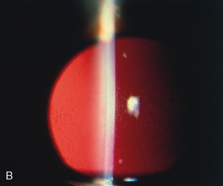
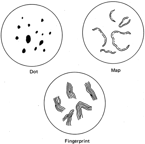
of Peters' anomaly. Eye 12:299, 1998.

60. Ozeki H, Shirai S, Nozaki M et al: Ocular and systemic features of Peters' anomaly. Graefes Arch ClinExp Ophthalmol 238:833, 2000.

61. Streeten B, Karpik A, Spitzer K: Posterior keratoconus associated with systemic abnormalities. Arch Ophthalmol 101:616, 1983.

62. Krachmer J, Rodrigues M: Posterior keratoconus. Arch Ophthalmol 96:1867, 1978.

63. Rao S, Padmanabhan P: Posterior keratoconus. An expanded classification scheme based on corneal topography. Ophthalmology 105:1206, 1998.

64. Alward WL: Axenfeld-Rieger syndrome in the age of molecular genetics. Am J Opthalmol 130:107, 2000.

65. Riise R, Storhaug K, Brondum-Nielsen K: Rieger's syndrome is associated with PAX6 deletion. Acta Ophthalmol Scand 79:201, 2001.

66. Shields MB, Buckley E, Klintworth GK: Axenfeld-Rieger syndrome. A spectrum of developmental disorders. Surv Ophthalmol 29:387, 1985.

67. Burian H, Braley A, Braley AL: Visibility of the ring of Schwalbe and the trabecular zone: An interpretation of the posterior corneal embryotoxon and the so-called congenital hyalin membranes on the posterior corneal surface. Arch Ophthalmol 53:767, 1955.

68. Ozeki H, Shirai S, Majima A et al: Clinical evaluation of posterior embryotoxon in one institution. Jpn J Ophthalmol 41:422, 1997.

69. Loewenstein A: “Knobs” at the periphery of Descemet's membrane. Br J Ophthalmol 34:246, 1950.

70. Shields M, Buckley E, Klintworth G et al: Axenfeld-Rieger syndrome. A spectrum of developmental disorders. Surv Ophthalmol 29:387, 1985.

71. Shields M: Axenfeld-Rieger syndrome: A theory of mechanism and distinctions from the iridocorneal endothelial syndrome. Trans Am Ophthalmol Soc 81:736, 1983.

72. Chisholm I, Chudley A: Autosomal dominant iridogoniodysgenesis with associated somatic anomalies: Four-generation family with Rieger's syndrome. Br J Ophthalmol 67:529, 1983.

73. Schanzlin D, Robin J, Erickson G et al:, Histopathologic and ultrastructural analysis of congenital corneal staphyloma. Am J Ophthalmol 95:506, 1983.

74. Leff S, Shields JAugsburger J et al: Congenital corneal staphyloma: Clinical, radiological and pathologic correlation. Br J Ophthalmol 70:427, 1986.

75. Pallikaris I, Kymionis G, Astyrakakis N: Corneal ectasia induced by laser in situ keratomileusis. J Cataract Refract Surg 27:1796, 2001.

76. Elliott J, Feman S, O'Day D et al: Hereditary sclerocornea. Arch Ophthalmol 103:676, 1985.

77. Waring GO3rd , Rodrigues MM: Ultrastructure and successful keratoplasty of sclerocornea in Mietens' syndrome. Am J Opthalmol 90:469, 1980.

78. Kim T, Cohen EJ, Schnall BM et al: Ultrasound biomicroscopy and histopathology of sclerocornea. Cornea 17:443, 1998.

79. Kasner L, Mietz H, Green WR: Agenesis of Bowman's layer. A histopathological study of four cases. Cornea 12:163, 1993.

80. Moriarty AP, Kerr-Muir MG: Sclerocornea and interstitial deletion of the short arm of chromosome 6—(46XY del[6 [p22, p24). J Pediatr Ophthalmol Strabismus 29:177, 1992.

81. Lindor NM, Michels VV, Hoppe DA et al: Xp22.3 microdeletion syndrome with microphthalmia, sclerocornea, linear skin defects, and congenital heart defects. Am J Med Genet 44:61, 1992.

82. Cox TC, Cox LL, Ballabio A: A very high density microsatellite map (1 STR/41 kb) of 1.7 Mb on Xp22 spanning the microphthalmia with linear skin defects (MLS) syndrome critical region. 7Eur J Hum Genet 6:406, 1998.

83. Henkind P, Marinoff G, Manas A et al: Bilateral corneal dermoids. Am J Ophthalmol 76:972, 1973.

84. Mann I: Developmental Anomalies of the Eye, p 357. Philadelphia, Lippincott, 1957.

85. Sugar H: The oculoauriculo-vertebral syndrome of Goldenhar. Am J Ophthalmol 62:678, 1966.

86. Mandelcorn M, Merin S, Cardarelli J: Goldenhar's syndrome and phocomelia: Case report and etiologic considerations. Am J Ophthalmol 72:618, 1966.

87. Margolis S, Aleksic S, Charles N: Retinal and optic nerve findings in Goldenhar-Gorlin syndrome. Ophthalmology 91:1327, 1984.

88. Jones B: Thygeson's superficial punctate keratitis. Trans Ophthalmol Soc UK 89:22, 1963.

89. Goldberg DB, Scanlon D, Brown S: Management of Thygeson's superficial punctate keratitis. Am J Ophthalmol 89:22, 1980.

90. Goto E, Shimmura S, Shimazai, J et al: Treatment of superior limbic keratoconjunctivitis by application of autologous serum.Cornea 20:807, 2001.

91. Theodore F, Ferry A: Superior limbic keratoconjunctivitis: Clinicopathologic correlations. Arch Ophthalmol 84:481, 1970.

92. Vastine D: Viral Diseases: Adenovirus and miscellaneous viral infections. In Smolin G, Thoft R (eds): Cornea, p 216. Boston, Little, Brown and Company, 1987.

93. Hammer L, Perry H, Donnenfeld E et al: Symblepharon formation in epidemic keratoconjunctivitis. Cornea 4:338, 1990

94. Laibson PR., Dhiri S, Oconer J: Corneal infiltrates in epidemic keratoconjunctivitis. Response to double-blind corticosteroid therapy. Arch Ophthalmol 84:36, 1970.

95. T'ang F, Chang H, Wank K: Studies on the etiology of trachoma with special reference to isolation of viris in the chick embryo. Clini Med J 75:429, 1957.

96. Dawson C, Juster R, Marx R et al: Limbal disease in trachoma and other chlamydial infections: Risk factors for corneal neovascularization. Eye 3:204, 1989.

97. Hidayat A, Risco JM: Amyloidosis of corneal stroma in patients with trachoma. A clinicopathologic study of 62 cases. Ophthalmology 96:203, 1989.

98. Ffytche T: Leprosy: Hansen's disease. In Gold D, Weingeist T (eds): The Eye in Systemic Disease Philadelphia, Lippincott, 1990.

99. Alien J, Byers J: The pathology of leprosy. I. Cornea.Arch Ophthalmol 64:216, 1960.

100. Foulks G, Sanfilippo F, Locascio J: Histocompatibility testing for keratoplasty in high-risk patients. Ophthalmology 90:230, 1893.

101. Passo M, Rosenbaum J: Ocular syphilis in patients with human immunodeficiency virus infection. Am J Ophthalmol 97:281, 1988.

102. Duke-Elder S: Diseases of the outer eye. In Duke-Elder (ed): System of Ophthalmology, pp 236–242. St. Louis, Mosby, 1965.

103. Tamesis R, Foster C: Ocular syphilis.Ophthalmology 97:1281, 1990.

104. Waring GO3rd , Font R, Rodrigues MM et al: Alterations of Descemet's membrane in interstitial keratitis. Am J Ophthalmol. 81:773, 1976.

105. Scattergood K, Breen E, Hirst L: Scrolls of Descemet's membrane in healed syphilitic interstitial keratitis. Ophthalmology 90:1518, 1983.

106. Steere A, Malawista S, Snydman D et al: Lyme arthritis: An epidemic of oligoarticular arthritis in children and adults in three Connecticut communities. Arthritis Rheum 20: 7, 1977.

107. Winterkorn J: Lyme disease: Neurologic and ophthalmic manifestations. Surv Ophthalmol 35:191, 1990.

108. Suresh P, Campbell I, Herzig S et al: Myobacterium keratitis following hyperopic laser in situ keratomileusis. Can J Ophthalmol 36:194, 2001.

109. Lennarson P, Barney N: Interstitial keratitis as presenting ophthalmic sign of sarcoidosis in a child. J Pediatr Ophthalmol Strabismus 32:194, 1995.

110. Basanez M, Boussinesq M: Population biology of human onchocerciasis. Philos Trans R Soc Lond B Biol Sci 354:809, 1999.

111. Hubbard A, Centifanto-Fitzgerald Y: Variability among HSV-1 strains and its importance in disease. In Ninth International Herpes Virus Workshop. 1984. Seattle, 1984.

112. Collin B, Abelson M: Herpes simplex virus in humanArch Ophthalmol 94: 1726, 1976.

113. Green WR, Zimmerman L: Granulomatous reactions to Descemet's membrane. Am J Ophthalmol 64:555, 1967.

114. Naumann G, Gass J, Font R: Histopathology of herpes zoster ophthalmicus. Am J Opthalmol 65:533, 1968.

115. Allen N, Cox C, Cobo M et al: Use of immunosuppressive agents in the treatment of severe ocular and vascular manifestations of Cogan's syndrome. Am J Med 88:296, 1990.

116. Cobo L, Haynes B: Early corneal findings in Cogan's syndrome. Ophthalmology 91:903, 1984.

117. Blaustein B, Gurwood A: Recurrent phlyctenular keratoconjunctivitis: A forme fruse manifestation of rosacea. Optometry 72:179, 2001.

118. Jones D: A plan for antimicrobial therapy. Trans Am Acad Ophthalmol Otolaryngol 79:95, 1975.

119. Musch D, Sugar A, Meyer R: Demographic and predisposing factors in corneal ulceration. Arch Ophthalmol 101:1545, 1983.

120. Pavan-Langston D: Herpetic infections in the cornea. In Smolin G (ed): Cornea, pp 240–254. Boston, Little, Brown and Company, 1987.

121. Font R: Chronic ulcerative keratitis caused by herpes simplex. Arch Ophthalmol 90:382, 1973

122. Ishibashi Y, Kaufman HE: Corneal biopsy in the diagnosis of keratomycosis. Am J Ophthalmol 101:288, 1986.

123. Stehr-Green J, Bailey T, Visvesvara : The epidemiology of Acanthamoeba keratitis in the United States. Am J Ophthalmol 107:331, 1989.

124. Moore M, McCulley J, Kaufman HE et al: Radial keratoneuritis as a presenting sign of Acanthamoeba keratitis. Ophthalmology. 93:1310, 1986.

125. Wilhelmus K, Osato M, Font R: Rapid diagnosis of Acanthamoeba keratitis using Calcofluor white. Arch Ophthalmol 104:1309, 1986.

126. Kuwabara T, Ciccarelli E: Meesmann's corneal dystrophy. A pathological study. Arch Ophthalmol 71:676, 1964.

127. Nakanishi I, Brown SI: Ultrastructure of the epithelial dystrophy of Meesmann. Arch Ophthalmol 93:259, 1975

128. Irvine AD, Corden LD, Swensson O et al: Mutations in cornea-specific keratin K3 or K12 genes cause Meesmann's corneal dystrophy. Nat Genet 16:184, 1997.

129. Corden L, Swensson O, Swensson B et al: Molecular genetics of Meesmann's corneal dystrophy. Ancestral and novel mutations in keratin 12 (K12) and complete sequence of the human KRT12 gene. Exper Eye Res , 7041, 2000.

130. Rodrigues MM, Fine B, Laibson PR et al: Disorders of the corneal epithelium. A clinicopathologic study of dot, geographic and finger print patterns. Arch Ophthalmol 92:475, 1974.

131. Cogan D, Donaldson D, Kuwabara T et al: Microcystic dystrophy of the corneal epithelium. Trans Am Ophthalmol Soc 62:213, 1964.

132. Lisch W, Steuhl KP, Lisch C et al: A new band shaped and whorled microcystic dystrophy of the corneal epithelium. Am J Ophthalmol 114:35, 1992.

133. Cibis G, Tripathi R, Harris D: Mucolipidosis I. Birth Defects 18:359, 1982.

134. Lisch W, Buttner A, Oeffner F et al: Lisch corneal dystrophy is genetically distinct form Meesmann corneal dystrophy and maps to xp22.3. Am J Ophthalmol 130:461, 2000.

135. Grayson M, Wilbrandt H:, Dystrophy of the anterior limiting membrane of the cornea (Reis-Bucklers type). Am J Ophthalmol 61:345, 1966.

136. Kuchle M, Green WH, Volcker H: Reevaluation of corneal dystrophies of Bowman's layer and anterior stroma (Reis-Bucklers and Thiel-Behnke types): A light and electron microscopic study of eight corneas and a review of the literature. Cornea 14:333, 1995.

137. Perry H, Fine B, Caldwell D: Reis-Bucklers' dystrophy. Arch Ophthalmol 97:644, 1979.

138. Munier F, Korvatska E, Djemai A et al: Kerato-epithelin mutations in four 5q31-linked corneal dystrophies. Nat Genet 15:247, 1997.

139. Yee R, Sullivan L, La H et al: Linkage mapping of Thiel-Behnke corneal dystrophy (CDB2) to chromosome 10q23-q24. Genomics 46:152, 1997.

140. Streeten BW, Qi Y, Klintworth GK et al: Immunolocalization of beta ig-h3 protein in 5q31-linked corneal dystrophies and normal corneas. Arch Ophthalmol 117:67, 1999.

141. Kocak-Altintas A, Kocak-Midillioglu I, Alakreu A et al: BIGH3 gene analysis in the differential diagnosis of corneal dystrophies. Cornea 20:64, 2001.

142. Stone E, Mathers W, Rosenwasser G et al: Three autosomal dominant corneal dystrophies map to chromosome 5q. Nat Genet 6:47, 1994.

143. Rodrigues MM, McGavic J: Recurrent corneal granular dystrophy: A clinicopathologic study. Trans Am Ophthalmol Soc 73:306, 1975.

144. Stuart J, Mund M, Iwamoto T et al: Recurrent granular corneal dystrophy. Am J Ophthalmol 79:18, 1975.

145. Herman S, Hughes W: Recurrence of hereditary corneal dystrophy following keratoplasty. Am J Ophthalmol 75:689, 1973.

146. Lanier J, Fine M, Togni B: Lattice corneal dystrophy. Arch Ophthalmol 94:921, 1976.

147. Klintworth GK: Proteins in ocular disease. In GarnerA, Klintworth GK (eds): Pathobiology of Ocular Disease. A Dynamic Approach, pp 999–1000. New York, Marcel Dekker, 1982.

148. Klintworth GK: Advances in the molecular genetics of corneal dystrophies. Am J Ophthalmol 128:747, 1999.

149. Haddad AM, Font R, Fine B: Unusual superficial variant of granular dystrophy of the cornea. Am J Ophthalmol 83:213, 1977

150. Jones S, Zimmerman L: Histopathologic differentiation of granular, macular and lattice dystrophies of the cornea. Am J Ophthalmol 51:394, 1961.

151. Garner A: Histochemistry of corneal granular dystrophy.Br J Ophthalm 53:799, 1969.

152. Brownstein S, Fine B, Sherman M et al: Granular dystrophy of the cornea: Light and electron microscopic confirmation of recurrence in a graft. Am J Ophthalmol 77:701, 1974

153. Iwamoto T, Stuart J, Srinivasan B et al: Ultrastructural variation in granular dystrophy of the cornea.Graefes Archiv Clin Exp Ophthalmol 194:1, 1975.

154. Klintworth GK, Vogel F: Macular corneal dystrophy: An inherited acid mucopolysaccharide storage disease of the corneal fibroblast. Am J Pathol 45:565, 1981.

155. Thonar E, Meyer R, Dennis R et al: Absence of normal keratin sulfate in blood of patients with macular corneal dystrophy. Am J Ophthalmol 102:561, 1986.

156. Donnenfeld E, Cohen EJ, Ingraham H et al: Corneal thinning in macular corneal dystrophy. Am J Ophthalmol 101:112, 1986.

157. Dubord P, Krachmer JH: Diagnosis of early lattice corneal dystrophy. Arch Ophthalmol 100:788, 1982.

158. Stansbury F: Lattice type of hereditary corneal degeneration: A report of five cases, including one of a child of two years. Arch Ophthalmol 40:189, 1948.

159. Meretoja J: Familial systemic paramyloidosis with lattice dystrophy of the cornea, progressive cranial neuropathy, skin changes and various internal symptoms. I. A previously unrecognized heritable syndrome. Ann Clin Res 1:314, 1969.

160. Meretoja J: Comparative histopathological and clinical findings in eyes with lattice corneal dystrophy of two different types. Ophthalmologica 165:15, 1972.

161. Kivela T, Tarkkanen A, Frangione B et al: Ocular amyloid deposition in familial amyloidosis, Finnish: an analysis of native and variant gelsolin in Meretoja's syndrome. Invest Ophthalmol Vis Sci 35:3759, 1994.

162. Seitz, Weidle EG, Naumann G: Unilateral type II (Hida) lattice stromal corneal dystrophy. Klin MonatsblAugenheilk 203:279, 1993.

163. Yanoff M, Fine B, Colosi N et al: Lattice corneal dystrophy.Arch Ophthalmol 95:651, 1977.

164. Freddo T, Polack FM, Leibowitz H: Ultrastructural changes in posterior layers of the cornea in Schnyder's crystalline dystrophy. Cornea 8:170, 1989.

165. Burns R, Connor W, Gipson I: Cholesterol turnover in hereditary corneal dystrophy of Schnyder. Trans Am Ophthalmol Soc 76:184, 1978.

166. Shearman A, Hudson T, Andersen J et al: The gene for Schnyder's crystalline corneal dystrophy maps to human chromosome 1p34. 1-p36. Hum Mol Genet 5:1667, 1996.

167. Kiskadon B, Campbell R, Waller R et al: Fleck dystrophy of the cornea: Case report. Ann Ophthalmol 12:700, 1980.

168. Nicholson D, Green WR, Cross H et al: A clinical and histopathologic study of Francois-Neetens speckled corneal dystrophy. Am J Ophthalmol 83:554, 1977.

169. Witschel H, Fine B, Grutzner P et al: Congenitial hereditary stromal dystrophy of the cornea. Arch Ophthalmol 96:1043, 1978.

170. Krachmer JH, Feder FS, Belin MW: Keratoconus and related noninflammatory corneal thinning disorders. Surv Ophthalmol 1984. 28: p. 293.

171. Rabinowitz W, Maumenee I, Lundergan M et al: Molecular genetic analysis in autosomal dominant keratoconus. Cornea 11:302, 1992.

172. Morrison D, Rosser E, Clouue C: Keratoconus associated with a chromosome 7,11 translocation. Eye 15:556, 2001.

173. Kenyon KR: Ocular manifestations and pathology of systemic mucopolysaccharidoses. Birth Defects 12:133, 1976.

174. Gahl W, Bashan N, Tietze F et al:, Cystine transport is defective in isolated leukocyte lysosomes from patients with cystinosis. Science 217:1263, 1982.

175. Group TCCR: Linkage of the gene for cystinosis to markers on the short arm of chromosome 17. Nat Genet 10:246, 1995.

176. Sanderson P, Kuwabara T, Stark W: Cystinosis: A clinical, histopathologic and ultrastructural study. Arch Ophthalmol 19:270, 1974.

177. Kaiser-Kupfer M, Chan C, Rodrigues MM et al: Nephropathic cystinosis: Immunohistochemical and histologic studies of cornea, conjunctiva and iris. Curr Eye Res 6:617, 1987.

178. Frxier P, Wong V: Cystinosis. Arch Ophthalmol 80:87, 1968.

179. Li Q, Ashraf M, Shen D et al: The role of apoptosis in the pathogenesis of Fuchs' endothelial dystrophy of the cornea. Arch Ophthalmol 119: 1597, 2001.

180. Lipman R, Rubenstein J, Torczynski E: Keratoconus and Fuchs' corneal dystrophy in a patient and her family. Arch Ophthalmol 108:993, 1990.

181. Rodrigues MM, Krachmer JH, Hacket J et al: Fuchs' corneal dystrophy. A clinicopathologic study of the variation in corneal edema. Ophthalmology 93:789, 1986.

182. Eagle RC Jr, Laibson PR, Arentsen J: Epithelial abnormalities in chronic corneal edema: A histopathologic study. Trans Am Ophthalmol Soc 87:107, 1990.

183. Krachmer JH: Posterior polymorphous dystrophy: A disease characterized by epithelial-like cells which influence management and prognosis. Trans Am Ophthalmol Soc 83:413, 1985.

184. Laganowski H, Sherrard E, Uir M et al: Distinguishing features of the iridocorneal endothelial syndrome and posterior polymorphous dystrophy: Value of endothelial specular microscopy. Br J Ophthalmol 175:212, 1991.

185. Judisch GF, Maumenee I: Clinical differentiation of recessive congenital hereditary endothelial dystrophy and dominant hereditary endothelial dystrophy. Am J Ophthalmol 85:606, 1978.

186. Hand C, Harmon D, Kennedy S et al: Localization of the gene for autosomal recessive congenital hereditary endothelial dystrophy (CHED2) to chromosome 20 by homozygosity mapping. Genomics 61:1, 1999.

187. Callaghan M, Hand C, Kennedy S et al: Homozygosity mapping and linkage analysis demonstrate that autosomal recessive congenital hereditary endothelial dystrophy (CHED) and autosomal dominant CHED are genetically distinct. Br J Ophthalmol 83:115.

188. Fraunfelder F, Wright P, Tripathi R: Corneal mucus plaques. Am J Ophthalmol 83:191, 1977.

189. Nelson J, Havener V, Cameron J: Cellulose acetate impressions of the ocular surface. Dry eye states. Arch Ophthalmol 101:1869, 1983.

190. Dodds H Laibson PR: Filamentary keratitis following cataract extraction. Arch Ophthalmol 88:609, 1972.

191. Fox R, Howell F, Bone R: Primary Sjogren's syndrome: Clinical and immunologic features. Semin Arthritis Rheum 14:77, 1984.

192. Daniels T: Salivary histopathology in the diagnosis of Sjogren's syndrome. Scand J Rheumatol 61:36, 1986.

193. Turner K, Pflugfelder S, Ji Z et al: Interleukin-6 levels in the conjunctival epithelium of patients with dry eye disease treated with cyclosporine ophthalmic emulsion. Cornea 19:492, 2000.

194. Sall K, Stevenson O, Mundorf T et al: Two multicenter randomized studies of the efficacy and safety of cyclosporine ophthamic emulsion in moderate to sever dry eye disease. CsA Phase 3 Study Group. Ophthalmology 107:631, 2000.

195. Brown N, Bron A: Recurrent erosion of the cornea.Br J Ophthalmol , 1976. 60:p. 84.

196. Franceschetti A: Hereditare rezidivierende erosion der Hornhaut. Z Augenheilkd 66:309, 1928.

197. Kenyon KR: Recurrent corneal erosion: Pathogenesis and therapy. Int Ophthalmol Clin 19:169, 1979.

198. Judge D, Payant J, Frase S et al: Anterior stromal micropuncture electron microscopic changes in the rabbit cornea. Cornea 9:152, 1990.

199. Goldman J, Dohlman C, Dravit B: The basement membrane of the human cornea in recurrent epithelial erosion syndrome. Trans Am Acad Ophthalmol Otolaryngol 73:471, 1969.

200. Tripathi R, Bron : Ultrastructural study of non-traumatic corneal erosion. Br J Ophthalmol 56:73, 1972.

201. Smith R, Farrell T, Bailey T: Keratomalacia. Surv Ophthalmo 20:213, 1975.

202. Sommer A: Nutritional Blindness: Xerophthalmia and Keratomalacia, Vol 145. New York, Oxford University Press, 1982.

203. Sommer A, Sugana T, Djunaedi E et al: Vitamin A-responsive panocular xerophthalmia in a healthy adult. Arch Ophthalmol 96:1630, 1978.

204. Sommer A, Green WR, Kenyon KR: Clinical-histopathogic correlation in xerophthalmic ulceration and necrosis. Arch Ophthalmol 100:953, 1982.

205. Sommer A, Green WR, Kenyon KR: Bitot's spots responsive and nonresponsive to vitamin A: Clinicopathologic correlations. Arch Ophthalmol 99:2014, 1981.

206. Cogan D, Kuwabara T: Arcus senilis: Its pathology and histochemistry. Arch Ophthalmol 61:353, 1959.

207. Hill J, Maske : Pathogenesis of pterygium. Eye. 3:218, 1989.

208. Ioachim-Velogianni E, Tsironi E, Agnantis E: HLA-DR antigen expression in pterygium epithelial cells and lymphocyte subpopulations: An immunohistochemistry study. Ger J Ophthalmol 4:123, 1995.

209. Guyer D, Barraquer J, McDonnell P et al: Terrien's marginal degeneration: Clinicopathologic case reports.Graefes Arch Clini Exp Ophthalmol 225:19, 1987.

210. Pouliquen Y, Dhermy P, Renard G et al: Terrien's disease: Clinical and ultrastructural studies, five case reports. Eye 3:791, 1989.

211. Soong H, Fitzgerald J, Boruchoff S et al: Corneal hydrops in Terrien's marginal degeneration. Ophthalmology 93:340, 1986.

212. Brown SI: Mooren's ulcers. Histopathology and proteolytic enzymes of adjacent conjunctiva. Br J Ophthalmol 59:670, 1975.

213. Young R, Watson P: Light and electron microscopy of corneal melting syndrome (Mooren's ulcer). Br J Ophthalmol. 66:341, 1982.

214. Zhao J, Jin X: Immunological analysis and treatment of Mooren's ulcer with cyclosporin A applied topically. Cornea 12:481, 1993.

215. Foulks G, Hatchell D, Proia A et al: Histopathology of silicone oil keratopathy in humans. Cornea 10:29, 1991.

216. Binder P, Deg J, Kohl F: Calcific band keratopathy after intraocular chondroitin sulfate. Arch Ophthalmol 105:1243, 1987.

217. Cursino J, Fine B: A histologic study of calcific and non-calcific band keratopathies. Am J Ophthalmol 82:395, 1976.

218. Tabbara K: Climatic droplet keratopathy. Int Ophthalmol Clin 26:63, 1986.

219. Matta C, Tabbara K, Cameron JA et al: Climatic droplet keratopathy with corneal amyloidosis. Ophthalmology 98:192, 1991.

220. Garner A, Morgan G, Tripathi R: Climatic droplet keratopathy. II. Pathologic findings. Arch Ophthalmol 75:799, 1973.

221. Brownstein S, Rodrigues MM, Fine B et al: The elastotic nature of hyaline corneal deposits. Am J Ophthalmol 75:799, 1973.

222. Garner A, Fraunfelder F, Barras T et al: Spheroidal degeneration of cornea and conjunctiva. Br J Ophthalmol 60:473, 1976.

223. Ferry A: A “new” iron line of the superficial cornea: Occurrence in patients with filtering blebs. Arch Ophthalmol 79:142, 1968.

224. Rodman R, Burnstine M, Esmaeli B et al: Wilson's disease: Presymptomatic patients with Kayser-Fleischer rings. Ophthalmic Genet 18:79, 1997.

225. Ferry A, Zimmerman L: Black cornea: A complication of topical use of epinephrine. Am J Ophthalmol 58:205, 1964.

226. Calkins L: Corneal epithelial changes occurring during chloroquine (Aralyn) therapy. Arch Ophthalmol 60:981, 1958.

227. Slowik C, Samodi S, von Gruben C et al: Detection of morphological corneal changes caused by chloroquine thereapy using confocal in vivo microscopy. Ophthalmologe 94:549, 1997.

228. D'Amico DJ, Kenyon KR, Ruskin JN: Amiodarone keratopathy: Drug-induced lipid storage disease. Arch Ophthalmol 99:257, 1981.

229. Ciancaglini M, Carpineto P, Zuppardi E, Nubile M et al: In vivo confocal microscopy of patients with amiodarone-induced keratopathy. Cornea 20:368, 2001.

230. Mantyjarvi M, Tuppurainen K, Ikaheimo K: Ocular side effects of amiodarone. Surv Ophthalmol 42:360, 1998.

231. Dolan BJ, Flach AJ, Peterson JS: Amiodarone keratopathy and lens opacities. J Am Optom Assoc 56:468, 1985.

232. Pollak PT: Clinical organ toxicity of antiarrhythmic compounds: Ocular and pulmonary manifestations. Am J Cardiol 84:37R, 1999.

233. Macaluso DC, Shults WT, Fraunfelder FT: Features of amiodarone-induced optic neuropathy. Am J Opthalmol 127:610, 1999.

234. Fine B, Yanoff M: Ocular Histology: A Text and Atlas, pp 159–165, Vol 168, New York, Harper & Row, 1972.

235. Hamanaka T: Scleral spur and ciliary muscle in man and monkey. Jpn J Ophthalmol 33:221, 1989.

236. Dutton J, Anderson R, Schelper R et al: Orbital malignant melanoma and oculodermal melanocytosis: Report of two cases and review of the literature. Ophthalmology 91:497, 1984.

237. Sugar H, Ilgren E, Adams C: Nevus of Ota associated with meningeal melanosis and intracranial melanoma. Case report. J Neurosurg 58:280, 1983.

238. Mansour AM, Barber JC, Reinecke RD et al: Ocular choristomas. Surv Ophthalmol 33:330, 1989.

239. Hered R, Hiles D: Epibulbar osseous choristoma and ectopic lacrimal gland underlying a dermolipoma. J Pediatr Ophthalmol Strabismus 24:255, 1987.

240. Conway V, Brownstein S, Chisholm I: Lacrimal gland choristoma of the ciliary body. Ophthalmology 92:449, 1985.

241. Pittke E, Marquardt R, Mohr W: Cartilage choristoma of the eye. Arch Ophthalmol 101:1569, 1983.

242. Shields JA, Shields C, Eagle RC Jr et al: Ocular manifestations of the organoid nevus syndrome. Ophthalmology 104:549, 1997.

243. Khalil M: Subhyaloid hemorrhage in osteogenesis imperfecta tarda. Can J Ophthalmol 18:251, 1983.

244. Howard F: Lax ligament syndrome in children associated with blue sclera and bat ears. Br J Gen Pract 40:233, 1990.

245. Zlotogora J, BenEzra D, Cohen T et al: Syndrome brittle cornea, blue sclera, and joint hyperextensibility. Am J Med Genet 36:269, 1990.

246. Pouliquen Y, Dhermy P, Espinasse MA et al: Keratoglobe. J Fr Ophtalmol 8:43, 1985.

247. Zimmerman L, Fischer R, Winterhalter K et al: Comparative studies of collagens in normal and keratoconus corneas. Exp Eye Res 46:431, 1988.

248. Sussman M, Kelly T, Rosenbaum K et al: Abnormality of cartilage collagen in a patient with unclassified chondrodystrophy. J Orthop Res 2:339, 1984.

249. Roxburgh S: Atypical retinitis pigmentosa with hypophosphatasia. Trans Ophthalmol Soc UK 103:513, 1983.

250. Chan C, Green WR, de la Cruz Z et al: Ocular findings in osteogenesis imperfecta congenita. Arch Ophthalmol 100:1459, 1982.

251. Mori S, Komatsu H, Watari H: Spontaneous posterior bulbar perforation of congenital scleral coloboma and its surgical treatment: A case report. Ophthalmic Surg 16:433, 1985.

252. Gass J: Uveal effusion syndrome: A new hypothesis concerning pathogenesis and technique of surgical treatment. Trans Am Ophthalmol Soc 81:246, 1983.

253. Brockhurst R: Nanophthalmos with uveal effusion. Trans Am Ophthalmol Soc 72:371, 1974.

254. Hotz F, Boehmer H, Mechtersheimer G et al: Uveal non-Hodgkin's lymphoma with epibulbar extension simulating choroidal effusion syndrome. Retina 19:343, 1999.

255. Donoso L, Shields JA, Nagy R: Epibulbar lesions simulating extraocular extension of uveal melanomas. Ann Ophthalmol 14:120, 1982.

256. Manschot W: Senile scleral plaques and senile scleromalacia. Br J Ophthalmol 62:376, 1978.

257. Carroll C, Peyman G, Raichand M: Surgical management of senile scleromalacia. Ophthalmic Surg 11:719, 1980.

258. Klintworth GK: Radiographic abnormalities in eyes with retinoblastoma and other disorders. Br J Ophthalmol 62:365, 1978.

259. Friedman E, Ivry M, Ebert E et al: Increased scleral rigidity and age-related macular degeneration. Ophthalmology 96:104, 1989.

260. Norn T: Topography of scleral emissaries and sclera-perforating blood vessels. Acta Ophthalmol (Copenh) 63:320, 1985.

261. Pollak M, Chou Y, Cerda J et al: Homozygosity mapping of the gene for alkaptonuria to chromosome 3q2. Nat Genet 5:201, 1993.

262. Daicker B, Riede U: Histological and ultrastructural findings in alkaptonuric ocular ochronosis. Ophthalmologica 69:277, 1974.

263. Kampik A, Sani J, Green WR: Ocular ochronosis. Clinicopathological, histochemical, and ultrastructural studies. Arch Ophthalmol 98:1441, 1980.

264. Gaines J: The pathology of alkaptonuric chronosis. Hum Pathol 20:40, 1989.

265. Phinney R, Mondino B, Abrahim A: Corneal icterus resulting from stroma bilirubin deposition. Ophthalmology 96:1212, 1989.

266. Lascari A: Carotenemia. A review. Clin Pediatr 20:25, 1981.

267. Congdon P, Kelleher J, Edwards P et al: Benign carotenaemia in children. Arch Dis Child 56:292, 1981.

268. Angeloni V, Salasche S, Ortiz R: Nail, skin, and scleral pigmentation induced by minocycline. Cutis 40:229, 1987.

269. Henderson R, Lander R: Scleral discoloration associated with long-term prednisone administration. Cutis 34:76, 1984.

270. Watson P, Young R: Changes at the periphery of a lesion in necrotizing scleritis: Anterior segment fluorescein angiography correlated with electron microsocpy. Br J Ophthalmol 69:656, 1985.

271. Flach A, Lavoie P: Episcleritis, conjunctivitis, and keratitis as ocular manifestations of Lyme disease. Ophthalmology 97:973, 1990.

272. Zaidman GW: Episcleritis and symblepharon associated with Lyme keratitis. Am J Ophthalmol 109:487, 1990.

273. McCluskey P, Wakefield D, Penny R: Scleritis and the spectrum of external inflammatory eye disease. Aust N Z J Ophthalmol 13:159, 1985.

274. Kleiner R, Raber I, Passero F: Scleritis, pericarditis, and aortic insufficiency in a patient with rheumatoid arthritis. Ophthalmology 91:941, 1984.

275. Robertson D, Winkelman R: Ophthalmic features of necrobiotic xantogranuloma with paraproteinemia. Am J Ophthalmol 97:173, 1984.

276. Lyne A, Pitkeathley D: Episcleritis and scleritis: Association with connective tissue disease. Arch Ophthalmol 80:171, 1968.

277. McGavin D, Williamson J, Forrester J: Episcleritis and scleritis: A study of their clinical manifestations and association with rheumatoid arthritis. Br J Ophthalmol 60:192, 1976.

278. Wilhelmus K, Yokoyama C: Syphilitic episcleritis and scleritis. Am J Ophthalmol 104:595, 1987.

279. Verhoeff F, King M: Scleromalacia perforans: A report of a case in which the eye was examined microscopically. Arch Ophthalmol 20:1013, 1938.

280. Ashton N, Hobbs H: Effect of cortisone on rheumatoid nodules of the sclera (scleromalacia perforans). Br J Ophthalmol 36:373, 1976.

281. Ferry A: Histopathology of rheumatoid episcleral nodules: An extraarticular manifestation of rheumatoid arthritis. Arch Ophthalmol 82:77, 1969.

282. Fraunfelder F, Watson P: Evaluation of eyes enucleated for scleritis. Br J Ophthalmol 60:227, 1976.

283. Lebowitz M, Jakobeic F, Donnenfeld E et al: Bilateral epibulbar rheumatoid nodulosis. A new ocular entity. Ophthalmology 95:256, 1988.

284. Lindenmuth K, Sugar A, Kincaid M et al: Invasive squamous cell carcinoma of the conjunctiva presenting as necrotizing scleritis and scleral perforation and uveal prolapse. Surv Ophthalmol 33:50, 1988.

285. Kim R, Seiff S, Howes E et al: Necrotizing scleritis secondary to squamous cell carcinoma in acquired immunodeficiency syndrome. Am J Ophthalmol 109:231, 1990.

286. Kaufman L, Folk E, Miller M et al: Necrotizing scleritis following strabismus surgery for thyroid ophthalmopathy. J Pediatr Ophthalmol Strabismus 26:236, 1989.

287. Young R, Watson P: Microscopical studies of necrotizing scleritis. II. Collagen degradation in the scleral stroma. Br J Ophthalmol 68:781, 1984.

288. Rao N, Marak G, Hidayat A: Necrotizing scleritis. A clinicopathologic study of 41 cases. Ophthalmology 92:1542, 1985.

289. Young R, Watson P: Microscopical studies of necrotizing scleritis. I. Cellular aspects. Br J Ophthalmol 68:770, 1984.

290. Mamalis N, Johnson M, Haines J et al: Corneal-scleral melt in association with cataract surgrey and intraocular lenses: A report of four cases. J Cataract Refract Surg 16:108, 1990.

291. Finger P, Perry H, Packer S et al: Posterior scleritis, as an intraocular tumor. Br J Ophthalmol 74:21, 1990.

292. Benson W: Posterior scleritis. Surv Ophthalmol 32:297, 1988.

293. Singh G, Guthoff R, Foster C: Observations on long-term follow-up of posterior sceleritis. Am J Ophthalmol 101:570, 1986.

294. Bernauer N, Buchi E, Daicker B: Immunopathological findings in posterior scleritis. Int Ophthalmol 18:229, 1994.

295. Shields C, Shields JA, Varenhorst M: Transscleral leiomyoma. Ophthalmology 98:84, 1991.

Back to Top