|
|
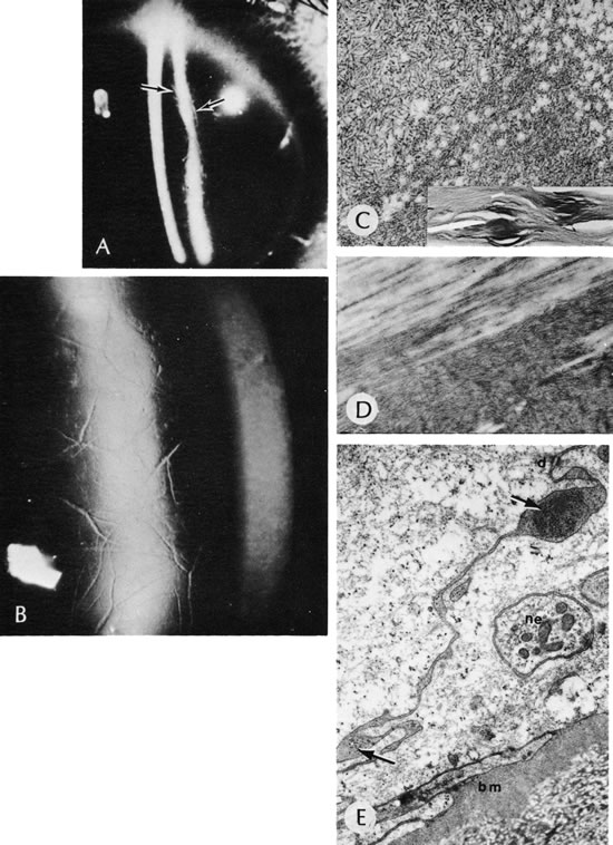
| Fig. 34. Lattice corneal dystrophy. A. Translucent branching lines of lattice corneal dystrophy are seen best by retroillumination (arrows). B. Appearance of the lattice network in the cornea. C. Hyaline lesions seen by light microscopy (inset) are composed of myriad individual filaments either in disarray (as in main figure), and therefore non-birefringent, or highly aligned (as in D), and therefore birefringent. E. Nonspecific alterations in the overlying epithelium. Note the loss of basal cell hemidesmosomes, accumulation of an abnormal quantity of thick homogeneous basement membrane (bm), and apparently similar material between the adjacent basal cells (arrows). d, desmosome; ne, intraepithelial neurite.(Courtesy of SEI Photoarchives.) |