|
|
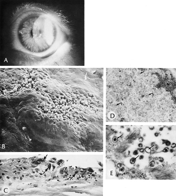
| Fig. 20. Herpes simplex. A. Typical dendritic ulcer. B. Scanning electron micrograph of a dendritic ulcer in the epithelium of a rabbit cornea. C. Many intranuclear inclusions (arrows) are present in the corneal epithelium near the edge of the ulcer. D. Virus particles (arrows) of herpes simplex are present in the nucleus. E. Virus particles also are present within the cytoplasm. Note the large size of the cytoplasmic virions. Some particles show empty capsids, whereas others are complete, containing nucleoids. (Courtesy of SEI Photoarchives.) (B Courtesy of Dr. R. C. Eagle Jr; C from Font RL: Chronic ulcerative keratitis caused by herpes simplex virus. Arch Ophthalmol 90:382, 1973.) |