|
|
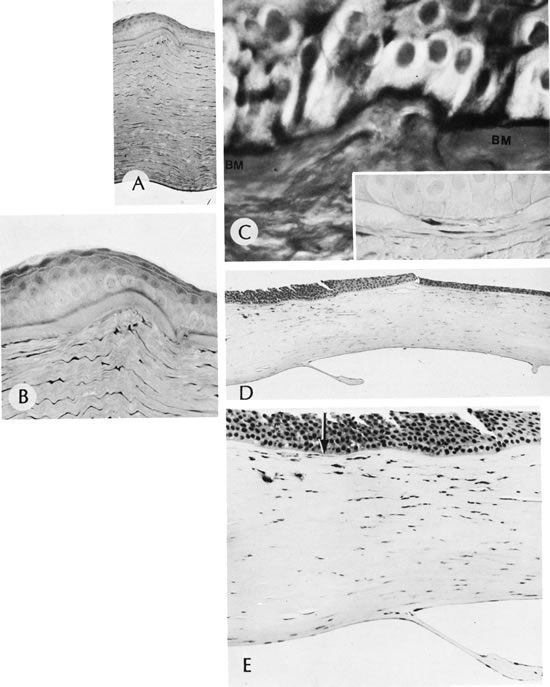
| Fig. 38. Keratoconus. A. Early changes consist of small breaks in Bowman's membrane and some irregularity of the nearby stromal lamellae. B. High magnification shows breaks in Bowman's membrane and stromal irregularity. C. Focal disruption of Bowman's membrane (BM) and accumulation along PAS-positive material beneath the epithelium. Inset shows focal destruction of Bowman's membrane replaced by cellular tissue. D. Late changes show disruption of Bowman's membrane, stromal scaring, and thinning and breaks in Descemet's membrane. E. High magnification shows an absence of Bowman's membrane (arrow), dense scarring of the thinned stroma, and a break in Descemet's membrane. (Courtesy of SEI Photoarchives.) (A–C modified from McTigue JW: The human cornea: A light and electron microscopic study of the normal cornea and its alterations in various dystrophies. Trans Am Ophthalmol Soc 65:591, 1967.) |