|
|
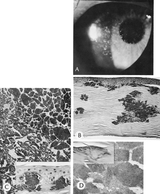
| Fig. 30. Granular dystrophy. A. Note the clear cornea between the stromal granules. B. Granules stain deeply with hematoxylin-eosin stain. C. Granules seen by light microscopy (inset) are shown by electron microscopy to consist of dense granules. Many granules are “apertured.” D. Close relationship of dense and apertured granules to packed, “folded” macromolecules (“filaments”). The latter are believed to be precursors of granule formation. Inset shows Congo red positivity at the periphery of the granule. (Courtesy of SEI Photoarchives.) |