|
|
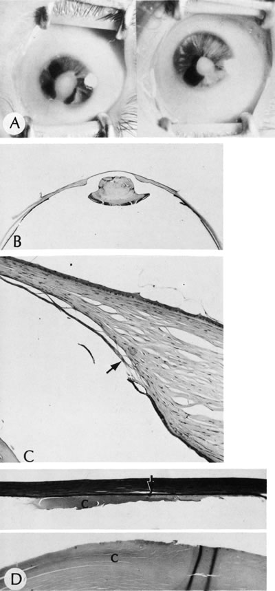
| Fig. 4. Peters' anomaly. A. Note the central corneal scar in the right and left eyes. The lens was adherent to the back of the corneal scar. Iris abnormalities also were present. B. The anterior segment shows a posterior corneal defect, a “top hat” appearance of the lens, and total adherence of the anterior surface of the iris to the cornea. C. High magnification shows termination of the endothelium and Descemet's membrane (arrow), corneal thinning, and localized absence of Bowman's membrane. The lens (lower left) is artifactually separated from the cornea. D. A PAS-positive membrane (lens capsule) is shown (top) adherent to the posterior corneal surface (arrow). The lens cortex (c) is artifactually separated from the rest of the lens (bottom). (Courtesy of SEI Photoarchives.) (B–D modified from Scheie HG, Yanoff M: Peter's anomaly and total posterior coloboma of retinal pigment epithelium and choroid. Arch Ophthalmol 87:525, 1972.) |