 |
 |
 |
 |
|
||||
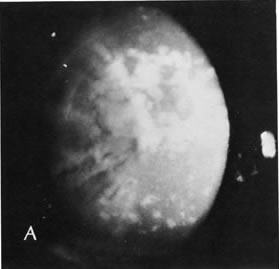
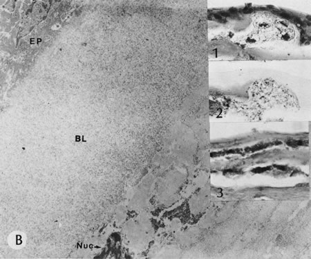
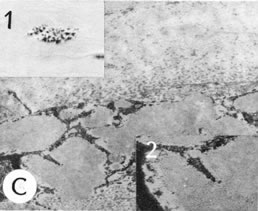
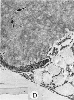
| Fig. 32. Macular dystrophy. A. Clinical appearance. No clear stroma is present between the opacities. B. A keratocyte beneath Bowman's layer (BL) is filled with vesicles containing acid mucopolysaccharide– positive (AMP) substance. There is a cluster of vacuolated cells beneath the epithelium (inset 1). The vacuoles are filled with AMP, stain blue with AMP stain (inset 2), and stain positive with PAS (inset 3). EP, epithelium; Nuc, keratocyte nucleus. (Courtesy of SEI Photoarchives.) |