|
|
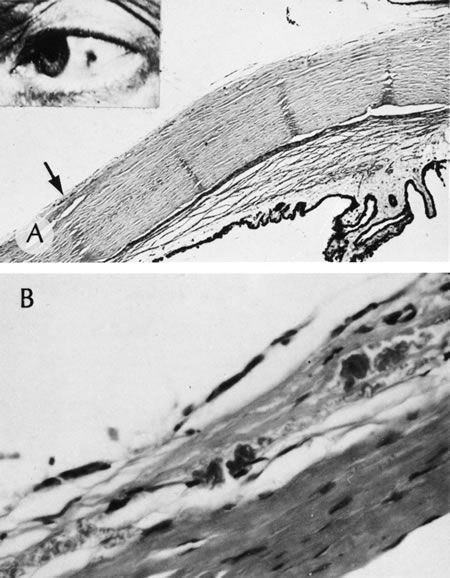
| Fig. 53. Ochronosis. A. Homogentisic acid deposit (arrow). B. Homogentisic cid deposit shown in high magnification. Note the typical “curlicues” within the superficial sclera and episclera. The clinical appearance of a pigmented spot of homogentisic acid deposition is shown in the inset. (Courtesy of SEI Photoarchives.) |