|
|
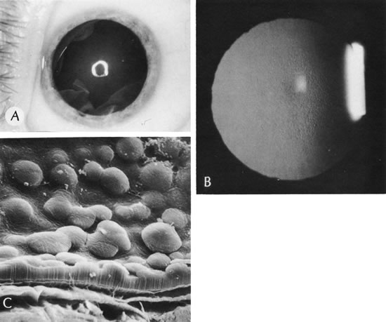
| Fig. 41. Cornea guttata. A. The clinical appearance with distortion of light reflex and central corneal haze. B. The fundus reflex shows the typical appearance of cornea guttata. C. Scanning electron micrograph shows a mushroom or anvil shape of the excrescences. The cut surface of Descemet's membrane is seen below. (Courtesy of SEI Photoarchives.) |