|
|
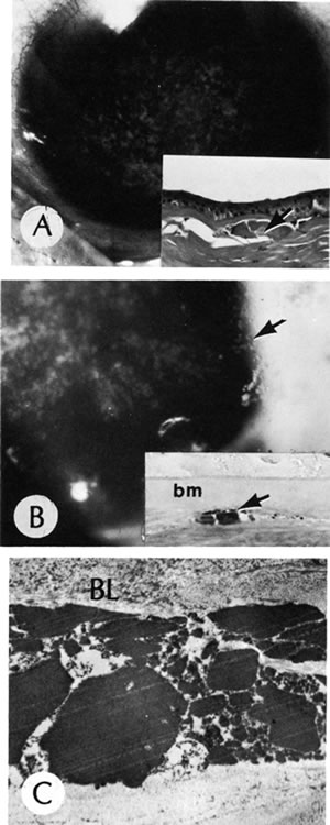
| Fig. 31. Granular dystrophy. A. Clinical appearance. Biopsy (inset) shows the presence of a typical stromal granule (arrow). B. Granular dystrophy recurred in full-thickness grafts several years later. The granules (arrow) are seen by side illumination. Biopsy (inset) shows a new lesion within the graft. A typical collection of granular material (arrow) lies within the stroma beneath Bowman's membrane (bm).C. These granules from a biopsy are typical of granular dystrophy. BL, Bowman's layer. (Courtesy of SEI Photoarchives.) (Brownstein S, Fine BS, Sherman ME et al: Granular dystrophy of the cornea—light and electron microscopic confirmation of recurrence in a graft. Am J Ophthalmol 77:701, 1974.) |