|
|
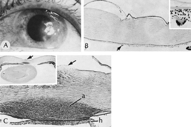
| Fig. 21. Herpes simplex. A. Clinical appearance of bullous keratopathy. B. Chronic condition shows development of bullous keratopathy. The anterior chamber inflammatory reaction contains multinucleated inflammatory giant cells (arrow), shown under high magnification in inset. C. Ulcerated bullous keratopathy (arrows). A corneal abscess (a) and hypopyon (h) are present. Note (inset) the subluxation of the lens to left, caused by the loss of zonula-lens attachments on the right, resulting in a “blunted” appearance of the right side of the lens. (Courtesy of SEI Photoarchives.) |