|
|
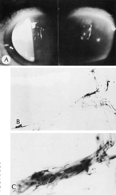
| Fig. 44. Filamentary keratitis. A. Clinical appearance of ropy secretions in the white beam (left) and blue beam (right—after fluorescein) of a slit lamp. B. Smear of ropy secretions from a patient who had keratitis sicca and filamentary keratitis. C. High magnification shows the ropy secretions composed of epithelial cells and mucus. (Courtesy of SEI Photoarchives.) |