Chapter 113C
Fluorescein Angiography in Retinal Vascular Diseases
JOSHUA D. STEIN , GARY C. BROWN and ALLEN C. HO
Main Menu   Table Of Contents

Search

RETINAL ARTERIAL OBSTRUCTIVE DISEASE
RETINAL VENOUS OBSTRUCTIVE DISEASE
OTHER RETINAL VASCULAR ABNORMALITIES
REFERENCES

This section describes the fluorescein angiographic features of retinal arterial obstructive disease, retinal venous obstructive disease, and other abnormalities of the retinal vasculature.
Back to Top
RETINAL ARTERIAL OBSTRUCTIVE DISEASE
The retinal vasculature can be affected by obstructive disease within the large arteries, such as the carotids, the ophthalmic artery, the central retinal artery, and the smaller retinal arteries. Starting with the larger vessels and progressing to the smaller ones, these disturbances are illustrated in the following discussion.

OCULAR ISCHEMIC SYNDROME

The ocular ischemic syndrome is the conglomeration of ocular symptoms and signs attributable to severe carotid artery obstructive disease.1 Rarely, it can be caused by obstruction of the innominate artery or chronic ophthalmic artery obstruction. Fluorescein angiographic signs of the ocular ischemic syndrome are listed in Table 1.


Table 1. Fluorescein Angiographic Signs of the Ocular Ischemic Syndrome


SignIncidence (%)
Delay, patchy choroidal filling60
Increased arteriovenous transit time95
Retinal vascular staining85
Macular edema17
(From Brown GC, Magargal LE: The ocular ischemic syndrome: Clinical, fluorescein angiographic and carotid angiographic features. Int Ophthalmol 11:239, 1988)


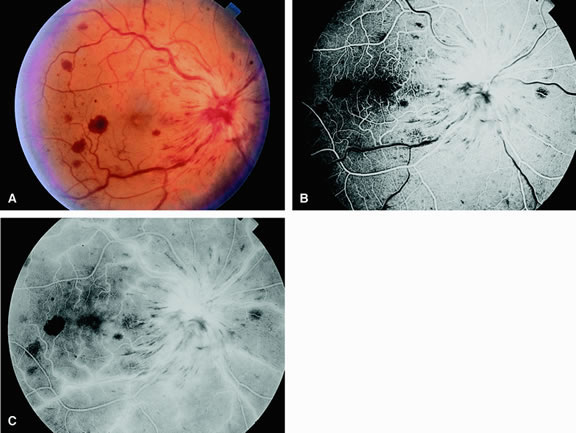
Normally, the choriocapillaris is completely filled with fluorescein dye within 5 seconds after the first appearance of dye within it. In eyes with the ocular ischemic syndrome, this filling can be delayed in extreme cases for 1 minute or longer. The posterior choroid is supplied by the temporal and nasal posterior ciliary arteries.2 In some normal eyes, but particularly in eyes with the ocular ischemic syndrome, delayed, asymmetric filling of the areas supplied by these vessels can be seen (Fig. 1).

Fig. 1. A. Fundus of a patient with the ocular ischemic syndrome. The retinal arteries appear narrowed with scattered intraretinal hemorrhages. Myelinated nerve fibers extend from the inferior margin of the disc. B. Fluorescein angiography of A demonstrates patchy choroidal filling within the peripapillary area and the presence of an abnormal leading edge of dye within a retinal artery.

Filling of the retinal arteries is often also delayed in eyes with the ocular ischemic syndrome, and this manifests as a delayed arm-to-retina circulation time. Although the retinal arteries usually start to fill within 15 seconds after an antecubital intravenous injection of sodium fluorescein dye, this time range varies according to several factors, including the site of injection, the rate of injection, and body circulation. A visible leading edge of dye (see Fig. 1) within a retinal artery is almost always abnormal after an intravenous injection and indicates diminished flow. In the Retina Vascular Unit at Wills Eye Hospital, Philadelphia, the upper-normal limit for retinal arteriovenous transit time (time from the first appearance of dye in the temporal retinal arteries of the arcades to the time when the corresponding veins are completely filled) is considered to be 10 to 11 seconds. The retinal arteriovenous transit time is usually prolonged in eyes with the ocular ischemic syndrome; in fact, this prolongation of time is the most common fluorescein angiographic feature in eyes with the ocular ischemic syndrome.

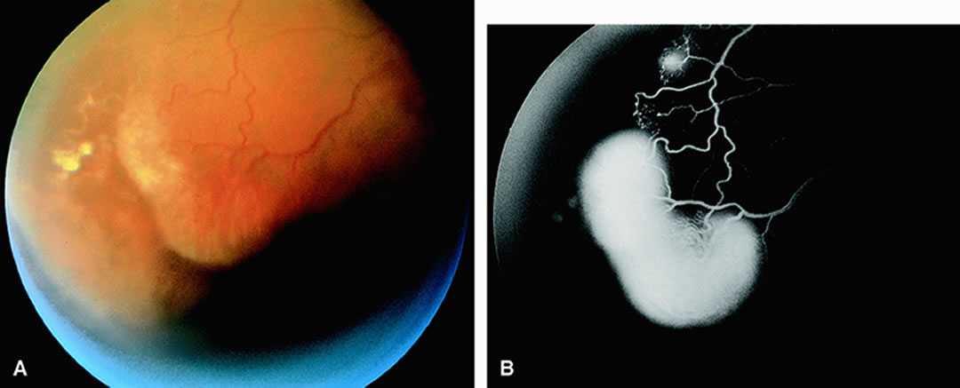
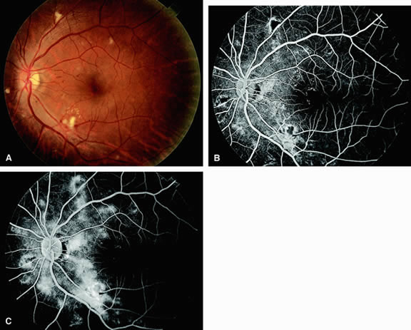
Leakage of fluorescein dye from the retinal vessels, particularly the arteries, occurs in 85% of eyes with the ocular ischemic syndrome (Fig. 2). Presumably, hypoxia and subsequent endothelial cell damage cause this leakage of dye. Leakage of fluorescein dye from the retinal vessels can be seen in the posterior pole and periphery. This hyperpermeability, combined with leakage of serum from microaneurysmal abnormalities, appears to account for the macular edema observed in some eyes with the ocular ischemic syndrome (see Fig. 2).

Fig. 2. A. Narrowing of the retinal arterioles and neovascularization of the optic disc in a patient with the ocular ischemic syndrome. B. Fluorescein angiogram of A shows a hyperfluorescent optic disc caused by neovascularization and leakage of dye. Dilated and telangiectatic capillaries can be observed temporal to the macula. C. As the study progresses, leakage of dye from the capillaries results in retinal edema. The optic disc remains significantly hyperfluorescent as well. D. At the end of the study, diffuse leakage of dye clouds the retina and underlying choroid.

Leakage of fluorescein dye from neovascularization of the disc is seen in approximately one third of eyes with the ocular ischemic syndrome (see Fig. 2). Hyperfluorescence resulting from leaking neovascularization of the retina is less common. Retinal capillary nonperfusion is visible on fluorescein angiography in some cases. Ischemic optic neuropathy is rarely observed. Iris neovascularization is found in approximately two thirds of cases at the time the diagnosis is made.

Fluorescein angiography is helpful in differentiating the ocular ischemic syndrome from conditions that can mimic it, including central retinal artery obstruction, mild central retinal vein obstruction, and diabetic retinopathy. Of these conditions, only the ocular ischemic syndrome has delayed choroidal filling present. Moreover, late staining of the retinal arteries is unusual with the other conditions. An increased arteriovenous transit time is usually present in eyes with the ocular ischemic syndrome but can also be seen in eyes with central retinal artery or vein obstruction and eyes with diabetic retinopathy and nonperfusion of the retinal capillary bed.

OPHTHALMIC ARTERY OBSTRUCTION

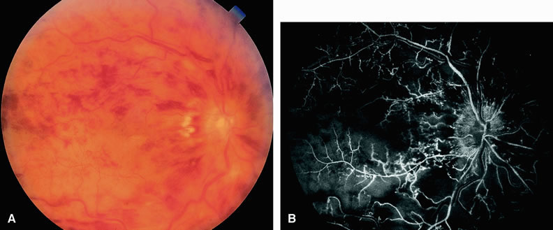
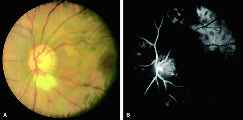
Clinically, acute ophthalmic artery obstructions differ from acute central retinal artery obstructions in that persons with the former often have no light perception and the retinal whitening appears more intense on examination.3 A cherry-red spot is often absent in eyes with acute ophthalmic artery obstruction, but its presence does not rule out the diagnosis (Fig. 3A). Electroretinography often reveals diminished amplitudes of both b- and a-waves that are caused by inner and outer retinal ischemia, respectively.3 In contrast, with a central retinal artery obstruction alone, the a-wave amplitude is usually normal and the b-wave amplitude is often diminished because of inner retinal ischemia.4

Fig. 3. A. Acute ophthalmic artery obstruction occurring secondary to a knife injury that severed the retinal and choroidal vessels posterior to the globe. Intense retinal whitening can be seen; a cherry-red spot is absent. B. Fluorescein angiogram of A taken at 55 seconds after injection reveals an absence of dye within the retina and most of the choroid. Mild peripapillary hyperfluorescence can be seen, presumably resulting from anastomoses between the episcleral, pial, and choroidal vessels in the vicinity of the optic disc. C. Increased peripapillary hyperfluorescence at 10.5 minutes after injection. (Brown GC, Magargal LE: Sudden occlusion of the retinal and posterior ciliary circulations in a youth. Am J Ophthalmol 88:690, 1979)

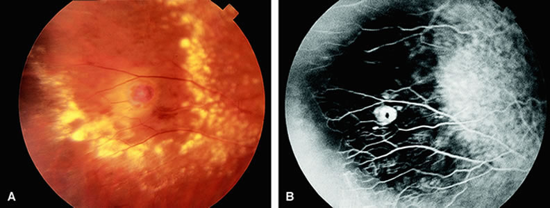
Fluorescein angiography of eyes with acute ophthalmic artery obstruction shows delayed filling of the retinal vessels and usually the choroidal vessels as well (see Fig. 3B and C). Focal, pinpoint areas of staining resulting from leakage of dye at the level of the retinal pigment epithelium can be seen in some instances. Diffuse staining is also occasionally observed. Prominent staining of the retinal vessels is usually absent with acute ophthalmic artery obstruction, although it can be seen with chronic ophthalmic artery obstruction.

RETINAL ARTERY OBSTRUCTION

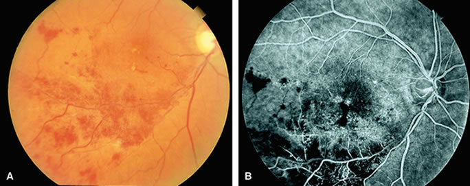
With acute central retinal artery obstruction, filling of the choroid is usually normal. Filling of the retinal arteries is often delayed, and in severe cases, a leading edge of dye can be seen (Fig. 4). A delay in retinal arteriovenous transit time is often noted.5 Box-carring or segmentation of the dye column can be seen in both the retinal arteries and veins when the obstruction is marked. In some cases, the flow appears normal because reperfusion of the blocked artery can occur fairly rapidly.6 Intraretinal leakage of dye in the late phases of the study, in a pattern consistent with macular edema, is generally not seen in eyes with acute central retinal artery obstruction. Fluorescein angiography can help identify eyes with acute central retinal artery obstruction in instances when the retinal whitening is subtle and the diagnosis is in question.

Fig. 4. A. Patient with a central retinal artery occlusion and neovascularization of the iris. Note the diffuse pallor of the retina. The retinal arteries appear narrow and poorly perfused with areas of box carring of the luminal blood columns. The classic cherry-red spot can be observed in the region of the macula. B. Fluorescein angiogram of A shows perfusion of the underlying choroid, and a leading edge of dye is present in the retinal arteries.

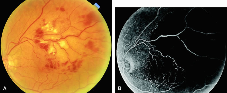
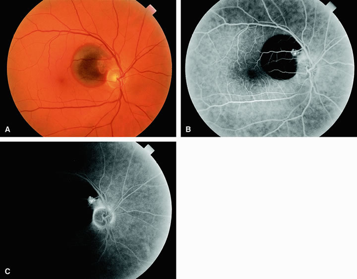
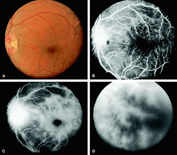
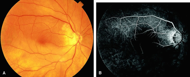
Approximately 10% of eyes with acute central retinal artery obstruction have a cilioretinal artery that supplies the retina in the papillomacular bundle and extends into the foveola.7 In more than 80% of these eyes, the visual acuity eventually improves to 20/50 or better. Fluorescein angiography typically shows earlier filling within the patent cilioretinal artery and the veins draining the area that it supplies compared with the filling of the remainder of the retina, which is supplied by the central retinal artery (Figs. 5 and 6).

Fig. 5. A. Fundus of a patient with occlusion of the central retinal artery with cilioretinal sparing. Most of the retina is pale and ischemic, and there is narrowing of the retinal arterioles and retinal edema. The cilioretinal artery distribution appears well perfused. B. Fluorescein angiogram of A shows perfusion of the cilioretinal artery and underlying choroid. Retrograde filling of the retinal arterioles inferiorly can be observed. The other vessels appear hypofluorescent as a result of nonperfusion.

Fig. 6. A. Fundus of a patient with a central retinal artery occlusion and sparing of the cilioretinal artery. Note that the arteries appear narrow and attenuated. The region of the retina from the optic disc to the macula appears well perfused. The rest of the retina appears pale and ischemic. B. Fluorescein angiogram of A, which shows fluorescence in the distribution of the cilioretinal artery and underlying choroid. The rest of the retinal vessels appear hypofluorescent.

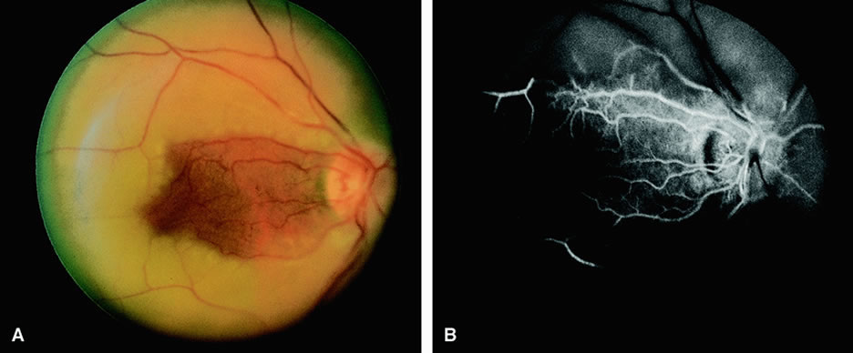
Fluorescein angiography of eyes with marked, acute branch retinal artery obstruction reveals a lack of filling of the retinal capillary bed within the distribution of the involved vessel. Retinal veins that normally drain the damaged area also demonstrate a delay in filling. With severe blockage, retrograde filling can be seen in the distal aspect of an obstructed branch retinal artery (Fig. 7).

Fig. 7. A. Patient with an inferotemporal branch retinal artery obstruction. The retina shows pallor in the distribution of the affected vessel. The other vessels appear to perfuse well. The area of pallor involves the inferior macula. B. Fluorescein angiogram of A shows retrograde filling of the affected vessel distally and retinal edema in the region of the occluded vessel.

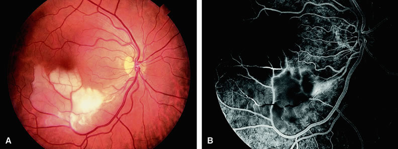
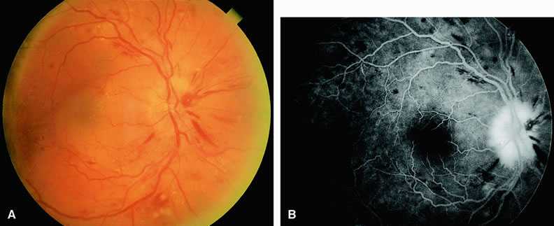
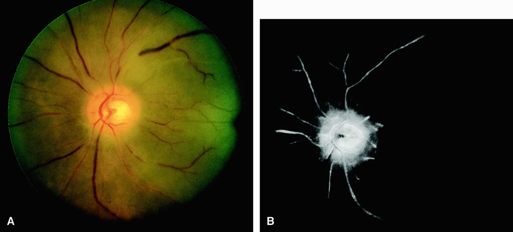
Cilioretinal artery obstruction is similar to branch retinal artery obstruction except that the former vessel usually emanates from the edge of the optic disc (Fig. 8). Cilioretinal artery obstructions can be seen as isolated fundus abnormalities or in association with central retinal vein obstruction or acute anterior ischemic optic neuropathy.8

Fig. 8. A. Fundus of an older adult patient with cilioretinal artery occlusion. The retina temporal to the optic disc corresponding to the distribution of the cilioretinal artery is edematous and white from nonperfusion. Moreover, the macula demonstrates drusen. B. Fluorescein angiogram of A demonstrates a hypofluorescent area in the distribution of the cilioretinal artery. The hypofluorescence is due to retinal artery and capillary nonperfusion.

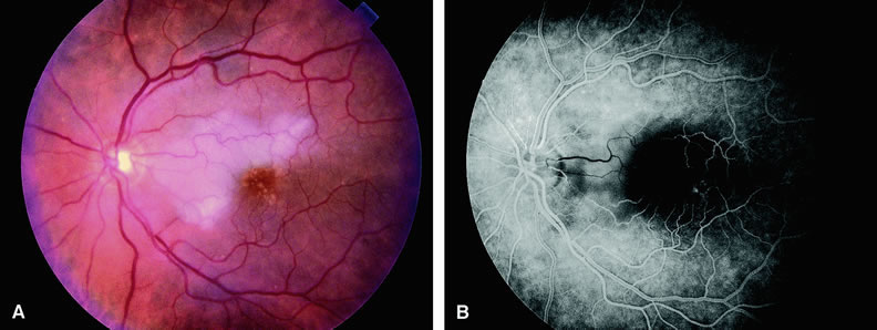
Cotton-wool spots are small areas of superficial retinal whitening that usually develop secondary to obstruction of axoplasmic flow caused by areas of focal retinal ischemia.9 Fluorescein angiography in these cases usually demonstrates relative hypofluorescence in the early and middle phase of the study (Fig. 9). Late staining of the cotton-wool spot can occur. As shown in Table 2, the differential diagnosis of cotton-wool spots is extensive; however, cotton-wool spots are most commonly observed in the setting of diabetic retinopathy, hypertensive retinopathy, collagen vascular disease, or hematologic abnormalities.

Fig. 9. A. Cotton-wool spots in the superior temporal arcade of a diabetic patient with nonproliferative retinopathy. B. In the mid-phase fluorescein angiogram of A, the cotton-wool spots are hypofluorescent. C. Late-phase angiogram demonstrates staining of the cotton-wool spots, numerous microaneurysms, and capillary leakage.


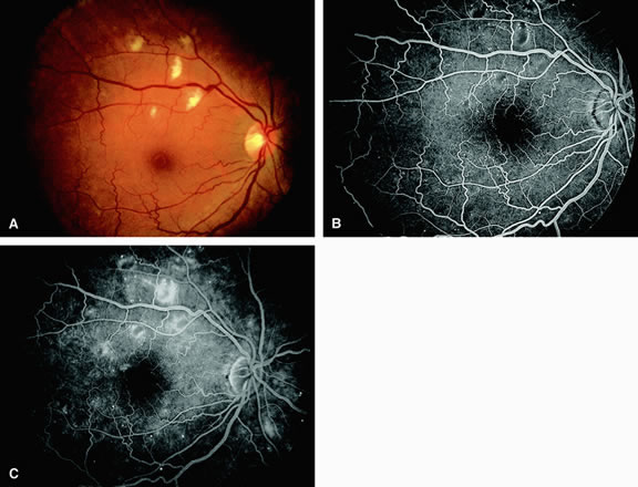
Table 2. Abnormalities Associated with Cotton-Wool Spots in the Fundus

  1. Diabetic retinopathy
  2. Systemic arterial hypertension
  3. Collagen-vascular disease
    1. Systemic lupus erythematosus
    2. Dermatomyositis
    3. Polyarteritis nodosa
    4. Scleroderma
    5. Giant cell arteritis

  4. Cardiac valvular disease
    1. Mitral valve prolapse
    2. Rheumatic heart disease
    3. Endocarditis

  5. Acquired immunodeficiency syndrome (AIDS)
  6. Central and branch retinal vein obstruction
  7. Partial central retinal artery obstruction
  8. Leukemia
  9. Trauma
  10. Radiation retinopathy
  11. Metastatic carcinoma
  12. Leptospirosis
  13. Rocky Mountain spotted fever
  14. High-altitude retinopathy
  15. Severe anemia
  16. Acute blood loss
  17. Papilledema
  18. Papillitis
  19. Carotid artery atherosclerosis
  20. Dysproteinemias
  21. Septicemia
  22. Aortic arch syndrome (pulseless disease)
  23. Intravenous drug abuse
  24. Acute pancreatitis
  25. Onchocerciasis
  26. Systemic alpha interferon administration
(From Sharma S, Brown GC: Retinal artery obstruction. In: Ryan SJ, Schachat AP [eds]. Retina. 3rd ed. Philadelphia: Mosby, 2001)


Back to Top
RETINAL VENOUS OBSTRUCTIVE DISEASE

CENTRAL RETINAL VEIN OBSTRUCTION

In eyes with central retinal vein obstruction, a thrombus is usually found in the obstructed vessel at or near the lamina cribrosa. Ophthalmoscopic signs of retinal vein obstruction include dilated tortuous veins, intraretinal hemorrhage, and ischemic retinal edema.10 Cotton-wool spots and a swollen optic disc are also often seen. Other less common signs of central retinal vein obstruction include vitreous hemorrhage and exudative retinal detachment. Several classifications have been employed to define central retinal vein obstructions. Central retinal vein obstructions can be divided into ischemic and nonischemic variants, depending in part on whether large areas of retinal capillary nonperfusion are present or absent. This classification is clinically useful because data from the Central Retinal Vein Occlusion Study Group11 and others12 suggest that laser panretinal photocoagulation can result in a regression of neovascularization of the iris, thereby preventing neovascular glaucoma.

Compared with eyes that have the ischemic type, eyes with nonischemic central retinal vein obstruction typically have a visual acuity of 20/200 or better, relatively few cotton-wool spots, and less marked retinal hemorrhage.13 Fluorescein angiography usually shows an increased retinal arteriovenous transit time secondary to delayed retinal venous filling. Delayed retinal arterial filling can also be present. The retinal capillary bed is generally well perfused (Fig. 10). The retinal veins appear as a hypofluorescent silhouette against the bright choroid. There is also variable staining of the retinal veins and variable leakage on the angiogram. Approximately 20% of nonischemic central retinal vein occlusions will progress to ischemic central retinal vein occlusions.

Fig. 10. A. Patient with a nonischemic central retinal vein occlusion. The fundus shows diffuse intraretinal hemorrhage. The veins appear dilated and tortuous. The optic disc is swollen and has diffuse retinal edema. Note that there are no visible areas of retinal ischemia. B. Fluorescein angiogram of A shows perfusion of the retinal arteries, arterioles, and capillaries. Hypofluorescence corresponds to areas of blockage from retinal hemorrhages. C. Late-phase angiogram demonstrates staining of the venous system, as well as the disc.

Ischemic central retinal vein obstructions generally reduce a patient's visual acuity to 20/200 or worse, most commonly to the counting-fingers or hand-motions range. Numerous cotton-wool spots and severe four-quadrant retinal hemorrhaging are often seen. Fluorescein angiography reveals confluent regions of retinal capillary nonperfusion (Fig. 11). Views of the posterior pole, as well as those of the four quadrants, may be necessary to demonstrate the capillary dropout. Magargal and associates12 quantitated the amount of retinal capillary nonperfusion in a posterior-pole 30-degree view in eyes with central retinal vein obstruction and calculated an ischemic index (area of retinal capillary nonperfusion/total area within the posterior 30-degree view). They found that when the index was 80% or greater, approximately 45% of eyes eventually developed rubeosis iridis and neovascular glaucoma.

Fig. 11. A. Fundus of a 35-year-old patient with an ischemic central retinal vein occlusion. The fundus has the classic “blood and thunder” appearance. Numerous intraretinal hemorrhages and retinal edema are present. The retinal veins appear dilated and tortuous. The optic nerve is swollen and hyperemic. B. Fluorescein angiogram of A shows fluorescence, indicating perfusion of the retinal arterioles. Filling of the tortuous retinal veins is delayed. Patchy areas of capillary nonperfusion indicating ischemia appear throughout the fundus.

In some eyes with central retinal vein occlusion, determining the degree of retinal capillary nonperfusion with fluorescein angiography is difficult. Worsening visual acuity and progressive intraretinal bleeding indicate that an eye may be progressing toward a more ischemic state.14 In eyes with central retinal vein obstruction and a visual acuity of 20/200 or worse, performing fluorescein angiography at the initial visit can be useful to evaluate perfusion of the retinal capillary bed. The presence of a large amount of intraretinal blood in the posterior pole is not necessarily a contraindication to performing the study, particularly because these eyes are probably more prone to ischemia. The retinal blood generally diminishes rapidly anterior to the posterior pole, facilitating angiographic evaluation of the peripheral retina.

BRANCH RETINAL VEIN OBSTRUCTION

Branch retinal vein obstruction usually occurs at the intersection of a branch retinal artery with a vein. In general, the artery overlies the vein, and the two vessels share a common adventitial sheath at the crossing. Retinal hemorrhages are present along with cotton-wool spots (Figs. 12 and 13). These hemorrhages usually are reabsorbed over a period of months. Numerous microvascular abnormalities generally remain, and macular edema may be persistent.

Fig. 12. A. A patient with a branch retinal vein occlusion. The fundus shows a large area of superficial hemorrhage and edema in the distribution of an inferotemporal venous arcade. The obstruction of the vein can be observed at the crossing of the artery and vein (AV crossing). A cotton-wool spot is present inferiorly. B. Fluorescein angiogram of A shows many telangiectatic vascular abnormalities and areas of hypofluorescence that are caused by capillary nonperfusion. Early venous-venous collaterals can be observed in the temporal macula.

Fig. 13. A. Fundus of a patient with a branch retinal vein occlusion. Note the presence of dot-and-blot hemorrhages and cotton-wool spots confined to the distribution of the superotemporal retinal vein. The vein itself appears dilated and mildly tortuous. Hemorrhage, cotton-wool spots, and retinal edema extend into the macula. The site of occlusion is at the crossing of the artery and vein (AV crossing). B. Fluorescein angiogram of A reveals a dilated, tortuous supertemporal retinal vein with areas of venous nonperfusion distal to the site of occlusion at the AV crossing. There are regions of hypofluorescence of the choroid resulting from the intraretinal hemorrhage and significant retinal capillary ischemia.

The Branch Retinal Vein Occlusion Study Group15 demonstrated that eyes with branch retinal vein obstruction and a visual acuity of 20/40 or worse may benefit from grid laser therapy to reduce macular edema. This study group recommended waiting at least 3 months after the onset of the obstruction to permit clearing of the retinal blood before performing fluorescein angiography. If the study shows that macular edema is responsible for the visual loss, grid laser therapy to the affected region within the vascular arcades can be considered. In cases in which foveal nonperfusion is responsible for the decrease in vision, laser therapy has not been shown to improve vision.

Eyes with branch retinal vein obstruction and retinal capillary nonperfusion can develop neovascularization of the retina and/or disc. New vessels on the retina or optic disc intensely leak fluorescein dye, whereas larger collateral vessels on the optic disc or in the retina usually do not; thus, fluorescein angiography is helpful in differentiating between neovascularization and collateral vessels. Proper identification of posterior-segment neovascularization in eyes with branch retinal vein obstruction is important because scatter laser treatment in such cases has been shown to reduce vitreous hemorrhage in these eyes.16 In addition, fluorescein angiography can be used to identify the areas of retinal capillary nonperfusion requiring therapy. Scatter sector laser photocoagulation should be delayed until the onset of retinal neovascularization.

Back to Top
OTHER RETINAL VASCULAR ABNORMALITIES

PERIPHERAL PROLIFERATIVE RETINOPATHIES

The most common cause of peripheral proliferative retinopathy is sickling hemoglobinopathies. They account for approximately one half of cases.17 Among patients with sickling hemoglobinopathy and peripheral proliferative retinopathy, hemoglobin SC is the most common variant. Other causes of peripheral proliferative retinopathy include retinal vein branch obstruction, diabetes mellitus, sarcoidosis, intravenous drug abuse, and Eales' disease.17

On fluorescein angiography, the posterior pole and the optic nerve often appear normal. Peripheral retinal capillary nonperfusion is seen in most eyes with peripheral extraretinal neovascularization.17,18 Shutdown of the larger peripheral retinal vessels is also often observed. Peripheral retinal neovascularization can assume several configurations, although the most common variant is that of the “sea fan” characteristic of sickle-cell retinopathy18 (Fig. 14). The angiogram demonstrates leakage of dye into the vitreous from these sites of neovascularization. In eyes in which scatter panretinal photocoagulation is being considered to control peripheral neovascularization, fluorescein angiography helps identify the regions of retinal capillary nonperfusion that require treatment.

Fig. 14. A. Midperiphery of the fundus of a patient with hemoglobin SC disease. Note the proliferation of new vessels producing a “sea fan” configuration. Inferiorly there is a vitreous hemorrhage. The patient has Stage 4 disease. B. Fluorescein angiogram of A shows a tortuous artery supplying the sea fan and a retinal vein draining the structure. Prominent hyperfluorescence of the sea fan is due to dye leakage into the vitreous. Also note the marked capillary nonperfusion surrounding this vascular anomaly.

COATS' DISEASE

Coats' disease19 is a retinal vascular abnormality of unknown etiology characterized by retinal telangiectatic formations in association with intraretinal and subretinal hard exudates (Fig. 15). Focal areas of dilation and narrowing of the larger retinal vessels are often seen. Fluorescein angiography20 demonstrates hyperfluorescence of the telangiectatic vessels early in the study along with hypofluorescence of proteinaceous exudates and mild hyperfluorescence of subretinal fluid. Enlargement of the retinal capillary bed and widened spaces between these small vessels is characteristic of Coats' disease. Retinal capillary nonperfusion is often seen, but associated retinal neovascularization is rare. Leakage of dye can occur from both the larger and smaller vessel abnormalities.

Fig. 15. A. The midperipheral fundus of a patient with Coats' disease demonstrates a retinal telangiectasia surrounded by yellow and white hard exudates and intraretinal edema, as well as hemorrhage. B. Fluorescein angiogram of A shows hyperfluorescent telangiectatic capillary beds with hypofluorescent regions between the capillary beds, consistent with capillary nonperfusion.

RETINAL ARTERIAL MACROANEURYSM

A retinal arterial macroaneurysm is an area of focal dilation, and often outpouching, along the course of an artery. First described by Robertson2 in 1973, the abnormality is often associated with systemic arterial hypertension and is found in older people, more commonly in women. More than one retinal arterial macroaneurysm may be present. Macroaneurysms most often occur in the superotemporal or the inferotemporal retinal arterial arcades, often after the second or third arteriolar branching. Local ocular complications include bleeding and the extravasation of serum and lipid into the surrounding retina.22 Blood from leaking macroaneurysms can extend into the vitreous cavity, retina, and/or subretinal space (Fig. 16).

Fig. 16. A. Fundus examination demonstrates a retinal artery macroaneurysm positioned at the 10 o'clock position relative to the optic disc. Accompanying the macroaneurysm is subretinal hemorrhage that extends peripherally from the optic disc up to but sparing the fovea. The patient's visual acuity was 20/20 -1. B. Fluorescein angiogram of A demonstrates a hyperfluorescent circular macroaneurysmal dilatation just distal to the second branch point of the superotemporal arcade. Subretinal hemorrhage has caused a large circular region of hypofluorescence of the choroidal vasculature with sparing of the retinal vasculature extending from the optic disc to just proximal to the macula. C. Late in the study, the macroaneurysmal abnormality remains hyperfluorescent.

Fluorescein angiography in eyes with an acquired retinal arterial macroaneurysm generally reveals relatively early and late hyperfluorescence of the abnormality. The hyperfluorescence usually increases as the study progresses. There is often narrowing of the vessels proximal and distal to the macroaneurysm. The retinal capillary bed surrounding the macroaneurysm is often dilated, and leakage from local telangiectatic and microaneurysmal abnormalities is often present. Identification of coexistent leaking vessels is important if laser therapy is being considered. The macroaneurysm is often obscured by an overlying hemorrhage. Occasionally, the macroaneurysm can thrombose and lead to obstruction of the retinal artery distal to it.

SYSTEMIC ARTERIAL HYPERTENSION

Chronic systemic arterial hypertension manifests in the fundus with focal and diffuse retinal arterial narrowing; retinal hemorrhages; cotton-wool spots; and in more severe or acute cases, lipid exudate23 (Fig. 17). The lipid exudates may be deposited in a radial pattern surrounding the macula, forming the so-called macular star. In extreme cases (malignant hypertension), optic disc swelling is also present. Small yellow spots at the level of the retinal pigment epithelium resulting from underlying damage of the choriocapillaris can also be seen.

Fig. 17. A. Fundus of a patient with acute hypertensive retinopathy showing diffuse retinal hemorrhages, optic disc edema, submacular fluid, early “macular star” (resulting from the presence of intraretinal lipid), and cotton-wool spots inferiorly. The arterioles appear narrow and attenuated. There are scattered lipid exudates adjacent to the superior arcades. The optic disc appears swollen, consistent with malignant hypertension. B. Fluorescein angiogram of A shows optic disc leakage and staining and numerous hypofluorescent densities corresponding to dot-and-blot retinal hemorrhages.

Fluorescein angiography of eyes with acute hypertensive changes shows irregular-appearing constricted arterioles and focal areas of hypofluorescence resulting from retinal capillary nonperfusion in areas corresponding to cotton-wool spots. Telangiectatic retinal vascular abnormalities and microaneurysms often surround the cotton-wool spots and leak dye into the retina as the study progresses. In some instances, large areas of retinal capillary nonperfusion are present. The optic disc is usually hyperfluorescent in eyes with nerve-head swelling and malignant hypertension (see Fig. 17). Focal areas of hyperfluorescence resulting from leakage at the level of the choriocapillaris can also be seen. These areas eventually resolve, leaving pigmented and depigmented foci (Elschnig spots) at the retinal pigment epithelial level.24 Despite findings on fluorescein angiography, ophthalmoscopic evaluation is more important in making the diagnosis. Fluorescein angiography is also not needed for treatment (that is, controlling blood pressure).

RADIATION RETINOPATHY

The ophthalmic community has long appreciated that radiation is an effective therapeutic option for many intraorbital, periorbital, and intracranial neoplasms. In 1933 in the first reported case of radiation retinopathy, Stallard25 described the potentially harmful effects of radiation on the retina. At that time, clinicians used large doses of radiation, and little effort was made to limit radiation exposure of nonpathologic tissue. In recent years, technologic advances have enabled clinicians to administer radiation in lower doses by using delivery systems that limit exposure of radiation to normal tissue. Although these new approaches have helped reduce the incidence of radiation retinopathy, cases of radiation retinopathy continue to be reported.

Radiation retinopathy slowly progresses and usually develops over months after exposure to ionizing radiation. External beam irradiation, 60Co plaque irradiation, and brachytherapy can all cause radiation retinopathy.26 The minimum amount of radiation capable of inducing retinopathy is controversial. Investigators have reported that as little as 1100 cGy can result in retinal damage.27 Amoaku and Archer28 recommend regularly screening all patients who have received at least 3000 cGy of radiation to the eye and closely observing all patients who have been exposed to 5000 cGy or more of radiation to the eye.

Brown and colleagues26 reviewed 32 cases of radiation-induced retinopathy. In this group, 20 eyes had been exposed to 60Co plaque irradiation and 16 eyes had been exposed to external beam radiation. As shown in Table 3, hard exudates, microaneurysms, and intraretinal hemorrhages are the most common ophthalmoscopic manifestations of radiation-induced retinopathy. Brown and associates observed a discrepancy in the percentages of eyes developing hard exudates between the two modalities of radiation exposure.


Table 3. Incidence of Retinopathic Signs in 36 Eyes with Radiation Retinopathy


Signs60Co PlaqueExternal Beam
 (N = 20)(N = 16)
Hard exudates17 (85%)6 (38%)
Microaneurysms15 (75%)13 (81%)
Intraretinal hemorrhages (streak, dot, and blot)13 (65%)14 (88%)
Telangiectases7 (35%6 (38%)
Cotton-wool spots6 (30%)6 (38%)
Vascular sheathing4 (20%)4 (25%)
(From Brown GC, Shields JA, Sanborn G et al: Radiation retinopathy. Ophthalmology 89:1495, 1982)


The fluorescein angiographic features of radiation retinopathy include capillary dilatation andclosure, formation of microaneurysms, and telangiectasia (Fig. 18). The dilated, tortuous fusiform capillaries develop vascular incompetence, and leakage of fluorescein can be observed as the retinopathy progresses. End-stage radiation retinopathy is characterized by generalized capillary disorganization resulting in retinal ischemia and cystoid macular edema.

Fig. 18. A. Fundus of a patient with radiation retinopathy. The fundus shows scattered cotton-wool spots, hard exudates, and dot-and-blot hemorrhages. Microaneurysms and macular edema are present in the region of the macula. B. Fluorescein angiogram of A shows diffuse areas of hyperfluorescence adjacent to the vascular arcades representing leakage of dye from retinal capillaries and decompensation of the retinal pigment epithelium (RPE). C. A later frame shows continued hyperfluorescence from retinal capillary leakage and more RPE leakage.

Back to Top
REFERENCES

1. Brown GC, Magargal LE: The ocular ischemic syndrome: Clinical, fluorescein angiographic and carotid angiographic features. Int Ophthalmol 11:239, 1988

2. Hayreh SS, Baines JAB: Occlusion of the posterior ciliary artery. 1. Effects on choroidal circulation. Br J Ophthalmol 56:719, 1972

3. Brown GC: Systemic associations of retinal arterial obstructive disease. Int Ophthalmol Clin 31:1–14, 1991

4. Johnson MA: Use of electroretinographic ratios in assessment of vascular occlusion and ischemia. In Heckenlively JR, Arden GB (eds): Principles and Practice of Clinical Electrophysiology of Vision. St Louis, CV Mosby, 1991, pp 617–618

5. Brown GC: Retinal arterial obstructive disease. In Ryan SJ (ed): Retina. Vol 2. St Louis, CV Mosby, 1989, p 403

6. Duker JS, Sivalingam A, Brown GC et al: A prospective study of acute central retinal artery obstruction. The incidence of secondary ocular neovascularization.Arch Ophthalmol 109:339, 1991

7. Brown GC, Shields JA: Cilioretinal arteries and retinal arterial occlusion. Arch Ophthalmol 97:84, 1979

8. Brown GC, Moffat K, Cruess A et al: Cilioretinal artery obstruction. Retina 3:182, 1983

9. McLeod D, Marshall J, Kohner EM et al: The role of axoplasmic transport in the pathogenesis of retinal cotton-wool spots. Br J Ophthalmol 61:177, 1977

10. Fong AC, Schatz H: Central retinal vein occlusion in young adults. Surv Ophthalmol 37:393, 1993

11. A randomized clinical trial of early panretinal photocoagulation for ischemic central vein occlusion. The Central Vein Occlusion Study Group N report. Ophthalmology 102:1434, 1995

12. Magargal LE, Brown GC, Augsburger JJ et al: Efficacy of panretinal photoocoagulation in preventing neovascular glaucoma following ischemic central retinal vein obstruction. Ophthalmology 89:780, 1982

13. Brown GC: Central retinal vein obstruction: Diagnosis and management. In Reinecke R (ed): Ophthalmology Annual. Norwalk, CT, Appleton-Century-Crofts, 1985, p 65

14. Minturn J, Brown GC: Progression of nonischemic central retinal vein obstruction to the ischemic variant. Ophthalmology 93:1158, 1986

15. Branch Retinal Vein Occlusion Study Group: Argon laser photocoagulation for macular edema in branch vein occlusion. Am J Ophthalmol 98:271, 1984

16. Branch Retinal Vein Occlusion Study Group: Argon laser scatter photocoagulation for prevention of neovascularization and vitreous hemorrhage in branch vein occlusion. Arch Ophthalmol 104:34, 1986

17. Brown GC, Brown RH, Brown MM: Peripheral proliferative retinopathies. Int Ophthalmol 11:41, 1987

18. McLeod DS, Merges C, Fukushima A et al: Histopathologic features of neovascularization in sickle cell retinopathy. Am J Ophthalmol 124:455, 1997

19. Coats G: Forms of retinal disease with massive exudation. R Lond Ophthalmol Hosp Rep 17:440, 1908

20. Shields JA, Shields CL, Honavar SG et al: Clinical variations and complications of Coats disease in 150 cases: The Sanford Gifford Memorial Lecture. Am J Ophthalmol 131:561, 2001

21. Robertson DM: Macroaneurysms of the retina vessels. Trans Am Acad Ophthalmol Otolaryngol 77:55, 1973

22. Rabb MF, Gagliano DA: Retinal artery macroaneurysms. Surv Ophthalmol 33:73, 1988

23. Richard G, Soubrane G, Yannuzzi LA: Vascular disease. In: Fluorescein and ICG Angiography. New York, George Thieme Verlag, 1998

24. Klein BA: Ischemic infarcts of the choroid (Elschnig spots): A cause of retinal separation in hypertensive disease with renal insufficiency. A clinical and histopathologic study. Am J Ophthalmol 66:1069, 1968

25. Stallard HB: Radiant energy as (a) a pathogenic and (b) a therapeutic agent in ophthalmic disorders. Br J Ophthalmol 1:70, 1933

26. Brown GC, Shields JA, Sanborn G et al: Radiation retinopathy. Ophthalmology 89:1494, 1982

27. Elsas T, Thorud E, Jetne V et al: Retinopathy after low dose irradiation for an intracranial tumor of the frontal lobe. Acta Ophthalmol Copenh 66:65, 1988

28. Amoaku WM, Archer DB: Fluorescein angiographic features, natural course and treatment of radiation retinopathy. Eye 4(Pt 5):657, 1990

Back to Top