|
|
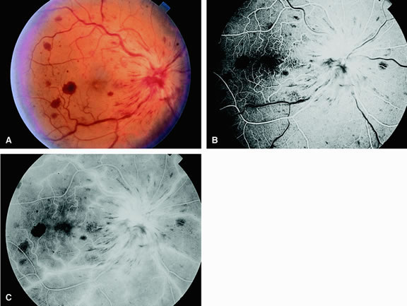
| Fig. 10. A. Patient with a nonischemic central retinal vein occlusion. The fundus shows diffuse intraretinal hemorrhage. The veins appear dilated and tortuous. The optic disc is swollen and has diffuse retinal edema. Note that there are no visible areas of retinal ischemia. B. Fluorescein angiogram of A shows perfusion of the retinal arteries, arterioles, and capillaries. Hypofluorescence corresponds to areas of blockage from retinal hemorrhages. C. Late-phase angiogram demonstrates staining of the venous system, as well as the disc. |