|
|
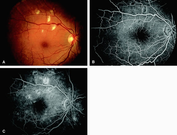
| Fig. 9. A. Cotton-wool spots in the superior temporal arcade of a diabetic patient with nonproliferative retinopathy. B. In the mid-phase fluorescein angiogram of A, the cotton-wool spots are hypofluorescent. C. Late-phase angiogram demonstrates staining of the cotton-wool spots, numerous microaneurysms, and capillary leakage. |