|
|
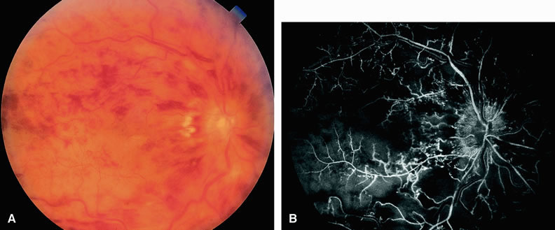
| Fig. 11. A. Fundus of a 35-year-old patient with an ischemic central retinal vein occlusion. The fundus has the classic “blood and thunder” appearance. Numerous intraretinal hemorrhages and retinal edema are present. The retinal veins appear dilated and tortuous. The optic nerve is swollen and hyperemic. B. Fluorescein angiogram of A shows fluorescence, indicating perfusion of the retinal arterioles. Filling of the tortuous retinal veins is delayed. Patchy areas of capillary nonperfusion indicating ischemia appear throughout the fundus. |