|
|
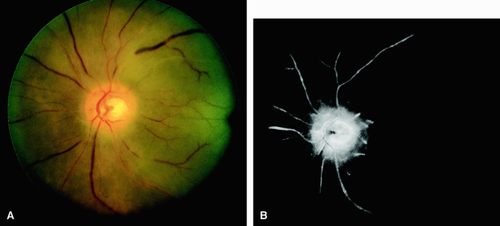
| Fig. 4. A. Patient with a central retinal artery occlusion and neovascularization of the iris. Note the diffuse pallor of the retina. The retinal arteries appear narrow and poorly perfused with areas of box carring of the luminal blood columns. The classic cherry-red spot can be observed in the region of the macula. B. Fluorescein angiogram of A shows perfusion of the underlying choroid, and a leading edge of dye is present in the retinal arteries. |