|
|
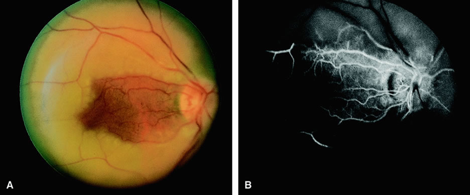
| Fig. 6. A. Fundus of a patient with a central retinal artery occlusion and sparing of the cilioretinal artery. Note that the arteries appear narrow and attenuated. The region of the retina from the optic disc to the macula appears well perfused. The rest of the retina appears pale and ischemic. B. Fluorescein angiogram of A, which shows fluorescence in the distribution of the cilioretinal artery and underlying choroid. The rest of the retinal vessels appear hypofluorescent. |