|
|
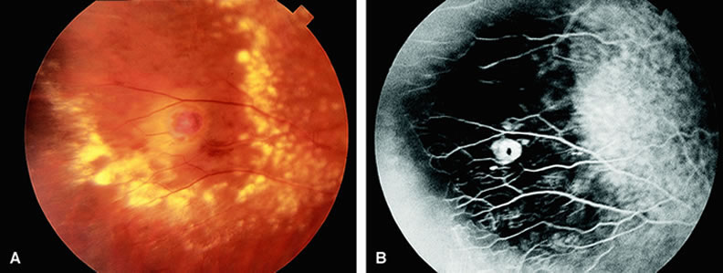
| Fig. 15. A. The midperipheral fundus of a patient with Coats' disease demonstrates a retinal telangiectasia surrounded by yellow and white hard exudates and intraretinal edema, as well as hemorrhage. B. Fluorescein angiogram of A shows hyperfluorescent telangiectatic capillary beds with hypofluorescent regions between the capillary beds, consistent with capillary nonperfusion. |