|
|
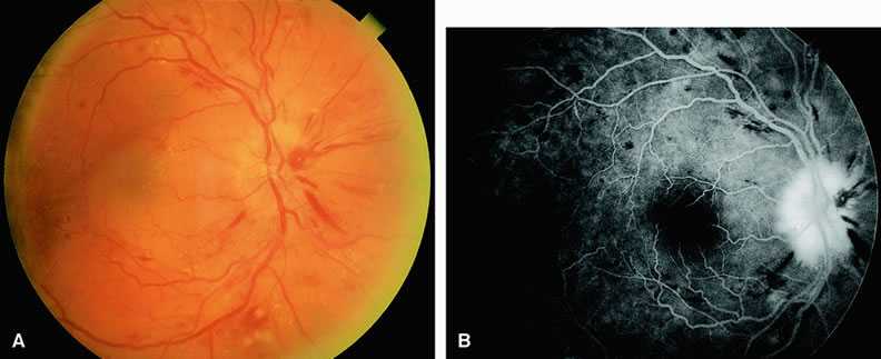
| Fig. 17. A. Fundus of a patient with acute hypertensive retinopathy showing diffuse retinal hemorrhages, optic disc edema, submacular fluid, early “macular star” (resulting from the presence of intraretinal lipid), and cotton-wool spots inferiorly. The arterioles appear narrow and attenuated. There are scattered lipid exudates adjacent to the superior arcades. The optic disc appears swollen, consistent with malignant hypertension. B. Fluorescein angiogram of A shows optic disc leakage and staining and numerous hypofluorescent densities corresponding to dot-and-blot retinal hemorrhages. |