|
|
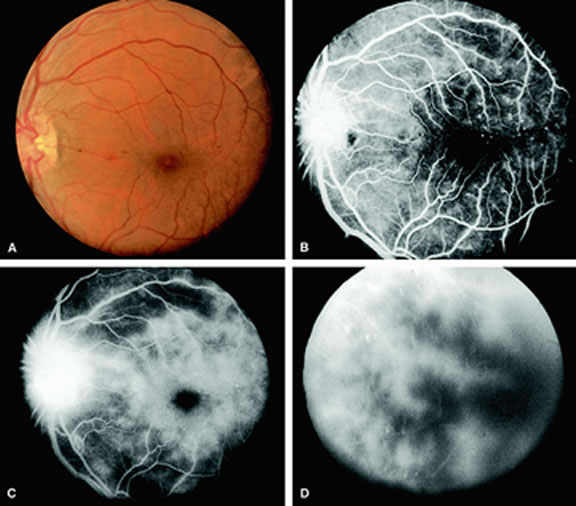
| Fig. 2. A. Narrowing of the retinal arterioles and neovascularization of the optic disc in a patient with the ocular ischemic syndrome. B. Fluorescein angiogram of A shows a hyperfluorescent optic disc caused by neovascularization and leakage of dye. Dilated and telangiectatic capillaries can be observed temporal to the macula. C. As the study progresses, leakage of dye from the capillaries results in retinal edema. The optic disc remains significantly hyperfluorescent as well. D. At the end of the study, diffuse leakage of dye clouds the retina and underlying choroid. |