|
|
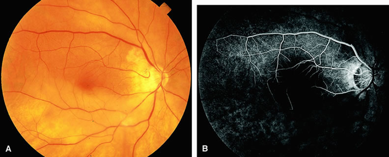
| Fig. 5. A. Fundus of a patient with occlusion of the central retinal artery with cilioretinal sparing. Most of the retina is pale and ischemic, and there is narrowing of the retinal arterioles and retinal edema. The cilioretinal artery distribution appears well perfused. B. Fluorescein angiogram of A shows perfusion of the cilioretinal artery and underlying choroid. Retrograde filling of the retinal arterioles inferiorly can be observed. The other vessels appear hypofluorescent as a result of nonperfusion. |