|
|
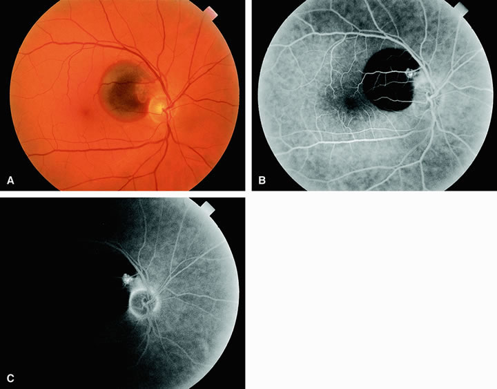
| Fig. 16. A. Fundus examination demonstrates a retinal artery macroaneurysm positioned at the 10 o'clock position relative to the optic disc. Accompanying the macroaneurysm is subretinal hemorrhage that extends peripherally from the optic disc up to but sparing the fovea. The patient's visual acuity was 20/20 -1. B. Fluorescein angiogram of A demonstrates a hyperfluorescent circular macroaneurysmal dilatation just distal to the second branch point of the superotemporal arcade. Subretinal hemorrhage has caused a large circular region of hypofluorescence of the choroidal vasculature with sparing of the retinal vasculature extending from the optic disc to just proximal to the macula. C. Late in the study, the macroaneurysmal abnormality remains hyperfluorescent. |