|
|
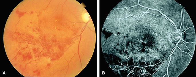
| Fig. 12. A. A patient with a branch retinal vein occlusion. The fundus shows a large area of superficial hemorrhage and edema in the distribution of an inferotemporal venous arcade. The obstruction of the vein can be observed at the crossing of the artery and vein (AV crossing). A cotton-wool spot is present inferiorly. B. Fluorescein angiogram of A shows many telangiectatic vascular abnormalities and areas of hypofluorescence that are caused by capillary nonperfusion. Early venous-venous collaterals can be observed in the temporal macula. |