Chapter 17
Sickle Cell Disease
DONALD A. GAGLIANO, LEE M. JAMPOL and MAURICE F. RABB
Main Menu   Table Of Contents

Search

GENETICS AND EPIDEMIOLOGY
PATHOPHYSIOLOGY
LABORATORY EVALUATION
CLINICAL MANIFESTATIONS
MANAGEMENT
THE FUTURE
REFERENCES

The first description of the clinical manifestations of sickle cell anemia was published by Herrick1 in 1910. It was not until 1930, however, that the first report of retinal changes in sickle cell disease appeared, in which Cook2 described a retinal hemorrhage in a young patient. We now know much about the protean ocular manifestations of sickle cell disease, which may be seen in the conjunctiva, iris, retina, optic nerve, and choroid. These changes result from vascular occlusions caused by sickled erythrocytes and from the increased adhesion of these cells to the vascular endothelium. The visible vascular networks of the eye provide the clinician a unique opportunity to observe the vaso-occlusive process, as well as its secondary manifestations, directly. A brief review of the genetics and pathophysiology of sickle cell disease will be followed by a detailed description of the clinical manifestations and management of sickle cell eye disease.
Back to Top
GENETICS AND EPIDEMIOLOGY
The hemoglobin (Hb) molecule is composed of two pairs of polypeptide chains: α and β. Two α-genes and one β-gene, located respectively on chromosome 16 and chromosome 11, encode these chains. Since we inherit one chromosome from each parent, a hemoglobin admixture capable of encoding up to four α-chains and two β-chains may occur. Hemoglobin A (Hb A) and hemoglobin A2 (Hb A2) are the predominant types of hemoglobin found in postnatal life. Hb A contains two α-chains and two β-chains; Hb A2 contains two α-chains and two δ-chains. The α-chain has 141 residues and the β-chain 146 residues. Fetal hemoglobin (Hb F), predominant during prenatal life, contains two α-chains and two γ-chains. A developmental gene switch from expressing fetal γ-globin to postnatal β-globin occurs on the basis of gestational age. By the end of the first year, red blood cell (RBC) composition typically remains stable.3

In 1949, Pauling and colleagues4 discovered that sickle cell disease was caused by the presence of an abnormal hemoglobin, named sickle hemoglobin (Hb S) because of the sickle shape it imparts to deoxygenated RBCs. In 1956, it was reported that the formation of Hb S was caused by a single amino acid substitution (i.e., valine for glutamic acid) in the sixth position from the N-terminal end of the β-chain.5,6 This amino acid substitution results from a single DNA base point mutation in which thymine is substituted for adenine in the sixth codon of the β-gene (i.e., GAG to GTG). Although many other point mutations have been described, another important variant is hemoglobin C (Hb C), caused by the substitution of lysine for glutamic acid in the sixth codon of the β-chain.

Thalassemias are genetic defects that result in abnormal hemoglobin chain synthesis. They are classified according to the amount of globin chain present. For example, β-thalassemia may have no β-chain present (β0-thalassemia) or a reduced amount of β-chain present (β+ -thalassemia). Synthesis of either of the two types of polypeptide chains in the hemoglobin molecule may be affected, resulting in either α- or β-thalassemia.7

The term hemoglobinopathy describes disorders associated with structurally abnormal hemoglobin. Sickle cell is the most common hemoglobinopathy affecting humans, with a gene frequency as high as 20% in some areas of Africa.8 Approximately 8% of black Americans carry the gene for Hb S. Hemoglobinopathies occur in a heterozygous or homozygous form. The most common form of sickle hemoglobinopathy is the heterozygous state, known as sickle cell trait (Hb AS), in which the RBC contains normal Hb A together with abnormal Hb S.7

The pathologic condition associated with sickle cell hemoglobinopathy is known as sickle cell disease. Sickle cell disease has a variable clinical presentation because expression of the disease depends on the genetic type, the amount of Hb F present in postnatal life, the presence or absence of α-thalassemia, and possibly the presence of other point mutations in the hemoglobin gene or other genes. The gene regions closely associated with the locus for the hemoglobin β-chain may be capable of modulating the expression of the gene.7

Sickle cell trait is generally excluded from the definition of sickle cell disease because of its mild clinical manifestations. Although the systemic manifestations of sickle cell trait are usually mild or absent, patients with sickle cell trait may occasionally develop ocular complications.

The hemoglobinopathies that produce sickle cell disease are Hb SS, Hb SC, and Hb S—thalassemia (β0 or β+). Hb SS produces homozygous sickle cell anemia (also known as SS disease). It is associated with the most severe systemic manifestations and often early morbidity. In the United States, the incidence of sickle cell anemia at birth is approximately 1 in 625. Hb SC produces SC disease (also known as sickle cell-hemoglobin C disease). Although it is associated with milder systemic manifestations than sickle cell anemia, SC disease is associated with more serious ocular disease. The prevalence of SC disease in black Americans is 1 in 1500.8 The most significant sickle cell-thalassemia disease is sickle cell-β-thalassemia, the clinical symptoms of which depend on the amount of β-chain that is absent.

Three other less common genotypes manifesting the features of sickle cell disease include Hb SD Punjab, Hb SO Arab, and Hb S Lepore Boston. Hb D has a glutamine residue instead of a glutamic acid residue at position 121 on the β-chain. Hb O has a lysine instead of glutamic acid, also at position 121 on the β-chain.9 Restriction endonuclease analysis suggests that the Hb S gene arose from three geographically independent mutations in equatorial Africa.10,11 Surprisingly, the sickle gene frequency has remained relatively stable.11,12 One theory put forth to explain the evolutionary survival of a gene mutation with such devastating clinical manifestations and early morbidity is called balanced polymorphism. According to this theory, the negative effects of a genetic mutation are balanced by its protective benefits, which aid in natural selection. In this case, the Hb S gene offers some benefit in terms of protection against malaria.13,14 The greatest prevalence of the Hb S gene exists in the malarial regions of Africa, and children with sickle cell trait (true heterozygotes) are afforded some degree of protection against malaria, particularly the type caused by Plasmodium falciparum.15

Like the Hb S gene, the Hb C gene is also highly concentrated in West Africa and offers some protection against malaria.11 Approximately 2% of black Americans carry one gene for Hb C, mostly as C trait (Hb AC). Homozygous C (Hb CC) is rare, occurring in 0.016% of black Americans.

Back to Top
PATHOPHYSIOLOGY
RBCs containing Hb S acquire a sickle-shaped deformity upon deoxygenation. This is due to the formation of intracellular aggregates of long polymers aligned in a crystalline gel.7 In contrast, deoxygenated Hb A, Hb A2, and Hb F do not form this crystalline gel and actually provide an inhibitory effect on gelation. Hb C can participate in gel formation, but only in the presence of Hb S.

Intracellular polymerization and gel formation produce poor erythrocyte deformability, and if recurrent, also cause distortion and damage to the RBC membrane. Damaged RBC membranes lead to potassium loss and intracellular dehydration, which further potentiates Hb S polymerization. Eventually the erythrocyte membrane is no longer capable of assuming the normal biconcave shape upon reoxygenation, thus forming an irreversibly sickled cell (ISC).

Oxygen is the most important determinant of Hb S polymerization. Very small changes in arterial oxygen tension, even with an oxygen saturation greater than 90%, can result in sickling.16 Other factors that influence polymer formation are cellular Hb S concentration, pH, and temperature.7 In the terminal arteriole and capillary circulation, oxygen availability decreases and the pH drops, enhancing Hb S polymerization.

Rheologic impairment underlies the complications of sickle cell disease.17 Rheologic factors include the presence of sickled cells, the vessel diameter, and the hematocrit. Vascular beds with low flow and high oxygen extraction are more prone to sickling and secondary vascular occlusion. The peripheral retina and macula appear to be the most susceptible to vascular occlusion.18 Interestingly, the terminal capillary bed in each of these zones borders on an avascular area and thins to a two-dimensional capillary bed.19

Small changes in blood vessel diameter dramatically affect the blood flow because flow resistance is inversely proportional to the fourth power of the vessel radius.20 The vascular occlusions of sickle cell retinopathy occur in the arterioles rather than in the capillaries, perhaps because the sphincters of the precapillary arterioles are narrower than the true capillaries.21,22

Compared with normal RBCs, those containing Hb S demonstrate greater vascular endothelium adherence. This adherence property is not exhibited by ISCs because their rigidity renders them unable to form a large surface contact with the endothelial cells. Thus, patients who generate a large number of ISCs have vascular occlusions at the precapillary arterioles because the rigid blood cells cannot enter the capillaries. Patients with fewer ISCs and more deformable cells have blood flow compromise in the capillaries, where RBCs containing Hb S readily adhere to the vascular endothelium.

Paradoxically, there is an inverse relationship between the severity of systemic disease and the severity of sickle cell retinopathy in homozygous (SS) versus doubly heterozygous (SC) sickle cell disease. Patients with homozygous sickle cell disease have more systemic complications, with multiple vaso-occlusive events and secondary organ damage. Patients with doubly heterozygous sickle cell disease have fewer systemic complications but a greater frequency and earlier onset of retinal neovascularization, resulting in more severe sickle cell retinopathy and more visually disabling ocular complications (Tables 1 and 2).


TABLE 1. Prevalence of Proliferative Sickle Retinopathy (PSR)


Genotype% with PSR
SC32.8%*
S-Thal14.0%†
SS2.6%‡

* Condon PI, Serjeant GR: Ocular findings in hemoglobin SC disease in Jamaica. Am J Ophthalmol 74:921, 1972
†Condon PI, Serjeant GR: Ocular findings in sickle cell thalassemia in Jamaica. Am J Ophthalmol 74:1105, 1972
‡Condon PI, Serjeant GR: Ocular findings in homozygous sickle cell anemia in Jamaica. Am J Ophthalmol 73:533, 1972


TABLE 2. Influence of Age on the Prevalence of Proliferative Sickle Retinopathy (PSR) in the Different Genotypes


 % with PSR
Genotype8 – 17 yrsOver 40 yrs
SC11.0%*68.0%
S-ThalNA 
SS0.4%14.0%

* Penman AD, Talbot JF, Chuang EL et al: New classification of peripheral retinal vascular changes in sickle cell disease. Br J Ophthalmol 78:681, 1994
NA = not available.


This paradox may be explained by a discussion of the hematologic factors involved. Accelerated destruction of RBCs combined with reduced erythropoietin production results in a state of marked anemia (homozygous sickle cell anemia).23,24 Since the viscosity of blood is proportional to the hematocrit, the reduced viscosity associated with homozygous sickle cell anemia may protect against vascular occlusion. In contrast, patients with Hb SC and Hb S-β-thalassemia hemoglobinopathies have higher hematocrits, causing higher whole blood viscosity and a relatively increased frequency of vascular occlusions in the retina. Analysis of hematologic factors, however, has so far provided only a partial explanation for the genotypic differences in the development of proliferative sickle retinopathy (PSR). In a group of patients with homozygous sickle cell anemia, a significant relationship was found between the development of retinal neovascularization and high Hb levels and low Hb F levels in men, but not in women.25 In patients with SC disease, a significant relationship was demonstrated between the development of neovascularization and high mean cell volume in men and a low Hb F level in both men and women.26 Further analysis comparing whole blood and plasma viscosity, together with RBC filterability, failed to reveal any differences in patients with SC disease, although some differences occurred in patients with homozygous sickle cell anemia.27,28

Genotypic differences in the incidence of retinal neovascularization cannot be explained by the frequency of vascular occlusions alone: the more severe systemic complications of patients with homozygous sickle cell anemia are, in fact, a result of an increased frequency of vascular occlusions. α-Globin gene number does appear to reduce the extent of peripheral retinal vessel closure, but has no apparent influence on the development of neovascularization in patients with homozygous sickle cell anemia.29,30 In a study of the Jamaican Sickle Cohort, consisting of 173 Jamaican children with SC disease, 315 with homozygous sickle cell anemia, and 250 age- and sex-matched normal (Hb AA) controls recruited between 1973 and 1981, Talbot and associates31–33 found that the peripheral retina demonstrates earlier vascular occlusions in sickle cell anemia than in SC disease. Comparing the peripheral retinal vascular bed with that of a normal cohort, however, they discovered that a significantly larger proportion of SC disease subjects had an abnormal peripheral vascular pattern, which they were able to correlate with the subsequent development of neovascularization. The authors concluded that a normal border, even if undergoing a posterior regression, results from a gradual modification of the capillary bed and indicates a low risk for PSR, whereas an abnormal border occurs as a radical alteration of retinal perfusion.34

This cohort study also revealed that in homozygous sickle cell anemia, vascular closures occurred more frequently with low Hb F levels, low mean total hemoglobin levels, high reticulocyte counts, and high ISC counts. In patients with SC disease, closure was associated with high reticulocyte counts and lower height and weight.31,32

The theory of ischemia versus infarction provides another explanation for the more severe PSR associated with SC disease:

  The retinal vascular occlusions caused by sickled cells in homozygous sickle cell anemia might result in a more complete vascular occlusion (infarction), causing necrosis of the retinal tissue.
  The less severe vascular occlusions in SC disease might result in an ischemic retina, which may provide an environment in which an angiogenic response is continuously stimulated.

This theory is supported by the development of neovascular tissue in other disease processes associated with retinal ischemia, such as diabetes mellitus and central retinal vein occlusion. Additional support of this theory is provided by the regression of neovascular tissue achieved by photocoagulating the ischemic retina around a neovascular membrane.35

Peachey and co-workers36,37 studied the electroretinographic response with neovascularization and correlated this with the degree of peripheral retinal capillary nonperfusion as determined by fluorescein angiography. They found significant correlations between reductions in electroretinographic amplitudes and the extent of retinal capillary nonperfusion in patients both with and without neovascularization. Patients with neovascularization also had prolonged generation of a maximum-amplitude response, similar to that seen in central retinal vein occlusion and diabetic retinopathy, suggesting that neovascularization is associated with the presence of ischemia in sickle cell retinopathy. Possibly, the more frequent and complete occlusions of homozygous sickle cell anemia are less likely to be associated with ischemia, and thus neovascularization is less likely to develop in these patients.

Back to Top
LABORATORY EVALUATION
The laboratory testing of patients with sickle cell disease are divided broadly into testing for the presence of Hb S, identifying the major genotypes, and subdividing the genetic characteristics of the major genotypes. For clinical purposes, it is not necessary for the ophthalmologist to subdivide the major genotypes; however, it is helpful to identify the major genotypes because they have different systemic and ocular clinical characteristics and prognosis.

The solubility test (Sickledex: Orthodiagnostics, Raritan, NJ) and the sickle prep (metabisulfite slide test) are used to identify the presence of Hb S. The solubility test is based on the insolubility of deoxygenated Hb S, and the sickling test is based on the morphologic change of erythrocytes containing deoxygenated Hb S. The solubility test is simpler to perform and has gained widespread acceptance as a screening test. Both tests are not sensitive enough to detect the lower levels of Hb S present at birth, but their major limitation is their inability to differentiate sickle cell trait from the clinically significant homozygous (SS) and heterozygous (SC) sickle cell disease. Therefore, a positive sickle prep test or solubility test must be followed by quantitative hemoglobin electrophoresis. The electrophoretic pattern of Hb SS and Hb S- β0-thalassemia are similar; however, they can be differentiated by quantification of Hb A2, which is elevated in Hb S-β0-thalassemia. Sickle cell trait (Hb AS) can be distinguished from Hb S-β+ thalassemia because more than 50% of the hemoglobin is Hb S in the latter disease. DNA analysis by restriction endonuclease is the most widely used method for antenatal diagnosis of sickle cell disease.

Back to Top
CLINICAL MANIFESTATIONS
Although the retinal manifestations of sickle cell disease are the most important, anterior segment and other ocular changes do occur.

ANTERIOR SEGMENT

Conjunctival Sickle Sign

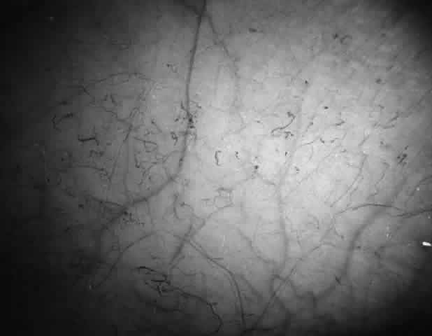
Abnormalities of the bulbar conjunctival blood vessels provide direct evidence of the vaso-occlusive process and were one of the earliest reported ocular changes.38–43 These abnormalities are believed to be the result of flow obstruction or impedance by sickled cells. The severity of the conjunctival changes ranges from linear dilatations to isolated groups of truncated, comma-shaped segments. These changes have been correlated with the ISC count, Hb S concentration, and the intraerythrocytic hemoglobin concentration (Fig. 1).44–47 Although they are known as the conjunctival sickle sign, these vascular abnormalities are not completely pathognomonic of sickle cell disease: in rare cases they are seen in patients with AIDS, chronic myelogenous leukemia, and other vaso-occlusive diseases.47–49

Fig. 1. Conjunctival vascular abnormalities in a patient with homozygous sickle cell anemia demonstrating interrupted, dilated, and truncated vascular segments.

Iris Atrophy and Neovascularization

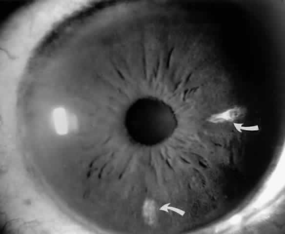
Occlusions of the iris vessels can result in atrophy, and patients may present with asymptomatic white patches of the iris.50,51 The area of atrophy may be extensive (Fig. 2) and may be associated with pupillary irregularity. Iris neovascularization may develop in eyes with chronic retinal detachment or major arteriole occlusions and can in rare cases cause a secondary neovascular glaucoma.52

Fig. 2. Iris infarcts in a patient with SC disease (arrows).

Hyphema

Sickle cell patients, including patients with sickle cell trait, are susceptible to developing central retinal artery occlusions and optic atrophy secondary to elevated intraocular pressure.53,54 Therefore, the potential for permanent ocular damage is an important consideration in sickle cell patients with even minimal hyphema from surgery or anterior segment trauma.55–57 The environment in the anterior chamber promotes Hb S polymerization and secondary impairment of outflow from blockage of the trabecular meshwork by sickled cells.58–63

It is warranted to order a sickle screen for every black American patient with hyphema and for every patient with hyphema associated with elevated intraocular pressure.64 If Hb S is present, the intraocular pressure should be closely monitored and should not be allowed to remain higher than 25 mmHg for more than 24 hours.56,57 Medical management should be restricted, if possible, to topical β-blockers and possibly oxygen (100%) inhalation or oxygen goggles, which deliver oxygen through the cornea to the aqueous humor.65 A judicious trial of 25 mg of oral methazolamide twice daily may be included. Methazolamide has been found to reduce intraocular pressure without altering renal bicarbonate secretion and causing systemic acidosis. Preferably, other carbonic anhydrase inhibitors and osmotic agents should be avoided because they may induce hemoconcentration and acidosis, causing further Hb S polymerization. If the intraocular pressure cannot be controlled by medical means, surgical intervention with anterior chamber lavage, repeated as necessary, is indicated.66

POSTERIOR SEGMENT

Disc Sign

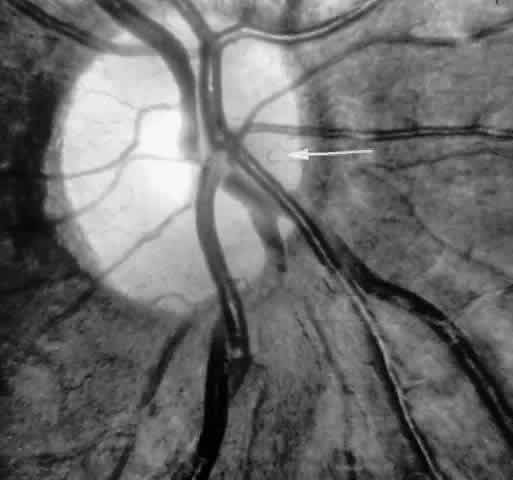
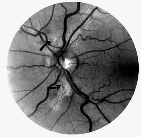
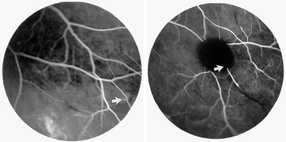
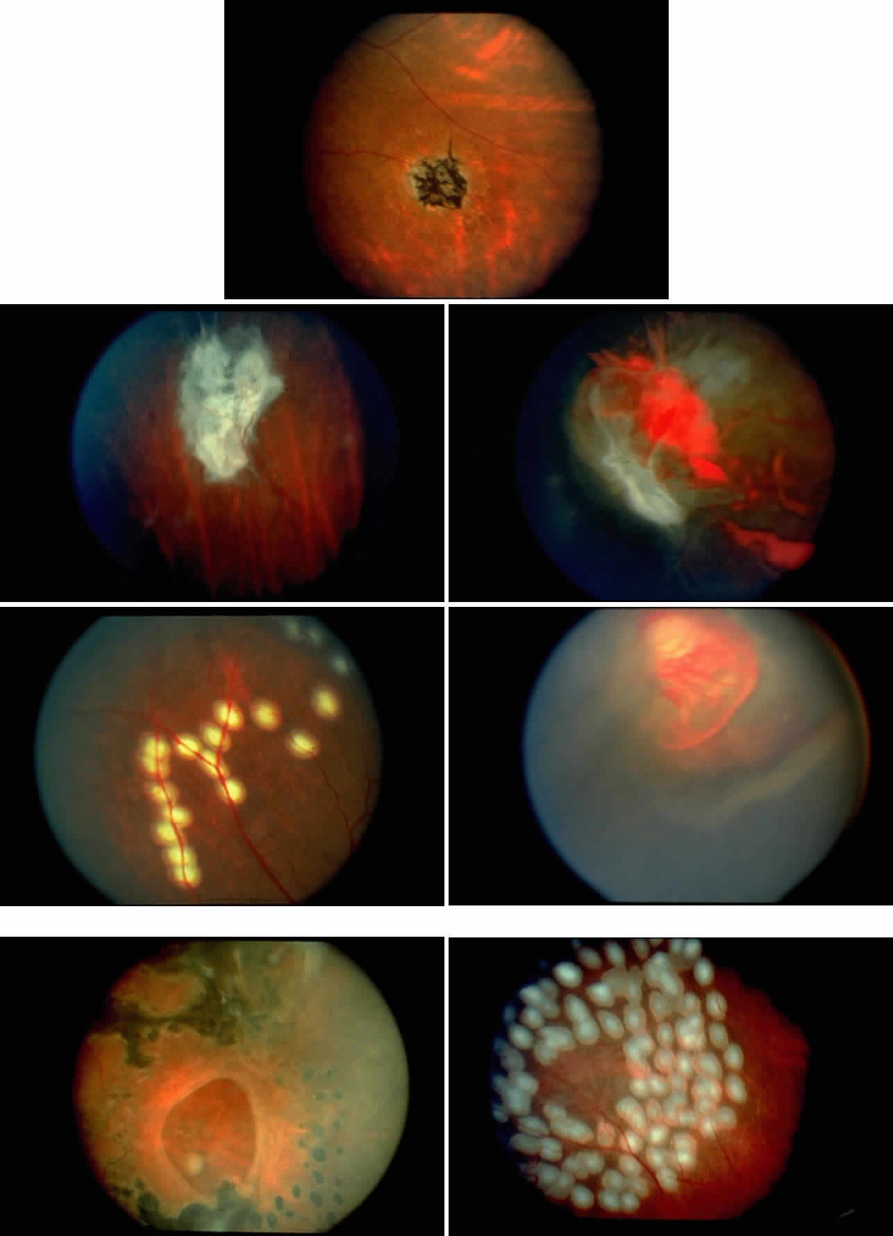
Transient dark red spots (similar to conjunctival commas), representing plugs of sickled erythrocytes within superficial capillaries, may be seen on the surface of the optic disc (Fig. 3 and Color Plate 1A). These disc changes are not associated with any functional or anatomic abnormalities. They are found in 11% of all patients with sickle cell disease, but appear to be more common in patients with homozygous sickle cell anemia, occurring in 29% of these patients.67 The disc sign correlates with the presence of conjunctival commas and ISCs.

Fig. 3. Disc sign showing dilated loops (arrow) and multiple comma-shaped vascular segments.

Color Plate 1 .A. Photograph of the disc in the same patient as in Fig. 16A, demonstrating the disc sign with multiple dilated vascular segments. B. Salmon-patch hemorrhage with preretinal blood obscuring the retinal vasculature. (B through G; Gagliano DA, Goldberg MF: The evolution of salmon-patch hemorrhages in sickle cell retinopathy. Arch Ophthalmol 107:1814, 1989.) C. Two weeks later, the hemorrhage shown in B has a central grayish white color as it begins to resolve. D. Two years later, the hemorrhage shown in B and C has resolved, and an iridescent spot remains. E. Salmon-patch hemorrhage with pre-, intra-, and sub-retinal blood. F. Two months later, the hemorrhage shown in E has resolved, and a lightly pigmented area surrounded by a depigmented halo is seen. G. Four years later, a well-pigmented black sunburst adjacent to the arteriole remains from the salmon-patch hemorrhage shown in E. H. Iridescent spot with refractile copper-colored granules.

Unlike some retinal vaso-occlusive diseases, sickle cell retinopathy is rarely associated with optic disc neovascularization.68 In our extensive experience with sickle cell disease, we have seen only one such case. The low incidence of optic disc neovascularization may be due to the peripheral location of the ischemia and to the localized changes, much of the retina not being significantly affected by the ischemia.69 Peripheral retinal scatter photocoagulation is effective in stimulating regression of optic disc neovascularization.

Vascular Tortuosity

Dilation and tortuosity of the retinal veins was one of the first recognized abnormalities of sickle cell eye disease. Although it is not pathognomonic of sickle cell disease, it reportedly occurs in up to 47% of patients with homozygous sickle cell anemia and 32% of patients with SC disease (Fig. 4).70 The significance of this venous tortuosity is unknown, and the incidence does not appear to be related to age.71

Fig. 4. A. Generalized vascular tortuosity, predominantly venous, in a patient with homozygous sickle cell anemia. B. Localized macular venous tortuosity in a patient with SC disease.

Angioid Streaks

Angioid streaks occur in association with sickle cell disease, with an overall incidence of less than 6%.72–75 The changes are more common in patients with homozygous sickle cell anemia and are age-dependent, occurring in 2% of sickle cell anemia patients less than 40 years of age versus 22% in those who are more than 40 years of age (Fig. 5).76

Fig. 5. A 45-year-old man with homozygous sickle cell anemia and angioid streaks (arrows).

Unlike the angioid streaks seen in patients with pseudoxanthoma elasticum, choroidal neovascularization and disciform disease are uncommon in association with sickle cell disease. Elastic tissue degeneration, as is seen in pseudoxanthoma elasticum, has not been demonstrated in the skin biopsy specimens of sickle hemoglobinopathy patients with angioid streaks.73,75 Initially, the etiology of angioid streaks in sickle cell disease was hypothesized to be secondary to iron deposition due to chronic hemolysis, causing brittleness of Bruch's membrane. Histopathologic examination of angioid streaks in a patient with homozygous sickle cell anemia, however, revealed heavy calcification of Bruch's membrane without evidence of iron or hemosiderin.77

Epiretinal Membranes

Epiretinal membranes may produce visual loss in patients with sickle cell disease. Macular epiretinal membranes are seen more frequently in eyes with retinal neovascularization, retinal tears, and vitreous hemorrhage, as well as in eyes that have had laser treatment or surgery of the retina or vitreous.78 Progressive visual loss from macular distortion has been reported in up to 32% of these patients over a 2.5-year period.79 Peripheral neovascularization may stimulate formation of epiretinal membranes by transudation of plasma and erythrocytes into the vitreous, disrupting the vitreous cortex and inducing posterior vitreous detachment. Successful treatment of the neovascular tissue reduces the risk of epiretinal membrane development by approximately 30%.79 Although spontaneous separation of epiretinal membranes following treatment of peripheral neovascularization has been observed, surgical removal may be considered when patients exhibit moderate to severe visual loss (Fig. 6).80,81

Fig. 6. A. Preoperative photograph of the left eye of a 64-year-old woman with SC disease, who received cryotherapy and scatter photocoagulation therapy for proliferative sickle retinopathy, resulting in the development of an epiretinal membrane. At this point, visual acuity was 20/400. B. Postoperative photograph after vitrectomy and removal of the epiretinal membrane. Visual acuity was improved to 20/60. This patient received preoperative exchange transfusions and postoperative oxygen therapy.

Traction across the macula from peripheral neovascularization is thought to contribute to the formation of macular holes in sickle cell retinopathy.82

Retinal Artery Occlusions

Occlusions of the central retinal artery and major arteriolar branches are probably most frequent in young patients with homozygous sickle cell anemia; however, they may also occur with other sickling genotypes (Fig. 7).39,83,84 They may cause permanent or transient visual loss and can occur simultaneously in both eyes.85–87 Arterial occlusion has also been reported to occur as a complication of retrobulbar anesthesia and following compression of the eye during photocoagulation (Fig. 8).88

Fig. 7. Transient perimacular arteriolar occlusions in a 32-year-old patient with SC disease, who presented with decreased vision in the right eye (20/40) after being tackled while playing football. A. Photograph of right macula showing a white, edematous retina and a cherry red spot due to multiple arteriolar occlusions. B. Fluorescein angiogram shows multiple avascular areas, particularly at the temporal raphe (arrowheads), and an irregular perifoveal capillary network (open arrows).

Fig. 8. A 27-year-old woman with homozygous sickle cell anemia and stage III sickle cell retinopathy. A. Three days after scatter photocoagulation to the right eye, a photograph of the right eye shows retinal arteriolar occlusions causing a white, edematous macula, a cherry red spot, and a cotton-wool spot superior to the macula. B. Fluorescein angiogram clearly shows the occluded arteriole superiorly, but no occlusion in the perifoveal or temporal macula. C. However, a fluorescein angiogram taken 1 year later demonstrates an irregular perifoveal capillary network with areas of capillary nonperfusion (arrows). D. Of interest, the left eye simultaneously developed an area of capillary nonperfusion, demonstrated by a cotton-wool spot nasal to the fovea. E. Two years later, there is resolution of the cotton-wool spot in the left eye, but a retinal depression sign remains, as demonstrated by an abnormal light reflex in the area nasal to the fovea (arrows).

Retinal artery occlusion has also been reported in patients with sickle cell trait secondary to airplane travel,70 with elevated intraocular pressure following blunt trauma,53,54 with extreme dehydration,89 and in association with tuberculosis and systemic lupus erythematosus.90

Macular Small Vessel Occlusions

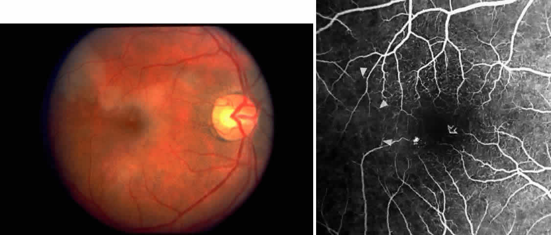
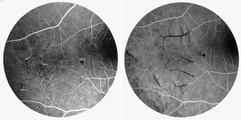
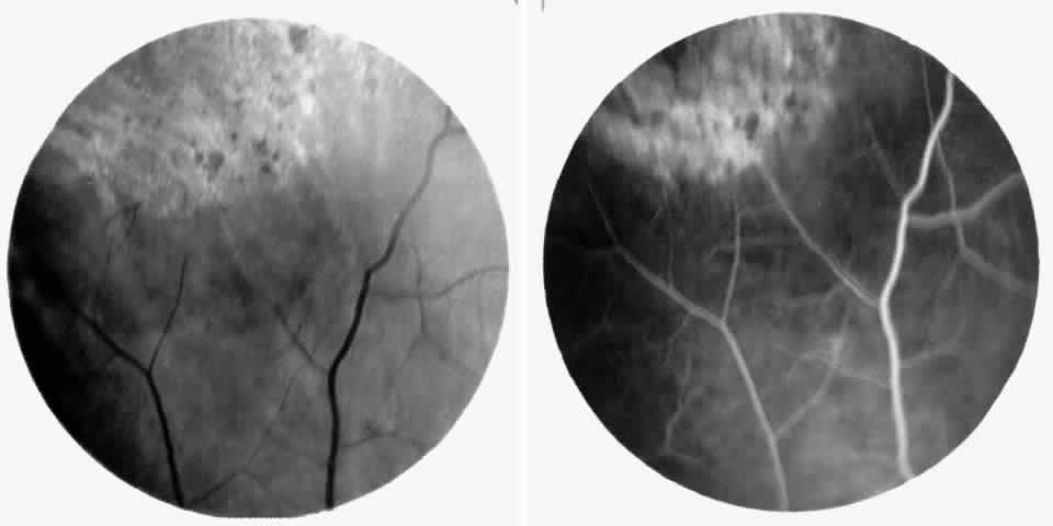
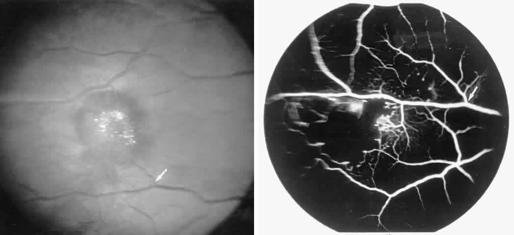
Occlusions of the fine vasculature of the macular and perimacular area have been reported in 10% to 40% of patients with sickle cell disease.18,83,91–99 In the acute phase, the occluded vessel will have a dark red appearance and may appear as a dark line on fluorescein angiography (Fig. 9). Nerve fiber layer infarcts (cotton-wool spots) are seen (see Fig. 8D and E;Fig. 10).100

Fig. 9. A 27-year-old man with homozygous sickle cell anemia. A. Fluorescein angiogram of the right eye shows multiple arteriolar occlusions temporal to the fovea (arrows). B. Same area 6 months later shows more extensive occlusions. The black arrowheads (A and B) identify corresponding arteriolar bifurcation.

Fig. 10. A 33-year-old woman with SC disease and stage III sickle cell retinopathy. A. Photograph of the right eye shows a cotton-wool spot with a dark segment identifying the occluded vessel (arrow). B. Fluorescein angiogram demonstrates nonfilling of the occluded vessel (arrow). C. Eighteen months later, the occluded vessel is still visible (arrow). D. Fluorescein angiogram demonstrates that there is still nonfilling of the vessel (arrow).

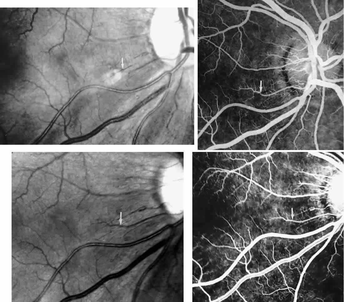
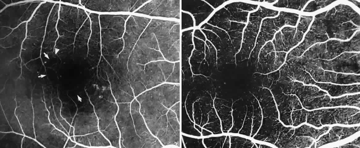
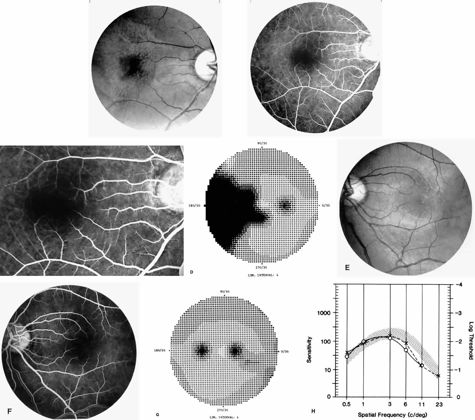
Other macular and perimacular changes include microaneurysm-like dots, dark and enlarged segments of arterioles, hairpin-shaped venular loops, pathologic avascular zones, and widening and irregularities of the foveal avascular zone (Figs. 11 and 12). In the Jamaican cohort study evaluating children with homozygous sickle cell anemia and SC disease between the ages of 5.0 and 7.5 years of age, no pathologic avascular zones could be identified despite a high incidence of peripheral vascular closure.31 In evaluating patients with homozygous sickle cell anemia, no relationship between ISC counts and macular abnormalities or visual acuity could be found.101 Using fluorescein angiography, investigators have found the foveal avascular zone to be significantly larger in eyes with clinical evidence of sickle cell maculopathy as compared with normal eyes and eyes without clinical evidence of sickle cell maculopathy.102–104

Fig. 11. A. A 40-year-old woman with homozygous sickle cell anemia. A fluorescein angiogram demonstrates multiple microaneurysm-like dots with fluorescein leakage, hairpin loop (arrowhead), pathologic avascular zones (arrows), and a widened, irregular foveal avascular zone (FAZ). B. Fluorescein angiogram of a 30-year-old woman with homozygous sickle cell anemia demonstrates multiple microaneurysm-like dots and a widened, irregular FAZ.

Fig. 12. A. Fluorescein angiogram of the left eye of a 40-year-old man with homozygous sickle cell anemia, demonstrating an irregular foveal avascular zone (FAZ), hairpin loops, and loss of the temporal capillary network. B. Fluorescein angiogram of the right macula of a 38-year-old woman with homozygous sickle cell anemia, showing an abnormal FAZ, hairpin loop (arrowhead), and pathologic avascular zones (arrows).

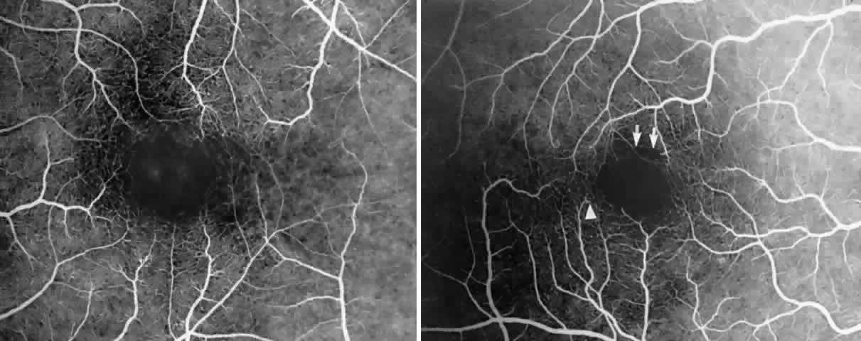
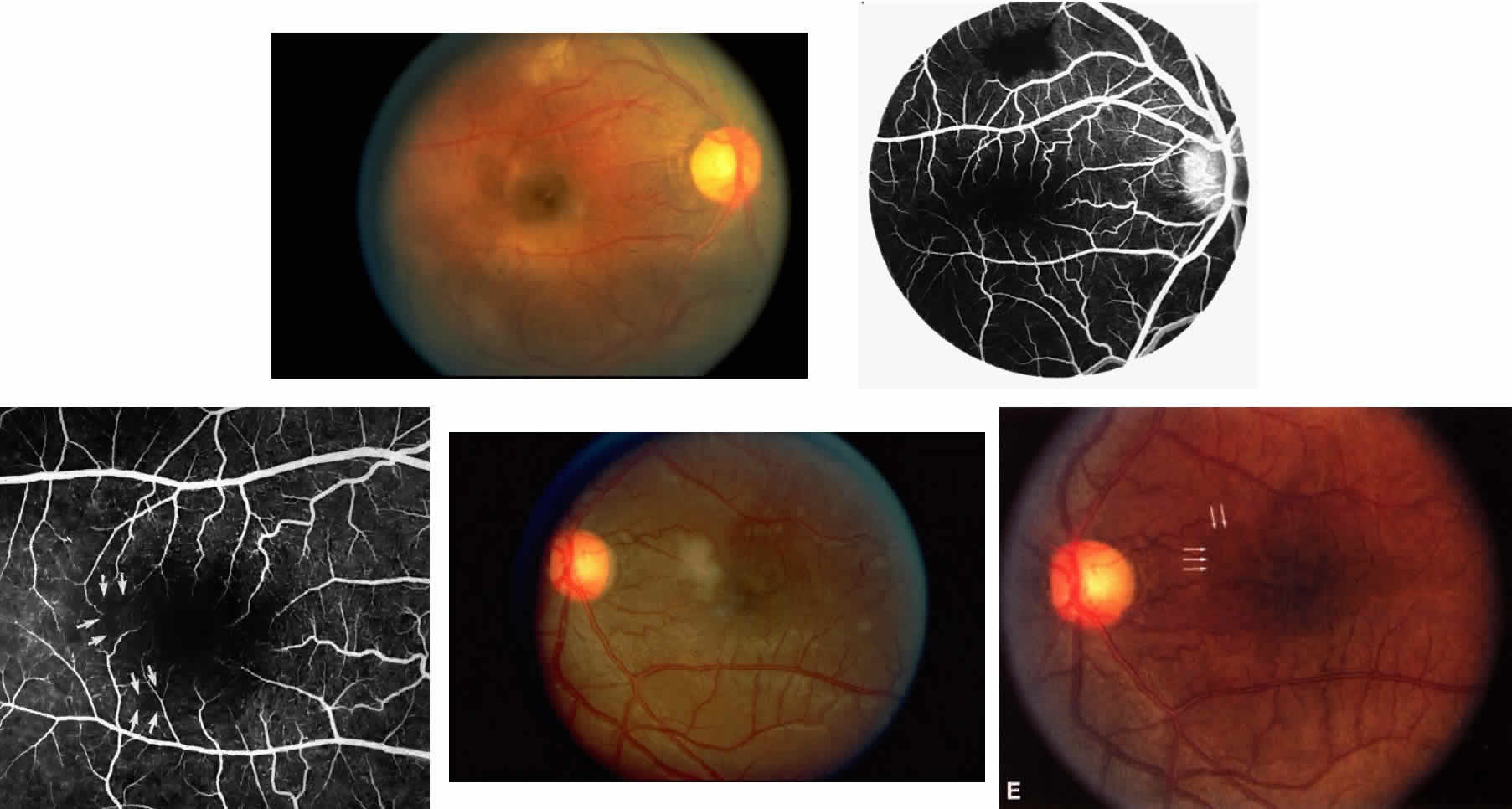
Careful examination by fluorescein angiography, looking for areas of capillary dropout and other capillary abnormalities, is often necessary to identify the macular changes. These changes may be transient, and the macula may appear normal on subsequent fluorescein angiograms (Fig. 13). Although fluorescein angiography may or may not demonstrate reperfusion of a previously occluded capillary bed, a loss of the inner retinal layers results in an ophthalmoscopic focal concavity with an abnormal reflex (retinal depression sign) (see Fig. 8E).105,106 These changes are usually permanent. The retinal depression sign is not pathognomonic of sickle cell disease and may be seen with other arteriolar occlusive diseases, such as embolic retinopathy, vasculitis, and hypertension.

Fig. 13. A. A 32-year-old man with homozygous sickle cell anemia and proliferative sickle retinopathy. A fluorescein angiogram shows temporal occlusions and a pathologic avascular zone (PAZ) temporal to the foveal avascular zone. B. Seven years later, there is filling of the previously noted PAZ.

Macular Function Testing in Sickle

Cell MaculopathyThe visual acuity in patients with sickle cell disease is often normal, despite the presence of an enlarged foveal avascular zone or other evidence of sickle cell maculopathy (Fig. 14). In addition, patients with sickle cell maculopathy have a remarkable absence of visual complaints. Although 55% of patients with homozygous sickle cell anemia had abnormal contrast sensitivity, no significant relationship was demonstrated between contrast sensitivity and macular vascular abnormalities.101 Automated visual field analysis has demonstrated significantly larger scotomas in patients with abnormally enlarged foveal avascular zone.102 Color vision testing has revealed a greater incidence of blue-yellow defects in patients with sickle cell retinopathy; however, no significant correlation has been demonstrated between color vision defects and the presence of sickle cell maculopathy.98,107

Fig. 14. A 25-year-old woman with Hb S-β-thalassemia and bilateral proliferative sickle retinopathy (visual acuity 20/25 OD and 20/15 OS). A. Photograph of the right eye demonstrates an irregular macular reflex believed to represent a macular depression sign. B and C. Fluorescein angiogram shows loss of capillary filling corresponding to the area of irregular reflex. D. Octopus perimetry of the right eye shows a large nasal visual field defect corresponding to the area of retinal nonperfusion. E. Photograph of the left eye demonstrates an irregular retinal reflex from the temporal macula, believed to represent a retinal depression sign. F. Fluorescein angiogram shows loss of capillary filling corresponding to the area of irregular reflex. G. Octopus perimetry of the left eye shows a scotoma corresponding to the area of nonperfusion. H. Contrast sensitivity test demonstrates decreased high spatial frequency thresholds (O—O = OD, X- - -X = OS).

Retinal Venous Occlusions

Retinal venous occlusions are surprisingly uncommon in sickle cell disease.108 We have seen a patient with homozygous sickle cell anemia and a branch vein occlusion, but this patient had hypertension. We have not seen central vein occlusion. An underlying systemic disease associated with a higher incidence of venous occlusions (e.g., hypertension) should be suspected when a venous occlusion occurs in a patient with sickle cell disease. The anemia and low blood pressure present in sickle cell patients may actually protect against venous occlusions.

Choroidal Vascular Occlusions

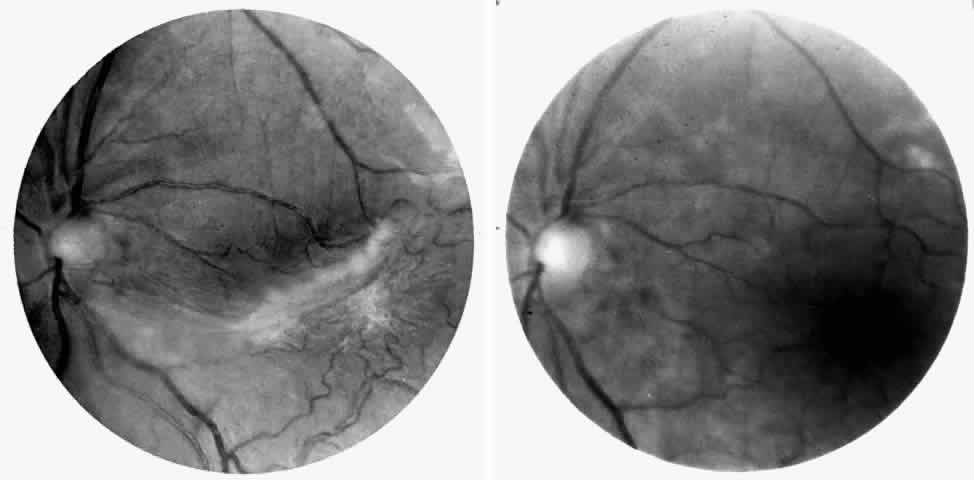
Choroidal vascular occlusions may occur focally at the level of the choroidal precapillary arteriole or capillary bed (Elschnig's spots) or from posterior ciliary artery occlusion. Although focal precapillary arteriole occlusions have not been specifically identified with sickle cell disease, clinical and histopathologic evidence of spontaneous posterior ciliary artery occlusions have been reported in sickle cell disease.109,110 The findings are similar to those described following compression of the eye during general anesthesia and after heavy peripheral photocoagulation.111,112 In the acute phase, the occlusions appear as white, circumscribed, triangular patches at the level of the retinal pigment epithelium and outer retina. Over the following weeks, the white lesions fade and retinal pigment epithelial mottling develops (Fig. 15). Since patients with acute ciliary artery occlusions may be asymptomatic and the diagnosis is often based solely on the appearance of peripheral pigment mottling, the frequency of this complication remains uncertain.

Fig. 15. A 25-year-old man with homozygous sickle cell anemia and proliferative sickle retinopathy. A. Photograph demonstrating nonperfusion and wedge-shaped pigment mottling representing a choroidal infarction. B. Fluorescein angiogram demonstrating hyperfluorescence at the level of the retinal pigment epithelium.

Focal areas of atrophy of the choriocapillaris have been seen histopathologically on postmortem examination of patients with homozygous sickle cell anemia. Mild sclerosis of the choriocapillaris, focal peripheral photoreceptor loss, and areas of choroidal neovascularization also have been noted.110 It has been suggested that choroidal ischemia plays a role in the development of angioid streaks in sickle cell disease. The one histopathologic report of angioid streaks indicated that the basement membrane of the underlying choriocapillaris endothelial cells was slightly thickened and that sickled erythrocytes were found within patent lumina of the choriocapillaris.77 No specific evidence exists to support a relationship between choroidal ischemia and angioid streaks.

Retinal Hemorrhages, Iridescent Spots, and Black Sunbursts

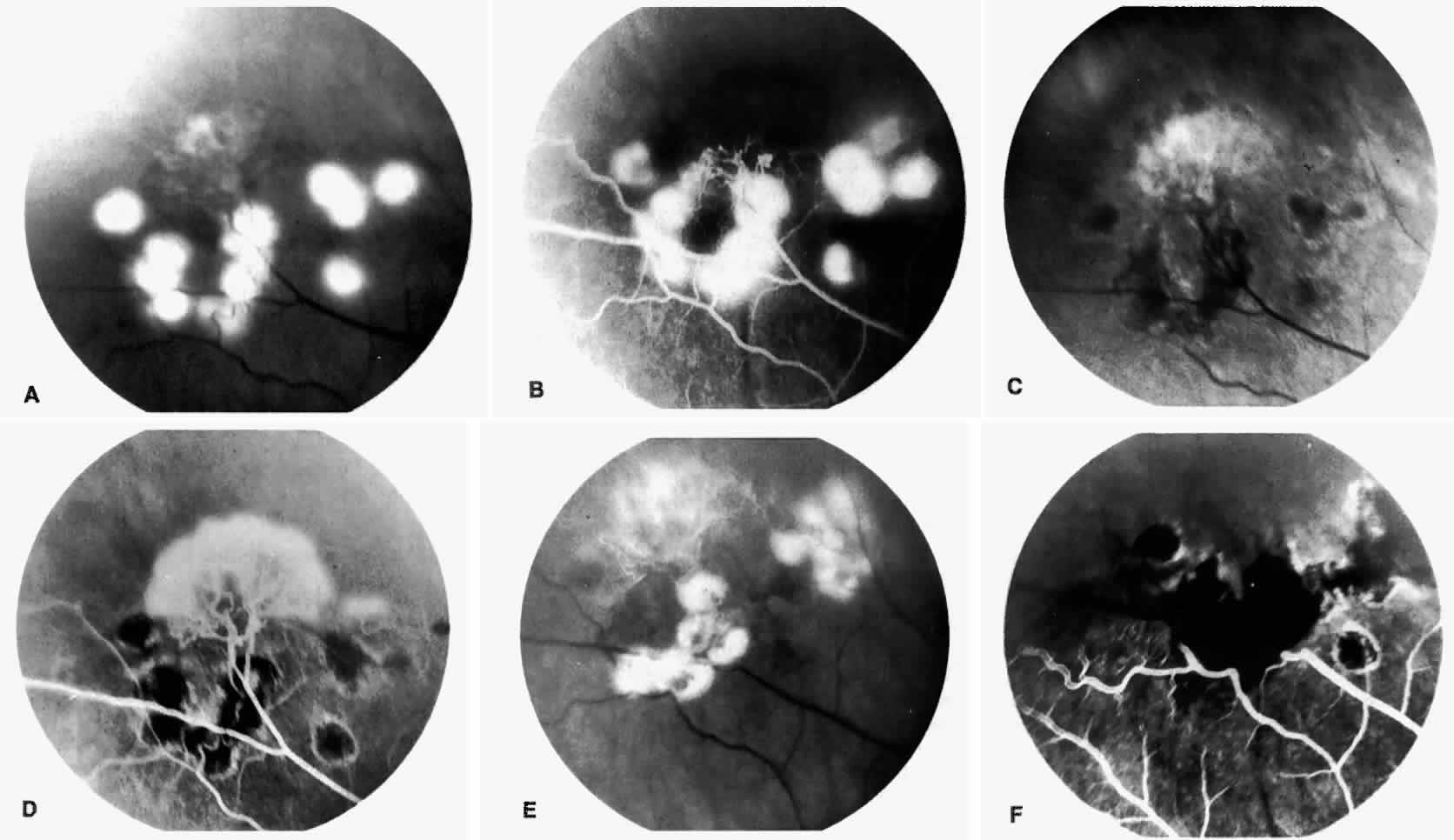
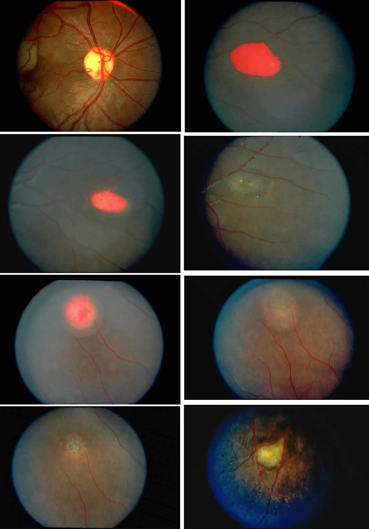
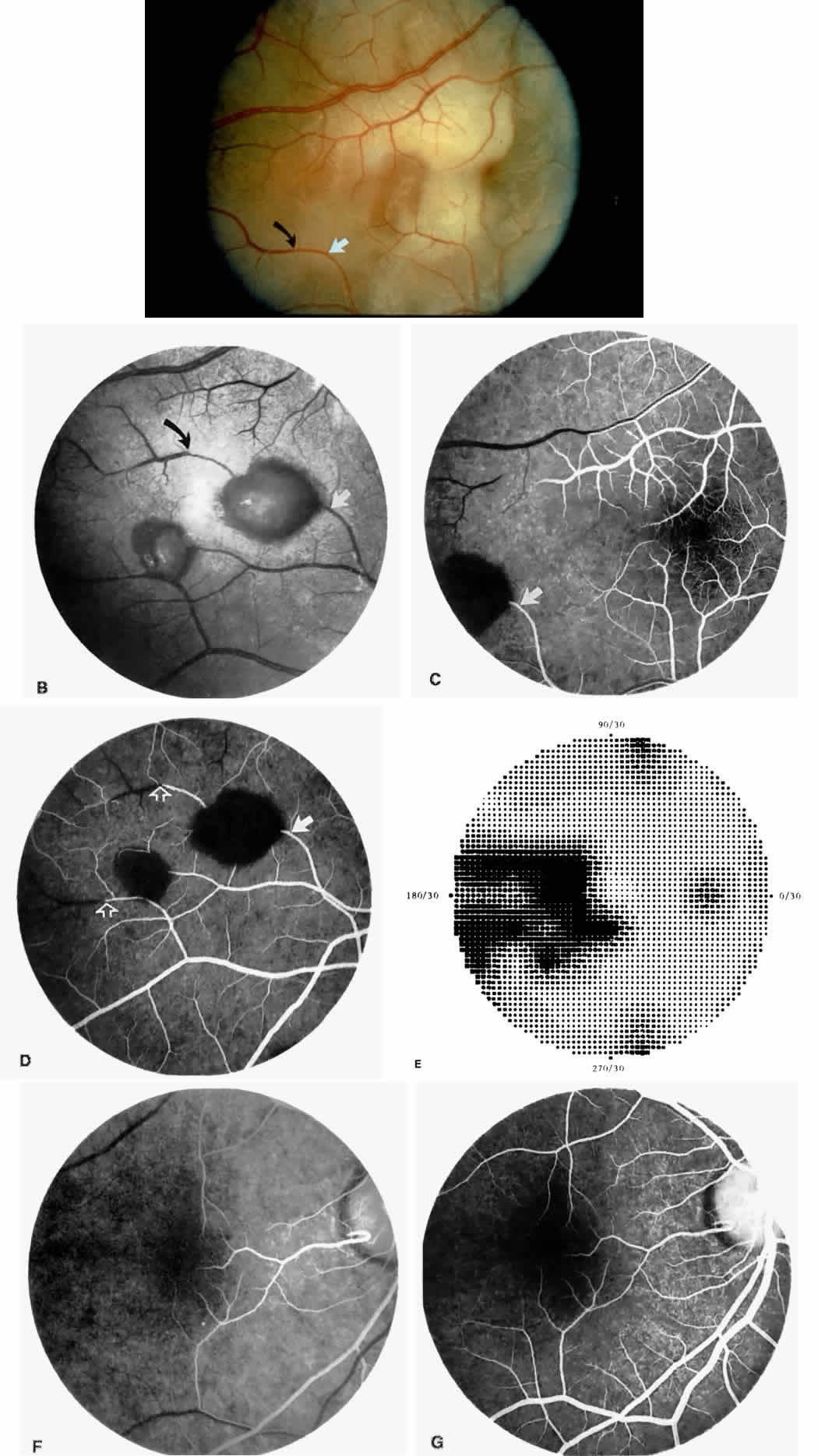
Retinal hemorrhages (“salmon patches”), found most commonly in the equatorial periphery, can be observed after an abrupt occlusion and rupture of an intermediate-sized retinal arteriole (Fig. 16).113 Because the hemorrhages typically appear adjacent or distal to an intraluminal obstruction, it is likely that ischemic necrosis causes a weakening of the vessel wall and that reperfusion of the vessel causes a rupture of the damaged vessel wall, resulting in a hemorrhage (Fig. 17).100 Acutely, these hemorrhages are bright red, but after several days, the partially degenerated blood acquires a characteristic orange-red color (hence the name salmon patch). In most cases, these hemorrhages are asymptomatic. The majority of these hemorrhages remain confined to the sensory retina; however, blood may leak through the internal limiting membrane into the vitreous or dissect deeper into the subretinal space (Fig. 18).114 Resolution occurs over days to weeks and may result in a focal area of atrophic split retina (a “schisis” cavity), a pigmented retinal scar, or a grayish-white vitreous deposit, depending on the location of the hemorrhage (Color Plate 1B through G).115 The blood is slowly cleared by macrophages.

Fig. 16. A 15-year-old boy with homozygous sickle cell anemia, stage II sickle cell retinopathy, and an ISC count of 23.8% noted an acute nasal visual field defect in the right eye. Visual acuity was 20/20 OU. A. Photograph of the right eye demonstrating arteriolar occlusions in the temporal macula, with white, edematous retina and cherry red spot in the fovea. Note the arteriolar occlusion inferiorly, with darkening of the vessel distal to the occlusion site (black arrow). (The closed white arrow identifies a corresponding arteriolar bifurcation in A through D.) B. Red-free photograph 6 days later showing two salmon-patch hemorrhages. The superior salmon-patch hemorrhage overlies the occlusion site noted in A. Note the distal movement of the occlusion site along the arteriole (black arrow). C. Early fluorescein angiogram, showing loss of capillary network in the temporal macula but preservation of the perifoveal network. D. Late phase of the fluorescein angiogram, showing salmon-patch hemorrhages temporally and the occlusions distal to the hemorrhages (open arrows).E. Visual field performed 9 months later shows persistent nasal field defect, but visual acuity has remained 20/20. F. Two years later, a fluorescein angiogram shows a cilioretinal artery perfusing the nasal macula. G. The late phase of the fluorescein angiogram demonstrates complete loss of the temporal macular capillary network.

Fig. 17. A 27-year-old man with homozygous sickle cell anemia. A. A small amount of fluorescein leakage is seen from an arteriole in the inferior nasal peripheral retina (arrow). B. Four months later, a salmon-patch hemorrhage is seen over the leakage site noted in A (arrow). Note the distal arteriolar occlusion, which probably caused increased intravascular pressure and vessel rupture at the area of leakage noted previously.

Fig. 18. Diagram of the evolution of salmon-patch hemorrhage and the development of black sunbursts from subretinal choroidal hemorrhage. (Courtesy of MG Goldberg, MD.)

Intraretinal blood breakdown products, either extracellular or within macrophages, may appear as refractile copper-colored granules (“iridescent spots”") (Color Plate 1H). Macular iridescent schisis lesions have not been described clinically, but they have been observed on histologic examination.114

The occluded vessels may reopen, and the capillary network in the area of a schisis cavity may appear normal; however, more commonly, the vessels will remain closed (Fig. 19). In rare cases, an area of retinal neovascularization may be found within a schisis cavity (Fig. 20).

Fig. 19. A. Iridescent spot in an area of a previous salmon-patch hemorrhage. B. Fluorescein angiogram of corresponding area, demonstrating retinal capillaries in the area of the iridescent spot but adjacent areas of capillary nonperfusion. Arrows in A and B indicate the same arteriolar bifurcation site.

Fig. 20. A. Photograph of an iridescent spot with neovascularization within the schisis cavity. B. Fluorescein angiogram reveals a neovascular membrane within the iridescent spot.

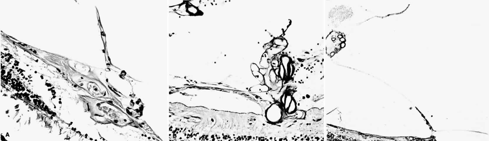
Black pigmented spiculate or stellate chorioretinal lesions (“black sunbursts”) are typically found around or anterior to the equator and adjacent to an arteriole.70 Occasionally, a pigmented lesion may be seen trailing from an arteriole or as a cuff of pigment overlying the vessel (Color Plate 2A).83 Additionally, the overlying arteriole may be occluded. Refractile deposits are often seen interspersed with the pigment. Black sunbursts are believed to be due to deep retinal blood stimulating pigment epithelial migration, hyperplasia, and hypertrophy.116,117 Histopathologic findings support this hypothesis,114 and the development of black sunbursts has been documented in an area of previous intraretinal and subretinal hemorrhage (see Color Plate 1E, F, and G).114,115 An alternative explanation for black sunbursts is the occurrence of choroidal ischemia and aborted choroidal neovascularization.22,118 A spontaneous chorioretinal neovascular membrane was shown to occur within a black sunburst in a 14-year-old girl with homozygous sickle cell anemia.119

Color Plate 2. A. Black sunburst with periarteriole pigment cuff. B. Elevated sea fan neovascularization with white fibroglial mantle. C. Untreated sea fan with localized and diffuse vitreous hemorrhage. D. Feeder vessel photocoagulation immediately after treatment, demonstrating segmented vessels over photocoagulation spots. E. Chorioretinal and choriovitreal neovascularization after feeder vessel photocoagulation. F. Peripheral retinal hole in a patient who was treated with feeder vessel photocoagulation applied to two adjacent sea fans. Scatter photocoagulation was placed around the hole. G. Scatter photocoagulation surrounding a perfused sea fan shown immediately after treatment. (D and G; Gagliano DA, Rabb MF: Sickle cell retinopathy. In Cowan CL (ed): Mediguide to Special Problems in Ophthalmology. New York, Lawrence Della Corte Publications, 1991.)

Dark- and White-Without-Pressure Fundus Lesions

Flat, geographic dark brown areas have been identified in the posterior pole or midperiphery of the retina in patients with sickle cell disease without any signs of a previous hemorrhage or definite evidence of retinal or choroidal vascular occlusion. These dark-without-pressure lesions are transient, changing shape or disappearing over weeks to months.120,121 Areas of white-without-pressure, which are possibly secondary to condensation of the overlying basal vitreous, also have been described.83,122 These two distinct fundus lesions have been noted in black American patients with sickle hemoglobinopathies, but their relationship to sickle hemoglobin, if any, remains uncertain.

Peripheral Retinal Manifestations

The retinal capillary network in the retinal periphery thins to a single layer approximately 1 mm from the ora serrata. A similar thinning of the retinal capillary network occurs around the foveal avascular zone.123–125 These two areas appear to be the most susceptible to vascular occlusions from sickle cell retinopathy. Repeated vascular closures and reopenings of the retinal periphery results in a dynamic remodeling and a centripetal recession of the vascularized border toward the posterior pole.100,126 Peripheral vascular occlusions are seen more frequently on the temporal side, tend to be more rapidly progressive in children and adolescents than in adults, and are significantly more common in homozygous sickle cell anemia than in SC disease.31,32

Redirection of blood flow results in the formation of arteriolar-venular anastomosis at the border of the perfused and nonperfused peripheral retina. This process of vascular closures and arteriolar-venular anastomosis formation is similar to that seen in the spleen, brain, and kidney.127,128 Perhaps unique to the retina, however, is the subsequent development of neovascularization near areas of arteriolar-venular anastomoses.129

The sequential changes of the peripheral retina form the basis of Goldberg's five-stage pathologic classification:

  Stage I: Peripheral arteriolar occlusions
  Stage II: Peripheral arteriolar-venular anastomoses
  Stage III: Preretinal neovascularization
  Stage IV: Vitreous hemorrhage
  Stage V: Retinal detachment (Table 3)130,131


TABLE 3. Pathogenetic Classification of Proliferative Sickle Retinopathy


StagePeripheral Retinal Findings
IPeripheral arteriolar occlusions
IIPeripheral arteriolar-venular anastomosis
IIINeovascular and fibrous proliferation
IVVitreous hemorrhage
VRetinal detachment
(Goldberg MF: Classification and pathogenesis of proliferative sickle retinopathy. Am J Ophthalmol 71:649, 1971.)


Fluorescein angiography remains the best method of identifying these peripheral retinal changes and documenting the presence of neovascularization.19

Examination of the Jamaican Sickle Cohort children with homozygous sickle cell anemia and SC disease, comparing their peripheral retinas with those of age- and sex-matched normal (Hb AA) controls, has revealed that the peripheral retina demonstrates a characteristic change that may allow identification of an increased risk of progressing to neovascularization. A classification scheme has been proposed that divides the peripheral retinal manifestations into qualitatively normal and qualitatively abnormal (Table 4). In the new classification scheme, if there is a continuous arteriolar-venular anastomosis at the margin of the perfused and nonperfused peripheral retina, it is considered qualitatively normal. An abnormal peripheral retinal vascular pattern has capillary stumps extending into nonperfused retina or an irregular capillary border.


TABLE 4. New Classification of Sickle Retinopathy


 Peripheral Retinal Vascular Pattern
Type IQualitatively normal; continuous arteriolar-venular loops with thinned capillary bed
Type IIQualitatively abnormal
 a. Capillary stumps extending into nonperfused retina
 b. Irregular capillary border without arteriolar-venular loops or capillary stumps
Type IIIIndeterminant
(Penman AD, Talbot JF, Chuang EL et al: New classification of peripheral retinal vascular changes in sickle cell disease. Br J Ophthalmol 78:681, 1994.)


Although Talbot and co-workers found that vascular occlusions occurred earlier in homozygous sickle cell disease (Hb SS) than in heterozygous sickle cell disease (Hb SC), a significantly larger proportion of subjects with SC disease versus sickle cell anemia had an abnormal peripheral vascular pattern.31–33 It appears that once an abnormal peripheral retinal vascular pattern appears, it remains qualitatively abnormal despite further loss of the capillary bed. An abnormal border probably occurs as a radical alteration of retinal perfusion and correlates with the subsequent development of neovascularization. A normal border, even if undergoing a posterior regression, results from a gradual modification of the capillary bed and indicates a low risk for PSR.34 However, caution must be exercised, and the entire extent of the peripheral retina must be examined: even though a part of the peripheral retina is qualitatively normal, there may be still be neovascularization in other areas.

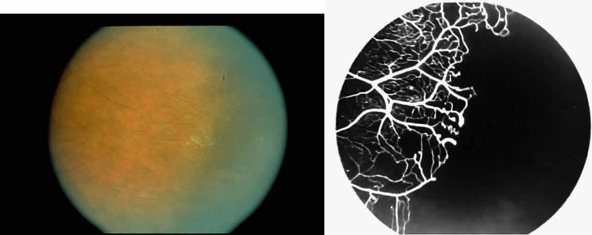
STAGE I: PERIPHERAL ARTERIOLAR OCCLUSIONS. This stage may be further subdivided into three grades: grade I, narrowing of the peripheral arterioles with tortuosity and abnormal looping of the peripheral venules; grade II, tortuosity, dilation, and microaneurysmal formation in the capillary network; and grade III, occlusion of the peripheral capillaries and arterioles.83

Histologic and trypsin digest studies support the theory of a sudden occlusion of the precapillary arteriolar circulation followed by degeneration of the occluded vessels and the distal nonperfused retina. The presence of focal areas of small vessel degeneration and vascular beading (but not typical retinal microaneurysms) also have been confirmed.132

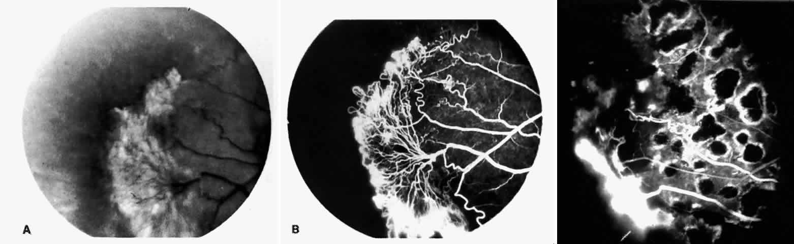
The occluded arterioles may be invisible or may have a “silver-wire” or chalk-white appearance, as first described by Goodman and colleagues39 (Fig. 21). Fluorescein angiography may demonstrate an abrupt complete occlusion at the interface between peripheral nonperfused and posterior perfused retina. Frequently, this occlusion will take place just distal to a branching vessel, giving the appearance of a freshly pruned rose bush. The nonperfused anterior peripheral retina will have a grayish brown appearance and on fluorescein angiography will appear blurred without clearly defined fundus markings.

Fig. 21. A. Photograph of the peripheral retinal vasculature shows sheathed vessels and absence of peripheral vascular perfusion. B. Fluorescein angiogram shows area of nonperfusion representing stage I sickle cell retinopathy. White arrow points to corresponding vascular bifurcation in A and B.

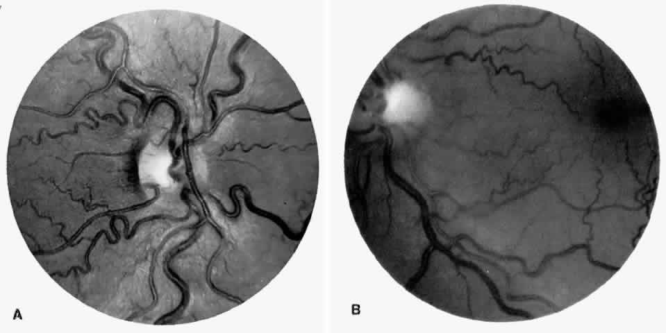
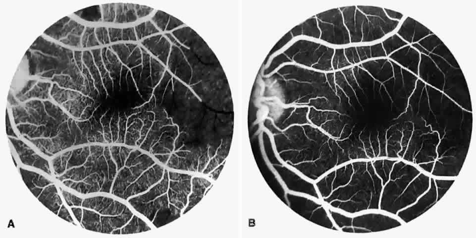
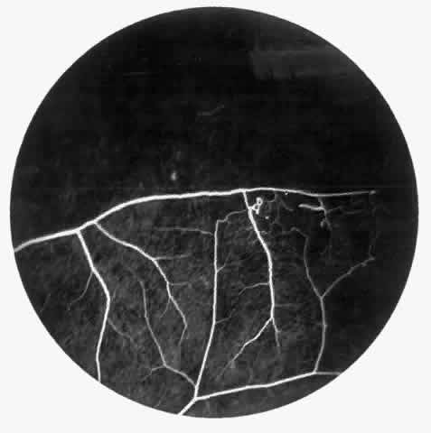
STAGE II: PERIPHERAL ARTERIOLAR-VENULAR ANASTOMOSES. Following occlusion of the terminal arterioles, anastomotic channels form to channel the blood from the occluded arteriole to the nearest venules. These anastomoses form at the interface between the perfused and nonperfused retina. Most likely, they are dilated preexisting capillaries rather than new vessels, since they do not leak on fluorescein angiography. The redirection of blood flow is probably due to hydrostatic forces (Figs. 22 and 23).

Fig. 22. A. Photograph of the peripheral retina, demonstrating capillary occlusions and exudate at the margin of perfused retina. B. Fluorescein angiogram of irregular capillary border, with capillary stumps extending into nonperfused retina and an arteriolar-venular anastomosis demonstrating stage II retinopathy.

Fig. 23. Fluorescein angiogram of continuous arteriolar-venular anastomosis demonstrating stage II retinopathy. (Note that this is the same eye demonstrating the qualitatively abnormal peripheral capillary border in Figure 25.)

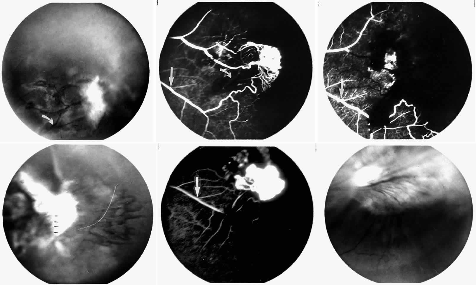
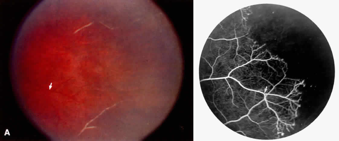
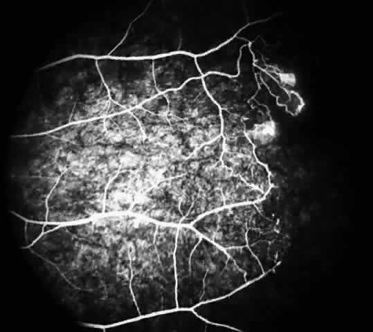
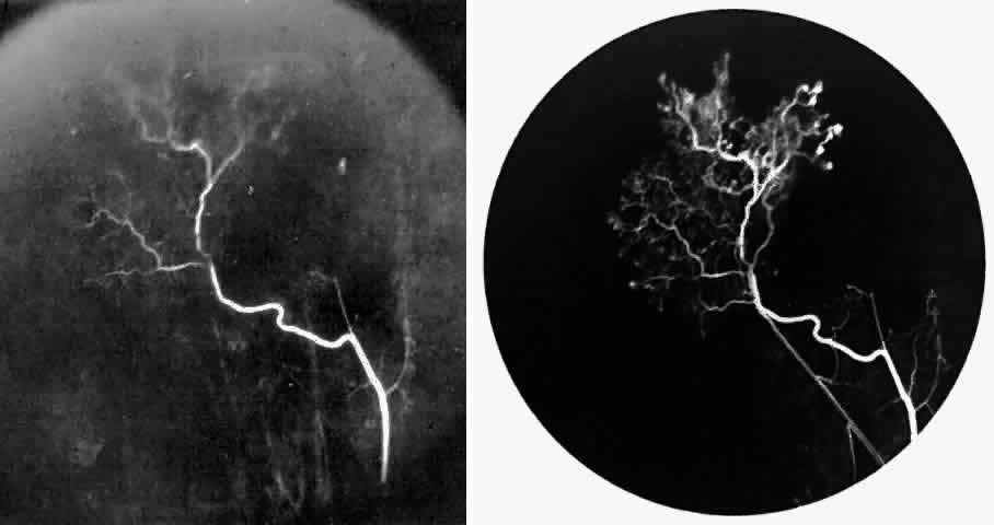
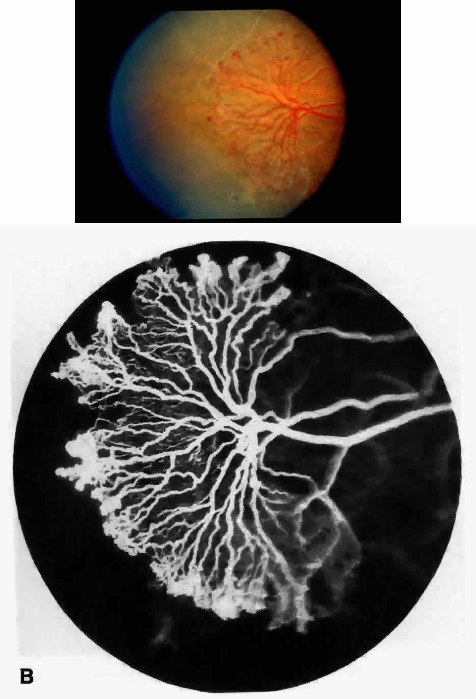
STAGE III: PRERETINAL NEOVASCULARIZATION (PROLIFERATIVE SICKLE RETINOPATHY). “Sea fan”-shaped neovascularization typically develops on the venular side of an arteriolar-venular anastomosis, mimicking the normal development of retinal capillaries (Fig. 24).125 A lowered oxygen tension and angiogenic factors released on the venular side may be the stimulus for neovascular growth.125,126 In most instances, the direction of growth is toward the ora serrata, from the perfused retina toward the nonperfused retina. Presumably, this represents an abortive attempt to revascularize the nonperfused retina, initiated by vasoproliferative factors.

Fig. 24. A. Photograph of the peripheral retina, demonstrating several small fibroglial membranes. B. Fluorescein angiogram corresponding to A, showing multiple arteriolar-venular anastomoses with early sea fan formation. C. Photograph of the same area 2 years later demonstrates more fibroglial membranes. D. Fluorescein angiogram corresponding to C shows new sea fans caused by an arteriolar-venular anastomosis (curved arrow). Large arrow (A through D) identifies corresponding arteriolar bifurcation.

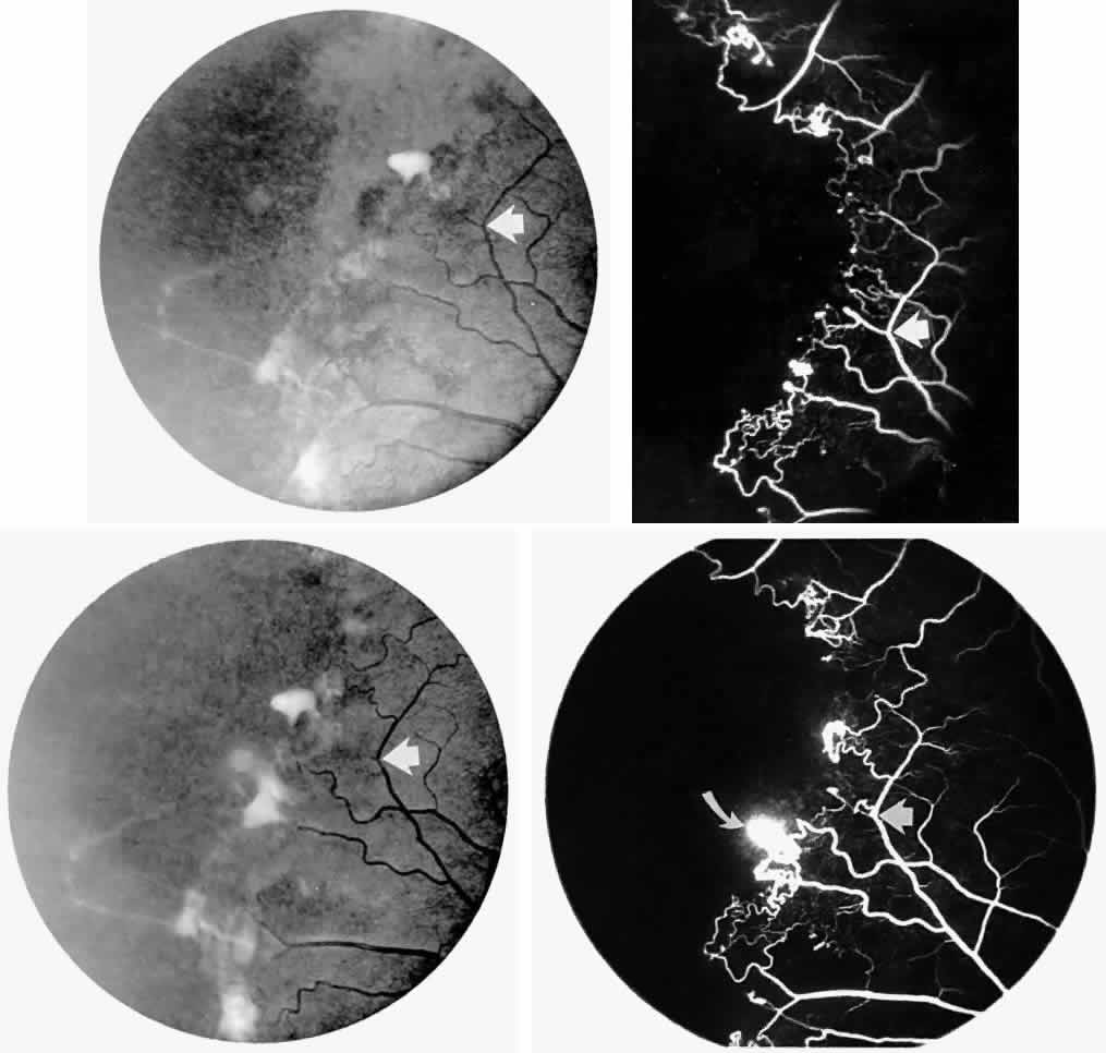
The characteristic neovascular lesions of PSR are called sea fans because they resemble the marine invertebrate Gorgonia flabellum.70 They tend to occur more commonly in the temporal periphery, but they have been reported to occur in the temporal macula in the presence of extensive nonperfusion.130,133 Initially they grow on the surface of the retina, but they often become elevated into the vitreous and adhere to a partially detached posterior hyaloid.114 It may be difficult to visualize small sea fans ophthalmoscopically; however, fluorescein angiography clearly demonstrates leakage of dye into the vitreous (Fig. 25). The feeding arteriole is usually more tortuous than the draining venule (Fig. 26). Early on, the neovascular lesion is fed by a single arteriole and drained by a single venule, but with time, additional arterioles and venules become arborized within the lesion (Fig. 27).129 Growth of the sea fan often occurs circumferentially, rather than radiallyÜmh- 1Ý, toward the ora serrata. Progressive circumferential growth may lead to neovascular lesions extending around the entire periphery. As it matures, a white fibroglial mantle often covers the neovascular tissue (Color Plate 2B).

Fig. 25. Fluorescein angiogram of early proliferative sickle retinopathy arising from an arteriolar-venular anastomosis in an area of irregular peripheral capillary border. Note that this area of qualitatively abnormal peripheral capillary border is in the same eye with a qualitatively normal peripheral retinal vasculature, as demonstrated in Figure 23.

Fig. 26. A. Arterial filling phase of the fluorescein angiogram of a sea fan demonstrates tortuosity of the feeding arteriole. B. Early arteriolar-venular filling phase demonstrates straightening of the draining venule. Note that this sea fan is adjacent to the qualitatively normal peripheral retinal vasculature demonstrated in Figure 23.

Fig. 27. A. Photograph of sea fan neovascularization with hemorrhages at the margins and a white line demarcating perfused and nonperfused retina. B. Fluorescein angiogram shows multiple feeding arterioles and draining venules.

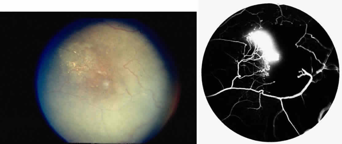
PSR is associated with the severe vision-threatening sequelae of sickle cell disease: vitreous hemorrhage (stage IV) and retinal detachment (stage V). These stages are believed to result from transudation of blood components into the vitreous through the incompetent neovascular tissue (Fig. 28). Vitreous fluorophotometry has quantified the leakage from the peripheral neovascularization.134 This leads to premature syneresis and collapse of the vitreous, inducing tractional forces on the retina that lead to vitreous hemorrhage, retinal tears, and tractional and rhegmatogenous retinal detachment. In rare cases, an exudative detachment may occur.

Fig. 28. A. Cross-section of retina shows vitreous adhesions to flat neovascular tissue (stage III proliferative sickle cell retinopathy [PSR]).Fig. 28 (continued).B. Cross-section of retina reveals large clump of neovascular tissue (stage III PSR) protruding from surface of retina into vitreous. C. Cross-section of retina and vitreous. Note large cluster of intravitreous neovascular tissue adherent to vitreous traction bands. (Romayananda N, Goldberg MF, Green WR: Histopathology of sickle cell retinopathy. Trans Am Acad Ophthalmol Otolaryngol 77:652, 1973.)

Spontaneous nonperfusion or autoinfarction, accompanied by regression of the neovascular lesion, occurs in 20% to 60% of eyes with PSR.135,136 The peak incidence of autoinfarction is 2 years after the development of PSR. It appears that autoinfarction occurs primarily as a result of (1) occlusion of the feeding arteriole due to traction on the neovascular lesion by contracting vitreous, or (2) occlusion by sickled RBCs. The latter probably is more common in homozygous sickle cell anemia, which is more commonly associated with autoinfarction and complete vascular occlusion.

Sea fans develop an average of 14 months after diagnosis of stage II, with an approximate incidence of 14% per year in patients with SC disease.129 In a series of selected patients with different hemoglobinopathies in Jamaica, the incidence of PSR was reported as follows: 2.6% with sickle cell anemia83; 14.0% with Hb S-β-thalassemia91; and 32.8% with SC disease (see Table 1).92 It is a common misconception that patients with homozygous sickle cell anemia have a very low risk of developing retinal neovascularization. Although the risk is recognizably lower, it must be remembered that the development of neovascularization is not only genotype-dependent but also age-dependent, increasing with age in both genotypes with the highest risk period of 20 to 34 years of age in SC disease and 40 to 50 years of age in homozygous sickle cell anemia.26,137 The incidence of PSR in patients with homozygous sickle cell anemia is 14% for patients older than 40 years and up to 29% in patients older than 50 years.25,76 In a series of 786 patients with homozygous sickle cell anemia, the prevalence of PSR was demonstrated to increase gradually, peaking in patients aged 30 years and older. The first cases were observed in patients in their late teens, the incidence rate increasing in patients who were 25 years and older.29 In the same study, a series of 533 patients with SC disease demonstrated that the prevalence of PSR reached a maximum in men in their late 20s and in women older than 40 years, whereas the incidence rates of PSR peaked in men in their early 20s and in women in their late 20s. The prevalence of PSR has been reported to be as high as 68% in SC disease patients 45 years of age and older.26 An ongoing cohort study in Jamaica will undoubtedly provide information on the evolution of PSR in patients with the various genotypes.

As mentioned previously, the otherwise benign condition of sickle cell trait can be associated in rare cases with ocular complications, including neovascularization. Nagpal and associates138 reported that their patients with sickle cell trait who had neovascularization also had associated underlying systemic diseases, including diabetes, hypertension, tuberculosis, syphilis, and sarcoidosis. The neovascularization in these cases was similar to that seen in sickle cell disease. They proposed that any patient with sickle cell trait and PSR be examined thoroughly for other underlying diseases. An additional consideration, and a frequent source of misdiagnosis, is the failure to perform a hemoglobin electrophoresis to clarify a positive sickle cell test. A sickle test can detect the presence of Hb S, but it cannot clarify whether a patient has sickle cell trait or one of the other genotypes associated with sickle cell eye disease. Sickle cell trait may be confused with Hb S-β+ -thalassemia unless a quantitative hemoglobin electrophoresis is done. This may account for some apparent cases of PSR with sickle cell trait. It is still unclear whether sickle cell trait alone can produce PSR.

Once PSR is established, the rates of progression vary greatly among patients. PSR does progress more rapidly in young patients with SC disease and homozygous sickle cell anemia.135

STAGE IV: VITREOUS HEMORRHAGE. Vitreous hemorrhage often complicates PSR. In a selected series of patients with untreated SC disease, vitreous hemorrhage was found in 28% at diagnosis and in 44% after 31 months.130 In the presence of neovascularization, the three risk factors for the development of vitreous hemorrhage include SC disease, more than 60° of perfused sea fans, and the presence of old blood in the eye.139 In a long-term follow-up of an untreated control group participating in a randomized clinical trial of feeder vessel photocoagulation for PSR, vitreous hemorrhage occurred in 45% of eyes and was recurrent in two thirds of these eyes.140

Transudation of plasma results in vitreous syneresis and fibrosis and induces collapse of the formed vitreous, which causes traction on the adherent neovascular tissue. The fragile elevated vessels in the neovascular membranes are prone to rupture, resulting in hemorrhage.141 The hemorrhage is frequently localized in the periphery near the sea fan, but diffuse hemorrhage does occur and may obscure fundus details (Color Plate 2C).

STAGE V: RETINAL DETACHMENT. Vitreous traction on the retina may cause tractional retinal detachments or retinal breaks that can result in localized or total rhegmatogenous retinal detachments.131,142 The retinal breaks are usually found adjacent to neovascular tissue and may be difficult to detect because of overlying hemorrhage. Exudative retinal detachments may occur in rare cases and reportedly have resolved after photocoagulation of the neovascularization.143

Back to Top
MANAGEMENT

TREATMENT OF NONPROLIFERATIVE SICKLE RETINOPATHY

Therapeutic intervention is not indicated for the peripheral manifestations of stage I or stage II sickle cell retinopathy. There is no proven benefit in treating these stages, nor is there any way at present to predict which patients will progress to the proliferative stage. For patients with sickle cell disease, particularly SC disease and Hb S-β+ thalassemia, frequent examinations of the peripheral retina with periodic fluorescein angiography are indicated in an effort to identify neovascular tissue in the early stages of evolution.

Treatment of vascular occlusions of the posterior pole should focus on promoting increased blood flow and preventing further sickling by initiating hydration and oxygenation. Hyperbaric oxygen increases inner retinal oxygen levels and could potentially prove useful in treating retinal vascular occlusions associated with sickle cell disease.65,144 Exchange transfusions have been used, but the true benefit of this is unproved because the natural history of these occlusions is not well documented. In view of the potential complications and variable outcome, routine use of exchange transfusions for retinal vascular occlusions in sickle cell disease is not recommended.87,145

TREATMENT OF PROLIFERATIVE SICKLE RETINOPATHY

A 10-year assessment of 120 patients with homozygous sickle cell anemia and 222 patients with SC disease demonstrated visual acuity loss (20/30 or less) in 10% of untreated eyes, which was strongly associated with PSR.146 The long-term evaluation of control patients enrolled in the feeder vessel treatment study demonstrated that despite auto- infarction, 80% of eyes with untreated PSR had persistent sea fans after 10 years.140 Therefore, treatment of PSR is necessary to prevent vision-threatening complications, including vitreous hemorrhage and retinal detachment.147

Treatment of PSR is aimed at ablating or inducing regression of the sea fan. Various modalities have been utilized, including diathermy, cryotherapy, xenon arc photocoagulation, and various techniques of laser photocoagulation, such as feeder vessel, local scatter, and peripheral circumferential scatter.134,140,147–160 Laser photocoagulation is the treatment modality most commonly used.

Feeder Vessel Photocoagulation

Randomized clinical trials demonstrating the efficacy of treatment utilizing the feeder vessel technique were performed on patients with PSR in Kingston, Jamaica, with xenon arc photocoagulation and in Chicago with argon blue-green laser photocoagulation.139,140,156

In these studies, the laser photocoagulation protocol was to use a spot size of 500 μm with a 0.2-second duration and a power high enough to cause closure of the feeding and draining blood vessels (Color Plate 2D). Treatment spots were first placed on the feeding arteriole, and after segmentation of the arteriole, the draining venule was treated in a similar fashion. When the arteriole cannot be segmented, retreatment can be performed 2 to 3 weeks later, when pigmentation has occurred in the area of treatment (Fig. 29). Because of possible complications, it may be prudent to approach the treatment as a two-stage process, allowing a lower power to be used and possibly reducing the complication rate. Typically, more than one sea fan requiring treatment will be identified during the initial treatment session.140 Future considerations for this technique may involve the use of dye yellow laser, which has a better hemoglobin absorption characteristic, or dye-enhanced photocoagulation.

Fig. 29. Feeder vessel photocoagulation. A. Photograph of feeder vessel photocoagulation applied to the feeding arterioles and draining venules of a sea fan. B. Fluorescein angiogram corresponding to A, showing nonperfusion of the sea fan. C. Photograph taken 2 weeks after treatment, demonstrating pigmented laser lesions and an increased fibrovascular appearance of the sea fan. D. Fluorescein angiogram corresponding to C, showing laser spots and perfusion of the previously closed sea fan. E. Photograph showing retreatment using the feeder vessel photocoagulation technique over the pigmented spots. F. Fluorescein angiogram corresponding to E, showing nonperfusion of the sea fan after retreatment.

Efficacy of treatment can be monitored with fluorescein angiography. Successful treatment is indicated by occlusion of the feeding arterioles as demonstrated by angiography, which is performed 4 to 6 weeks after the treatment to determine whether there is complete and permanent interruption of blood flow. Retreatment is often necessary to achieve complete closure. In addition, patients should be followed closely to identify any new areas of neovascularization. There does not appear to be any significant reduction in the development of new sea fans with the feeder vessel technique, since new sea fans developed in approximately 50% of both the untreated and treated patients during nearly a decade of observation.140

Potential complications of feeder vessel photocoagulation include chorioretinal/choriovitreal neovascularization (Fig. 30, Color Plate 2E), retinal breaks (Color Plate 2F), retinal detachment, retinal hemorrhages, choroidal hemorrhages, and peripheral choroidal ischemia.112,156,157,161–167 The complication rate with xenon arc photocoagulation is higher (61%) in comparison with argon laser photocoagulation (up to 32%)140,156,157,164; however, all the complications were found to have arisen within 6 months of treatment, and after a 10-year evaluation, no significant incidence of visual loss was found as a result of complications from feeder vessel laser photocoagulation.140

Fig. 30. Evolution of choriovitreal neovascularization (CVN) after feeder vessel photocoagulation. Straight arrow in A, C, and E identifies the same vascular bifurcation. A. Photograph of a sea fan in the left eye taken 2 weeks after feeder vessel photocoagulation. The treatment spots are seen to have pigmented. Curved arrow in A and B identifies the treatment location that will develop a CVN. B. Fluorescein angiogram corresponding to A reveals continued perfusion of the sea fan. C. Fluorescein angiogram 1 week after second feeder vessel photocoagulation treatment, demonstrating nonperfusion of the sea fan. D. Photograph of the sea fan taken 1 month later, demonstrating an elevated neovascular membrane proximal to the sea fan over the treatment on the feeding arteriole with vitreous hemorrhage. Black arrows identify the distal extent of the CVN membrane. E. Fluorescein angiogram corresponding to D, demonstrating rapid filling of the chorioretinal and choriovitreal neovascularization. F. Photograph of chorioretinal and choriovitreal neovascularization taken 6 weeks later demonstrates continued enlargement and elevation. (See Color Plate 2E for appearance 4 weeks later.)

Local Scatter Photocoagulation

This technique involves the placement of laser spots around the sea fan in a scatter fashion, 1 spot-diameter apart, and extending from 1 disc diameter anterior to 1 disc diameter posterior to the lesion, and 1 clock-hour to each side (Color Plate 2G). We use a spot size of 500 μm with a 0.1-second duration and enough power to produce a light gray spot. Additional treatment is applied when there is growth or reperfusion of neovascular tissue. No spots are placed directly on the vessels, but all neovascular lesions are treated.153 All previous studies have used argon blue-green or green laser, but krypton red or diode infrared laser treatment would probably be equally effective.

It must be remembered that the power level should be lowered in moving from perfused retina to the nonperfused, thinner retina to avoid the complication of applying excessively intense (hot) spots and risking rupture of Bruch's membrane. A useful technique when applying photocoagulation to the retina is to use low levels of power initially and to place the laser treatment in the area anticipated to have the best absorption, thus helping to reduce the occurrence of this potential complication.

A randomized, prospective clinical trial using argon blue-green laser and a localized scatter technique demonstrated a significant reduction in both prolonged vision loss and the incidence of vitreous hemorrhage.160 There were no complications reported, indicating that this technique is much safer than the feeder vessel technique. With scatter photocoagulation, as with all photocoagulation techniques, successful treatment is indicated by reduced perfusion or nonperfusion of the sea fan, as demonstrated by a lack of leakage on fluorescein angiography (Fig. 31). Complete closure of neovascularization was achieved in 30.2% of eyes and partial closure in 51%. An additional secondary benefit was a possible reduction in the development of new sea fans, which were demonstrated in 34.3% of treated eyes versus 41.3% of control eyes.

Fig. 31. Scatter photocoagulation. A. Photograph of a large sea fan with localized vitreous hemorrhage. B. Fluorescein angiogram corresponding to A, demonstrating multiple feeding arterioles and draining venules. C. Fluorescein angiogram, demonstrating closure of the sea fan with scatter photocoagulation treatment.

Another recent study demonstrated that complete infarction of lesions is more likely if the neovascularization is small (less than 15° of circumferential involvement) and if the patients are young (less than 25 years).168 When scatter photocoagulation does not induce sufficient regression of neovascular tissue, it may be necessary to perform feeder vessel photocoagulation.159

Peripheral Circumferential Scatter

In the peripheral circumferential scatter technique, scatter laser photocoagulation is placed over the entire 360° anterior peripheral zone of capillary nonperfusion.154,155 Although this technique has not been tested in a randomized clinical trial, investigators have reported complete regression of neovascularization in 33% of sea fans and partial regression in 46%. Interestingly, new sea fans developed in only 1.4% of eyes treated with this technique, compared with 34.3% of eyes treated with local scatter and more than 50% of eyes treated with the feeder vessel technique.140,154,155 However, treating 360° in every patient may not be necessary when local scatter can achieve adequate closure of neovascular tissue. The extensive photocoagulation of peripheral circumferential scatter may cause secondary complications related to photocoagulation, such as macular pucker. In the situation of unreliable follow-up, however, a 360° treatment may be preferable.159

Laser Vitreolysis

Neodymium-yytrium aluminum garnet (Nd:YAG) laser vitreolysis may be used to treat localized vitreous traction and tractional retinal detachments. The indications for using this technique are the presence of avascular vitreous bands located at least 3 mm from the retinal surface.169 Treatment closer than 3 mm to the retina has been reported to cause choroidal hemorrhage and retinal pigment epithelial lesions.

SURGICAL TREATMENT

Vitreoretinal surgery may be indicated for the treatment of tractional or rhegmatogenous retinal detachments, nonclearing vitreous hemorrhages, or visually disabling epiretinal membranes. Special consideration is warranted in sickle cell disease patients because of potentially devastating operative and postoperative pulmonary, cerebral, and ocular thromboembolic complications.

The thromboembolic ocular complications of vitrectomy and retinal detachment repair include anterior segment ischemia and intraoperative hemorrhage, with sickling and secondary glaucoma. Surgery is further complicated by difficulty visualizing peripheral retinal breaks, which are often hidden by overlying neovascular membranes, fibrous tissue, and hemorrhage. Because of thinning of the peripheral retina, iatrogenic breaks have been reported in up to 33% of cases.170 Additionally, the peripheral location of the neovascular membranes increases the risk of lens damage.158

A high incidence of anterior segment ischemia (up to 71% in SC disease patients undergoing scleral buckling procedures) has stimulated the use of preoperative prophylactic exchange transfusions or erythrophoresis in patients with sickle cell disease.172–175 Preoperative exchange transfusions may reduce the potential for intraoperative and postoperative complications related to elevated intraocular pressure caused by hyphema or posterior hemorrhage. The risk of acquired infections (e.g., HIV; non-A, non-B hepatitis) and other transfusion-related complications, however, has stimulated a reevaluation of the routine use of preoperative exchange transfusions.176 Use of perioperative oxygen, improved vitreoretinal surgical techniques, and other preoperative and intraoperative measures to reduce complications has resulted in an improvement in surgical and systemic outcome without transfusions that must be balanced against the risks of exchange transfusion.176–179

The goals of preoperative partial exchange transfusions, if elected, are to elevate Hb A levels to 40% to 60% and to achieve a final hematocrit of no higher than 35% to 39% to prevent elevated whole-blood viscosity. Other benefits of partial exchange transfusions are enhancement of perfusion and oxygen delivery and minimization of intraoperative and early postoperative sickling.170

The following are additional ways to reduce potential complications, particularly anterior segment ischemia: (1) using no sympathomimetics with local anesthesia; (2) using topical sympathomimetics minimally; (3) using supplemental 100% oxygen for 48 hours after surgery; (4) avoiding excessive manipulation of extraocular muscles; (5) using transscleral diathermy or cryotherapy minimally and avoiding the long ciliary arteries; (6) avoiding the use of wide encircling scleral buckling elements; (7) limiting the use of expansile concentrations of intraocular gases; (8) utilizing internal drainage of subretinal fluid; and (9) closely monitoring and treating elevated intraocular pressure, avoiding the use of carbonic anhydrase inhibitors.

Treatment of anterior segment ischemia is difficult. When available, oxygen by mask, especially under hyperbaric conditions, and oxygen delivery to the anterior segment via goggles may be helpful in preventing and treating anterior segment ischemia.65,144 We used this technique successfully to reverse anterior segment ischemia following scleral buckling surgery in an SC disease patient of ours.

Back to Top
THE FUTURE
In the last 25 years, many advances have reduced the incidence of vision loss due to sickle cell retinopathy. At present, it seems unlikely that breakthroughs in the use of laser or new vitreoretinal surgical techniques will improve the outcomes greatly. The development of techniques to detect risk factors that predict future adverse events (e.g., sea fan formation) may allow earlier detection and selective treatment of high-risk patients.

Further research is needed to determine the cellular and rheologic factors contributing to the microvascular occlusions in sickle cell disease, as well as therapeutic measures to reduce sickling and secondary occlusions. Potential modalities include increasing Hb F levels, replacing the abnormal sickle cell β-globin gene, stabilizing the RBC membrane, reducing ISC levels, decreasing sickled cell adhesiveness, and selective arteriolar vasodilatation.180–183 Several pharmacologic modalities have demonstrated efficacy in enhancing γ-globin synthesis (Hb F) in patients with homozygous sickle cell anemia, and this in turn has been shown to improve the filterability of RBCs and to reduce the number of dense ISCs. Most promising is the use of nontoxic metabolites, such as sodium butyrate and its analogues.184

Back to Top
REFERENCES

1. Herrick JB: Peculiar elongated and sickle-shaped red blood corpuscles in a case of severe anemia. Arch Intern Med 6:517, 1910

2. Cook WC: A case of sickle cell anemia with associated subarachnoid hemorrhage. J Med 11:541, 1930

3. Stamatoyannopoulos G, Nienhuis AW: Hemoglobin switching. In Stamatoyannopoulos G, Nienhuis AW, Majerus PW, Varmus H (eds): The Molecular Basis of Blood Diseases, 2nd ed, pp 107–155. Philadelphia, WB Saunders, 1994

4. Pauling L, Itano HA, Singer SJ, Wells IC: Sickle cell anemia: a molecular disease. Science 110:543, 1949

5. Ingram VM: A specific chemical difference between the globins of normal human and sickle-cell anaemia haemoglobin. Nature 178:792, 1956

6. Ingram VM: Gene mutations in human haemoglobin: the chemical difference between normal and sickle-cell haemoglobin. Nature 180:326, 1957

7. Lee GR, Bithell TC, Foerster J et al (eds): Wintrobe's Clinical Hematology, 9th ed, pp 1023–1145. Philadelphia, Lea & Febiger, 1993

8. Rucknagel DL: The genetics of sickle cell anemia and related syndromes. Arch Intern Med 133:595, 1974

9. Dean J, Schechter AN: Sickle cell anemia: molecular and cellular bases of therapeutic approaches. N Engl J Med 299:752, 1978

10. Nagel RL, Ranney HM: Genetic epidemiology of structural mutations of the β-globin gene. Semin Hematol 27:342, 1990

11. Kan YW, Dozy AM: Evolution of the hemoglobin S and C genes in world populations. Science 209:388, 1980

12. Chauhan PM, Kondlapoodi P, Natta CL: Pathology of sickle cell disorders. Pathol Annu 18:253, 1983

13. Raper AB: The incidence of sicklaemia. East Afr Med J 26:281, 1949

14. Allison AC: Protection afforded by sickle cell trait against subtertion malarial infection. Br Med J 1:290, 1954

15. Friedman MJ: Erythrocytic mechanism of sickle cell resistance to malaria. Proc Natl Acad Sci USA 75:1994, 1978

16. Green MA, Noguchi CT, Keidan AJ et al: Polymerization of sickle cell hemoglobin at arterial oxygen saturation impairs erythrocyte deformability. J Clin Invest 81:1669, 1988

17. Kurantsin-Mills J, Klug PP, Lessin LS: Vaso-occlusion in sickle cell disease: pathophysiology of the microvascular circulation. Am J Pediatr Hematol Oncol 10:357, 1988

18. Stevens TS, Busse B, Lee C et al: Sickling hemoglobinopathies: macular and perimacular vascular abnormalities. Arch Ophthalmol 92:455, 1974

19. Asdourian GK, Goldberg MF: The angiographic pattern of the peripheral retinal vasculature. Arch Ophthalmol 97:2316, 1979

20. Klug PP, Lessin LS, Radice P: Rheological aspects of sickle cell disease. Arch Intern Med 133:577, 1974

21. Horne MK: Sickle cell anemia as a rheologic disease. Am J Med 70:288, 1981

22. McLeod GS, Goldberg MF, Lutty GA: Dual-perspective analysis of vascular formations in sickle cell retinopathy. Arch Ophthalmol 111:1234, 1993

23. McCurdy PR, Sherman AS: Irreversibly sickled cells and red cell survival in sickle cell anemia: a study with both DF32P and 51Cr. Am J Med 64:253, 1978

24. Bunn HF, Forget BG: Hemoglobin: Molecular, Genetic and Clinical Aspects. Philadelphia, WB Saunders, 1986

25. Hayes RJ, Condon PI, Serjeant GR: Haematological factors associated with proliferative retinopathy in homozygous sickle cell disease. Br J Ophthalmol 65:29, 1981

26. Hayes RJ, Condon PI, Serjeant GR: Haematological factors associated with proliferative retinopathy in sickle cell-haemoglobin C disease. Br J Ophthalmol 65:712, 1981

27. Serjeant BE, Mason KP, Condon PI et al: Blood rheology and proliferative retinopathy in sickle cell-haemoglobin C disease. Br J Ophthalmol 68:325, 1984

28. Serjeant BE, Mason KP, Maude GH et al: Blood rheology and proliferative retinopathy in homozygous sickle cell disease. Br J Ophthalmol 70:522, 1986

29. Fox PD, Dunn DT, Morris JS, Serjeant GR: Risk factors for proliferative sickle retinopathy. Br J Ophthalmol 74: 172, 1990

30. Fox PD, Higgs DR, Serjeant GR: Influence of a thalassaemia on the retinopathy of homozygous sickle cell disease. Br J Ophthalmol 77:89, 1993

31. Talbot JF, Bird AC, Serjeant GR, Hayes RJ: Sickle cell retinopathy in young children in Jamaica. Br J Ophthalmol 66:149, 1982

32. Talbot JF, Bird AC, Rabb LM et al: Sickle cell retinopathy in Jamaican children: a search for prognostic factors. Br J Ophthalmol 67:782, 1983

33. Talbot JF, Bird AC, Maude GH et al: Sickle cell retinopathy in Jamaican children: further observations from a cohort study. Br J Ophthalmol 72:727, 1988

34. Penman AD, Talbot JF, Chuang EL et al: New classification of peripheral retinal vascular changes in sickle cell disease. Br J Ophthalmol 78:681, 1994

35. Jampol LM, Ebroon DA, Goldbaum MH: Peripheral proliferative retinopathies: an update on angiogenesis, etiologies and management. Surv Ophthalmol 38:519, 1994

36. Peachey NS, Charles HC, Lee CM et al: Electroretinographic findings in sickle cell retinopathy. Am J Ophthalmol 105:934, 1987

37. Peachey NS, Gagliano DA, Jacobson MS et al: Correlation of electroretinographic findings and peripheral retinal nonperfusion in patients with sickle cell retinopathy. Arch Ophthalmol 108:1106, 1990

38. Knisely MH, Bloch EH, Eliot TS, Warner L: Sludged blood. Science 106:431, 1947

39. Goodman G, Sallman L, Holland MG: Ocular manifestations of sickle-cell disease. Arch Ophthalmol 58:655, 1957

40. Geeraets WJ, Guerry D: Clinical observations on conjunctival capillaries with special reference to sickle cell disease. South Med J 53:949, 1960

41. Fink AI, Funahashi T, Robinson M, Watson RJ: Conjunctival blood flow in sickle-cell disease. Arch Ophthalmol 66:824, 1961

42. Paton D: The conjunctival sign of sickle-cell disease. Arch Ophthalmol 66:90, 1961

43. Paton D: The conjunctival sign of sickle-cell disease: further observations. Arch Ophthalmol 68:627, 1962

44. Serjeant GR, Serjeant BE, Condon PI: The conjunctival sign in sickle cell anemia. JAMA 219:1428, 1972

45. Nagpal KC, Asdourian G, Goldbaum M et al: The conjunctival sickling sign, hemoglobin S, and irreversibly sickled erythrocytes. Arch Ophthalmol 95:808, 1977

46. Roy MS, Rodgers GR, Podgor MJ et al: Conjunctival sign in sickle cell anaemia: an in-vivo correlate of the extent of the red cell heterogeneity. Br J Ophthalmol 69:629, 1988

47. Gagliano DA, Jacobson MS, Labatka R et al: The conjunctival sickling sign, red cell density, and irreversibly sickled cells in sickle cell hemoglobinopathy. Poster presentation, 93rd Meeting of the American Academy of Ophthalmology, New Orleans, LA, October 29-November 2, 1989

48. Teich SA: Conjunctival vascular changes in AIDS and AIDS-related complex. Am J Ophthalmol 103:332, 1987

49. Swartz M, Jampol LM: Comma-shaped venular segments of conjunctiva in chronic granulocytic leukemia. Can J Ophthalmol 10:458, 1975

50. Galinos SO, Rabb MF, Goldberg MF, Frenkel M: Hemoglobin SC disease and iris atrophy. Am J Ophthalmol 75:421, 1973

51. Chambers J, Puglisi J, Kernitsky R, Wise GN: Iris atrophy in hemoglobin SC disease. Am J Ophthalmol 77:247, 1974

52. Goldberg MF, Tso MOM: Rubeosis iridis and glaucoma associated with sickle cell retinopathy: a light and electron microscopic study. Ophthalmology 85:1028, 1978

53. Sorr EM, Goldberg RE: Traumatic central retinal artery occlusion with sickle cell trait. Am J Ophthalmol 80: 648, 1975

54. Wax MB, Ridley ME, Magargal LE: Reversal of retinal and optic disc ischemia in a patient with sickle cell trait and glaucoma secondary to traumatic hyphema. Ophthalmology 89:845, 1982

55. Goldberg MF: The diagnosis and treatment of sickled erythrocytes in human hyphemas. Trans Am Ophthalmol Soc 76:481, 1978

56. Goldberg MF: Sickled erythrocytes, hyphema, and secondary glaucoma: I. The diagnosis and treatment of sickled erythrocytes in human hyphemas. Ophthalmic Surg 10:17, 1979

57. Goldberg MF: The diagnosis and treatment of secondary glaucoma after hyphema in sickle cell patients. Am J Ophthalmol 87:43, 1979

58. Goldberg MF, Dizon R, Raichand M: Sickled erythrocytes, hyphema, and secondary glaucoma: II. Injected sickle cell erythrocytes into human, monkey, and guinea pig anterior chambers: The induction of sickling and secondary glaucoma. Ophthalmic Surg 10:32, 1979

59. Goldberg MF, Dizon R, Raichand M et al: Sickled erythrocytes, hyphema, and secondary glaucoma: III. Effects of sickle cell and normal human blood samples in rabbit anterior chambers. Ophthalmic Surg 10:51, 1979

60. Goldberg MF: Sickled erythrocytes, hyphema, and secondary glaucoma: IV. The rate and percentage of sickling of erythrocytes in rabbit aqueous humor, in vitro and in vivo. Ophthalmic Surg 10:62, 1979

61. Goldberg MF: Sickled erythrocytes, hyphema, and secondary glaucoma: V. The effect of vitamin C on erythrocyte sickling in aqueous humor. Ophthalmic Surg 10(4): 70, 1979

62. Goldberg MF, Dizon R, Moses VK: Sickled erythrocytes, hyphema and secondary glaucoma: VI. The relationship between intracameral blood cells and aqueous humor pH, pO2, and pCO2. Ophthalmic Surg 10:78, 1979

63. Goldberg MF, Tso MOM: Sickled erythrocytes, hyphema, and secondary glaucoma: VII. The passage of sickled erythrocytes out of the anterior chamber of the human and monkey eye: light and microscopic studies. Ophthalmic Surg 10(4):89, 1979

64. Greenwald MJ, Crowley TM: Sickle cell hyphema with secondary glaucoma in a non-black patient. Ophthalmic Surg 16:170, 1985

65. Jampol LM, Orlin C, Cohen SB et al: Hyperbaric and transcorneal delivery of oxygen to the rabbit and monkey anterior segment. Arch Ophthalmol 106:825, 1988

66. Deutsch TA, Weinreb RN, Goldberg MF: Indications for surgical management of hyphema in patients with sickle cell trait. Arch Ophthalmol 102:566, 1984

67. Goldbaum MH, Jampol LM, Goldberg MF: The disc sign in sickling hemoglobinopathies. Arch Ophthalmol 96: 1957, 1978

68. Ober RR, Michels RG: Optic disk neovascularization in hemoglobin SC disease. Am J Ophthalmol 85:711, 1978

69. Kimmel AS, Magargal LE, Tasman WS: Proliferative sickle retinopathy and neovascularization of the disc: regression following treatment with peripheral retinal scatter laser photocoagulation. Ophthalmic Surg 17:20, 1986

70. Welch RB, Goldberg MF: Sickle-cell hemoglobin and its relation to fundus abnormality. Arch Ophthalmol 75: 353, 1966

71. Friberg TR, Young CM, Milner PF: Incidence of ocular abnormalities in patients with sickle hemoglobinopathies. Ann Ophthalmol 18:150, 1986

72. Geeraets WJ, Guerry D III: Angioid streaks and sickle-cell disease. Am J Ophthalmol 49:450, 1960

73. Nagpal KC, Asdourian G, Goldbaum M et al: Angioid streaks and sickle haemoglobinopathies. Br J Ophthalmol 60:31, 1976

74. Clarkson JG, Altman RD: Angioid streaks. Surv Ophthalmol 26:235, 1982

75. Hamilton AM, Pope FM, Condon PI et al: Angioid streaks in Jamaican patients with homozygous sickle cell disease. Br J Ophthalmol 65:341, 1981

76. Condon PI, Serjeant GR: Ocular findings in elderly cases of homozygous sickle cell disease in Jamaica. Br J Ophthalmol 60:361, 1976

77. Jampol LM, Acheson R, Eagle RC et al: Calcification of Bruch's membrane in angioid streaks with homozygous sickle cell disease. Arch Ophthalmol 105:93, 1987

78. Carney MD, Jampol LM: Epiretinal membranes in sickle cell retinopathy. Arch Ophthalmol 105:214, 1987

79. Moriarty BJ, Acheson RW, Serjeant GR: Epiretinal membranes in sickle cell disease. Br J Ophthalmol 71:466, 1987

80. Sumers KD, Jampol LM, Goldberg MF, Huamonte FU: Spontaneous separation of epiretinal membranes. Arch Ophthalmol 98:318, 1980

81. Messner KH: Spontaneous separation of preretinal macular fibrosis. Am J Ophthalmol 83:9, 1977

82. Raichand M, Dizon RV, Nagpal KC et al: Macular holes associated with proliferative sickle cell retinopathy. Arch Ophthalmol 96:1592, 1978

83. Condon PI, Serjeant GR: Ocular findings in homozygous sickle cell anemia in Jamaica. Am J Ophthalmol 73:533, 1972

84. Chopdar A: Multiple major retinal vascular occlusions in sickle cell hemoglobin C disease. Br J Ophthalmol 59: 493, 1975

85. Acacio I, Goldberg MF: Peripapillary and macular vessel occlusions in sickle cell anemia. Am J Ophthalmol 75: 861, 1973

86. Condon PI, Whitelocke RAF, Bird AC et al: Recurrent visual loss in homozygous sickle cell disease. Br J Ophthalmol 69:700, 1985

87. Weissman H, Nadel AJ, Dunn M: Simultaneous bilateral retinal arterial occlusions treated by exchange transfusions. Arch Ophthalmol 97:2151, 1979

88. Klein ML, Jampol LM, Condon PI et al: Central retinal artery occlusion without retrobulbar hemorrhage after retrobulbar anesthesia. Am J Ophthalmol 93:573, 1982

89. Stein MR, Gay AJ: Acute chorioretinal infarction in sickle cell trait: report of a case. Arch Ophthalmol 84:484, 1970

90. Kabakow B, van Weimokly SS, Lyons HA: Bilateral central artery occlusion: occurrence in a patient with cortisone-treated systemic lupus erythematosus, sickle cell trait and active pulmonary tuberculosis. Ophthalmology 54:670, 1955

91. Condon PI, Serjeant GR: Ocular findings in sickle cell thalassemia in Jamaica. Am J Ophthalmol 74:1105, 1972

92. Condon PI, Serjeant GR: Ocular findings in hemoglobin SC disease in Jamaica. Am J Ophthalmol 74:921, 1972

93. Asdourian GK, Nagpal KC, Busse B et al: Macular and perimacular vascular remodeling in sickling haemoglobinopathies. Br J Ophthalmol 60:431, 1976

94. Condon PI, Marsh RJ, Maude GH et al: Alpha thalassaemia and the macular vasculature in homozygous sickle cell disease. Br J Ophthalmol 67:779, 1983

95. Knapp JW: Isolated macular infarction in sickle cell (SS) disease. Ophthalmology 73:857, 1972

96. Merritt JC, Risco JM, Pantell JP: Bilateral macular infarction in SS disease. J Pediatr Ophthalmol Strabismus 19: 275, 1982

97. Ryan SJ: Occlusion of the macular capillaries in sickle cell hemoglobin C disease. Am J Ophthalmol 77:459, 1974

98. Asdourian GK, Goldberg MF, Rabb MF: Macular infarction in sickle cell B+ thalassemia. Retina 2:155, 1982

99. Westrich DJ, Feman SS: Macular arteriolar occlusions in sickle cell beta thalassemia. Am J Ophthalmol 101:739, 1986

100. Goldberg MF: Retinal vaso-occlusion in sickling hemoglobinopathies. Birth Defects 12:474, 1976

101. Marsh RJ, Ford SM, Rabb MF et al: Macular vasculature, visual acuity, and irreversibly sickled cells in homozygous sickle cell disease. Br J Ophthalmol 66:155, 1982

102. Lee CM, Charles HC, Smith RT et al: Quantification of macular ischaemia in sickle cell retinopathy. Br J Ophthalmol 71:540, 1987

103. Kaplan GR, Van Houten PA, Goldberg MF et al: Computer assisted area analysis of macular ischemia in sickle cell retinopathy. Invest Ophthalmol Vis Sci 28(suppl): 111, 1987

104. Sanders RJ, Brown GC, Rosenstein RB, Magargal L: Foveal avascular zone diameter and sickle cell disease. Arch Ophthalmol 109:812, 1991

105. Jampol LM: Arteriolar occlusive diseases of the macula. Ophthalmology 90:534, 1983

106. Goldbaum MH: Retinal depression sign indicating a small retinal infarct. Am J Ophthalmol 86:45, 1978

107. Roy MS, Rodgers G, Gunkel R et al: Color vision defects in sickle cell anemia. Arch Ophthalmol 105:1676, 1987

108. Cohen SB, Fletcher ME, Goldberg MF, Jednock NJ: Diagnosis and management of ocular complications of sickle hemoglobinopathies: Part III. Ophthalmic Surg 17:184, 1986

109. Condon PI, Serjeant GR, Ikeda H: Unusual chorioretinal degeneration in sickle cell disease: possible sequelae of posterior ciliary vessel occlusion. Br J Ophthalmol 57: 81, 1973

110. Dizon RV, Jampol LM, Goldberg MF, Juarez C: Choroidal occlusive disease in sickle cell hemoglobinopathies. Surv Ophthalmol 23:297, 1979

111. Jampol LM, Goldbaum M, Rosenberg M, Bahr R: Ischemia of ciliary arterial circulation from ocular compression. Arch Ophthalmol 93:1311, 1975

112. Goldbaum MH, Galinos SO, Apple D et al: Acute choroidal ischemia as a complication of photocoagulation. Arch Ophthalmol 94:1025, 1976

113. Jampol LM, Condon PI, Dizon-Moore R et al: Salmon patch hemorrhages after central retinal artery occlusion in sickle cell disease. Arch Ophthalmol 99:237, 1981

114. Romayananda N, Goldberg MF, Green WR: Histopathology of sickle cell retinopathy. Trans Am Acad Ophthalmol Otolaryngol 77:652, 1973

115. Gagliano DA, Goldberg MF: The evolution of salmon-patch hemorrhages in sickle cell retinopathy. Arch Ophthalmol 107:1814, 1989

116. Asdourian G, Nagpal KC, Goldbaum M et al: Evolution of the retinal black sunburst in sickling haemoglobinopathies. Br J Ophthalmol 59:710, 1975

117. Okun E: Development of sickle cell retinopathy. Doc Ophthalmol 26:574, 1969

118. Lutty GA, Merges C, Crone S, McLeod DS: Immunohistochemical insights into sickle cell retinopathy. Curr Eye Res 13:125, 1994

119. Liang JC, Jampol LM: Spontaneous peripheral chorioretinal neovascularisation in association with sickle cell anemia. Br J Ophthalmol 67:107, 1983

120. Nagpal KC, Goldberg MF, Asdourian G et al: Dark-without-pressure fundus lesions. Br J Ophthalmol 59:476, 1975

121. Sugar HS, Wolff L: Geographic dark posterior fundus patches. Am J Ophthalmol 83:847, 1977

122. Nagpal KC, Huamonte F, Constantaras A et al: Migratory white-without-pressure retinal lesions. Arch Ophthalmol 94:576, 1976

123. Marquardt R: A contribution to the topography and anatomy of the retinal vessels of the human eye. Klin Monatsbl Augenheilkd 148:50, 1966

124. Toussaint D, Kuwabara TG, Cogan DG: Retinal vascular patterns: II. Human retinal vessels studied in three dimensions. Arch Ophthalmol 65:575, 1961

125. Michaelson IC: Retinal Circulation in Man and Animals, pp 74–139. Springfield, IL, Charles C Thomas, 1954

126. Galinos SO, Asdourian GK, Woolf MB et al: Spontaneous remodeling of the peripheral retinal vasculature in sickling disorders. Am J Ophthalmol 79:853, 1975

127. Diggs LW: Anatomic lesions in sickle cell disease. In Abramson H, Bertles JF, Wethers DL (eds): Symposium on Sickle Cell Disease: Diagnosis, Management, Education and Research. St. Louis, CV Mosby, 1973

128. Merkel KHH, Ginsberg PL, Parker JC Jr, Post MJD: Cerebrovascular disease in sickle cell anemia: a clinical, pathological and radiological correlation. Stroke 9:45, 1978

129. Raichand M, Goldberg MF, Nagpal KC et al: Evolution of neovascularization in sickle cell retinopathy: a prospective fluorescein angiographic study. Arch Ophthalmol 95: 1543, 1977

130. Goldberg MF: Natural history of untreated proliferative sickle retinopathy. Arch Ophthalmol 85:428, 1971

131. Goldberg MF: Classification and pathogenesis of proliferative sickle retinopathy. Am J Ophthalmol 71:649, 1971

132. Eagle RC, Yanoff M, Fine BS: Hemoglobin SC retinopathy and fat emboli to the eye: a light and electron microscopical study. Arch Ophthalmol 92:28, 1974

133. Frank RN, Cronin MA: Posterior pole neovascularization in a patient with hemoglobin SC disease. Am J Ophthalmol 88:680, 1979

134. Paylor RR, Carney MD, Ogura Y et al: Alteration of blood-retinal barrier and vitreous in sickle cell retinopathy. Int Ophthalmol Clin 9:103, 1986

135. Condon PI, Serjeant GR: Behaviour of untreated proliferative sickle retinopathy. Br J Ophthalmol 64:404, 1980

136. Nagpal KC, Patrianakos D, Asdourian GK et al: Spontaneous regression (autoinfarction) of proliferative sickle retinopathy. Am J Ophthalmol 80:885, 1975

137. Condon PI, Hayes RJ, Serjeant FR: Retinal and choroidal neovascularization in sickle cell disease. Trans Ophthalmol Soc UK 100:434, 1980

138. Nagpal KC, Asdourian GK, Patrianakos D et al: Proliferative retinopathy in sickle cell trait: report of seven cases. Arch Intern Med 137:325, 1977

139. Condon PI, Jampol LM, Farber MD et al: A randomized clinical trial of feeder vessel photocoagulation of proliferative sickle cell retinopathy: II. Update and analysis of risk factors. Ophthalmology 91:1496, 1984

140. Jacobson MS, Gagliano DA, Cohen SB et al: A randomized clinical trial of feeder vessel photocoagulation of sickle cell retinopathy: a long term follow-up. Ophthalmology 98:581, 1991

141. Ryan SJ: Role of the vitreous in the haemoglobinopathies. Trans Ophthalmol Soc UK 95:403, 1975

142. Goldberg MF: Retinal detachment associated with proliferative retinopathies. Ophthalmic Surg 2:222, 1971

143. Durant WJ, Jampol LM, Daily M: Exudative retinal detachment in hemoglobin SC disease. Retina 2:152, 1982

144. Jampol LM: Oxygen therapy and intraocular oxygenation. Trans Am Ophthalmol Soc 85:407, 1987

145. Khwarg SG, Feldman S, Ligh J, Straatsma BR: Exchange transfusion in sickling maculopathy. Retina 5:227, 1985

146. Moriarty BJ, Acheston RW, Condon PI, Serjeant GR: Patterns of visual loss in untreated sickle cell retinopathy. Eye 2:330, 1988

147. Goldberg MF: Treatment of proliferative sickle retinopathy. Trans Am Acad Ophthalmol Otolaryngol 75:532, 1971

148. Lee C, Woolf MB, Galinos SO: Cryotherapy of proliferative sickle retinopathy: Part I. Single freeze-thaw cycle. Ann Ophthalmol 7:1299, 1975

149. Goldberg MF, Acacio I: Argon laser photocoagulation of proliferative sickle retinopathy. Arch Ophthalmol 90:35, 1973

150. Condon PI, Serjeant GR: Photocoagulation and diathermy in the treatment of proliferative sickle retinopathy. Br J Ophthalmol 58:650, 1974

151. Hanscom TA: Indirect treatment of peripheral retinal neovascularization. Am J Ophthalmol 93:88, 1982

152. Jampol LM: New techniques in treating proliferative sickle cell retinopathy. In Fine S (ed): Current Concepts in Management of Retinal Vascular Disorders, pp 218–224. Baltimore, Williams & Wilkins, 1983

153. Rednam KRV, Jampol LM, Goldberg MF: Scatter retinal photocoagulation for proliferative sickle cell retinopathy. Am J Ophthalmol 93:594, 1982

154. Cruess AF, Stephens RF, Magargal LE, Brown GC: Peripheral circumferential retinal scatter photocoagulation for treatment of proliferative sickle retinopathy. Ophthalmology 90:272, 1983

155. Kimmel AS, Magargal LE, Stephens RF, Cruess AF: Peripheral circumferential retinal scatter photocoagulation for the treatment of proliferative sickle retinopathy: an update. Ophthalmology 93:1429, 1986

156. Condon PI, Serjeant GR: Photocoagulation in proliferative sickle retinopathy: results of a 5-year study. Br J Ophthalmol 64:832, 1980

157. Jampol LM, Condon P, Farber M et al: A randomized clinical trial of feeder vessel photocoagulation of proliferative sickle cell retinopathy: I. Preliminary results. Ophthalmology 90:540, 1983

158. Stephens RF: Proliferative sickle cell retinopathy: the diagnosis and a review of its management. Ophthalmic Surg 18:222, 1987

159. Jampol LM, Farber M, Rabb MF, Serjeant GR: An update on techniques of photocoagulation treatment of proliferative sickle cell retinopathy. Eye 5:260, 1991

160. Farber MD, Jampol LM, Fox P et al: A randomized clinical trial of scatter photocoagulation of proliferative sickle cell retinopathy. Arch Ophthalmol 109:363, 1991

161. Jampol LM, Goldberg MF: Retinal breaks after photocoagulation of proliferative sickle cell retinopathy. Arch Ophthalmol 98:676, 1980

162. Galinos SO, Asdourian GK, Woolf MB et al: Choroidovitreal neovascularization after argon laser photocoagulation. Arch Ophthalmol 93:524, 1975

163. Benson WE, Townsend RE: Choriovitreal and subretinal proliferation: complications of photocoagulation. Trans Am Acad Ophthalmol Otolaryngol 86:283, 1979

164. Condon PI, Serjeant GR: Choroidal neovascularization: an important complication of photocoagulation for proliferative sickle cell retinopathy. Trans Ophthalmol Soc UK 101:429, 1981

165. Condon PI, Jampol LM, Ford SM, Serjeant GR: Choroidal neovascularization induced by photocoagulation in sickle cell disease. Br J Ophthalmol 65:192, 1981

166. Dizon-Moore RV, Jampol LM, Goldberg MF: Chorioretinal and choriovitreal neovascularization: their presence after photocoagulation of proliferative sickle cell retinopathy. Arch Ophthalmol 99:842, 1981

167. Carney MD, Paylor RR, Cunha-Vaz JG et al: Iatrogenic choroidal neovascularization in sickle cell retinopathy. Ophthalmology 93:1163, 1986

168. Fox PD, Minninger K, Forshaw ML et al: Laser photocoagulation for proliferative retinopathy in sickle haemoglobin C disease. Eye 7:703, 1993

169. Hrisomalos NF, Jampol LM, Moriarty BJ et al: Neodymium-YAG laser vitreolysis in sickle cell retinopathy. Arch Ophthalmol 105:1087, 1987

170. Jampol LM, Green JL, Goldberg MF, Peyman GA: An update on vitrectomy surgery and retinal detachment repair in sickle cell disease. Arch Ophthalmol 100:591, 1982

171. Ryan SJ, Goldberg MF: Anterior segment ischemia following scleral buckling in sickle cell hemoglobinopathy. Am J Ophthalmol 72:35, 1971

172. Eagle RC, Yanoff M, Morse PH: Anterior segment necrosis following scleral buckling in hemoglobin SC disease. Am J Ophthalmol 75:426, 1973

173. Treister G, Machemer R: Results of vitrectomy for rare proliferative and hemorrhagic diseases. Am J Ophthalmol 84:394, 1977

174. Wilhelm JL, Zakov ZN, Hoeltge GA: Erythrophoresis in treating retinal detachments secondary to sickle-cell retinopathy. Am J Ophthalmol 92:582, 1981

175. Goldbaum MH, Peyman GA, Nagpal KC et al: Vitrectomy in sickling retinopathy: report of five cases. Ophthalmic Surg 7:92, 1976

176. Griffin TC, Buchanan GR: Elective surgery in children with sickle cell disease without preoperative blood transfusions. J Pediatr Surg 28:681, 1993

177. Morgan CM, D'Amico DJ: Vitrectomy surgery in proliferative sickle retinopathy. Am J Ophthalmol 104:133, 1987

178. Pulido JS, Flynn HW Jr, Clarkson JG, Blankenship GW: Pars plana vitrectomy in the management of complications of proliferative sickle retinopathy. Arch Ophthalmol 106: 1553, 1988

179. Freilich DB, Seelenfreund MH: Hyperbaric oxygen, retinal detachment, and sickle cell anemia. Arch Ophthalmol 90:90, 1973

180. Kurantsin-Mills J, Lessin LS: Cellular and rheological factors contributing to sickle cell microvascular occlusion. Blood Cells 12:249, 1986

181. Noguchi CT, Rodgers GP, Serjeant G, Schechter AN: Levels of fetal hemoglobin necessary for treatment of sickle cell disease. N Engl J Med 318:96, 1988

182. Roy MS, Rodgers GP, Noguchi CT, Schechter AN: Retroequatorial red retinal lesions in sickle cell anemia. Ophthalmologica 195:26, 1987

183. Rodgers GP: Recent approaches to the treatment of sickle cell anemia. JAMA 265:2097, 1991

184. Perrine SP, Ginder GD, Faller DV et al: A short-term trial of butyrate to stimulate fetal-globin-gene expression in the beta-globin disorders. N Engl J Med 328:81, 1993

Back to Top