|
|
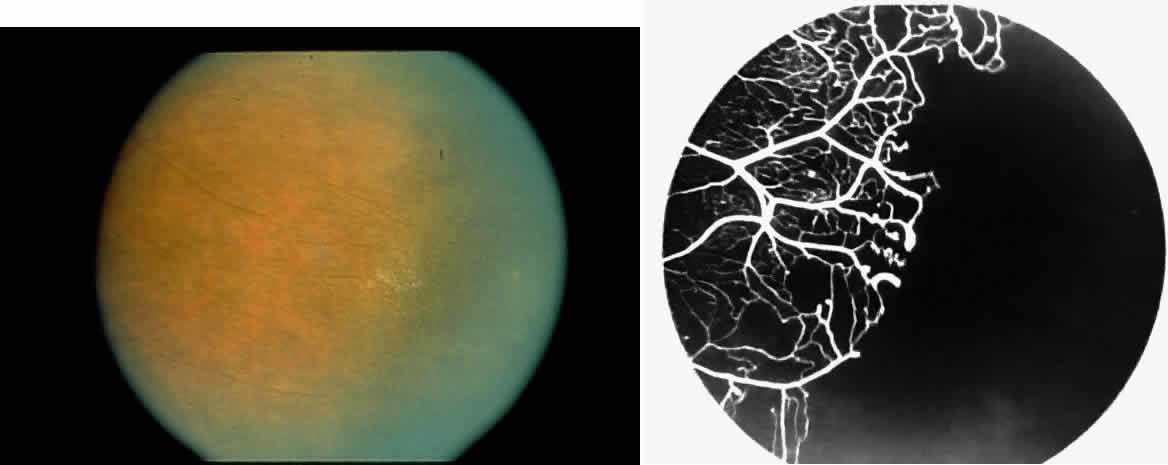
| Fig. 22. A. Photograph of the peripheral retina, demonstrating capillary occlusions and exudate at the margin of perfused retina. B. Fluorescein angiogram of irregular capillary border, with capillary stumps extending into nonperfused retina and an arteriolar-venular anastomosis demonstrating stage II retinopathy. |