|
|
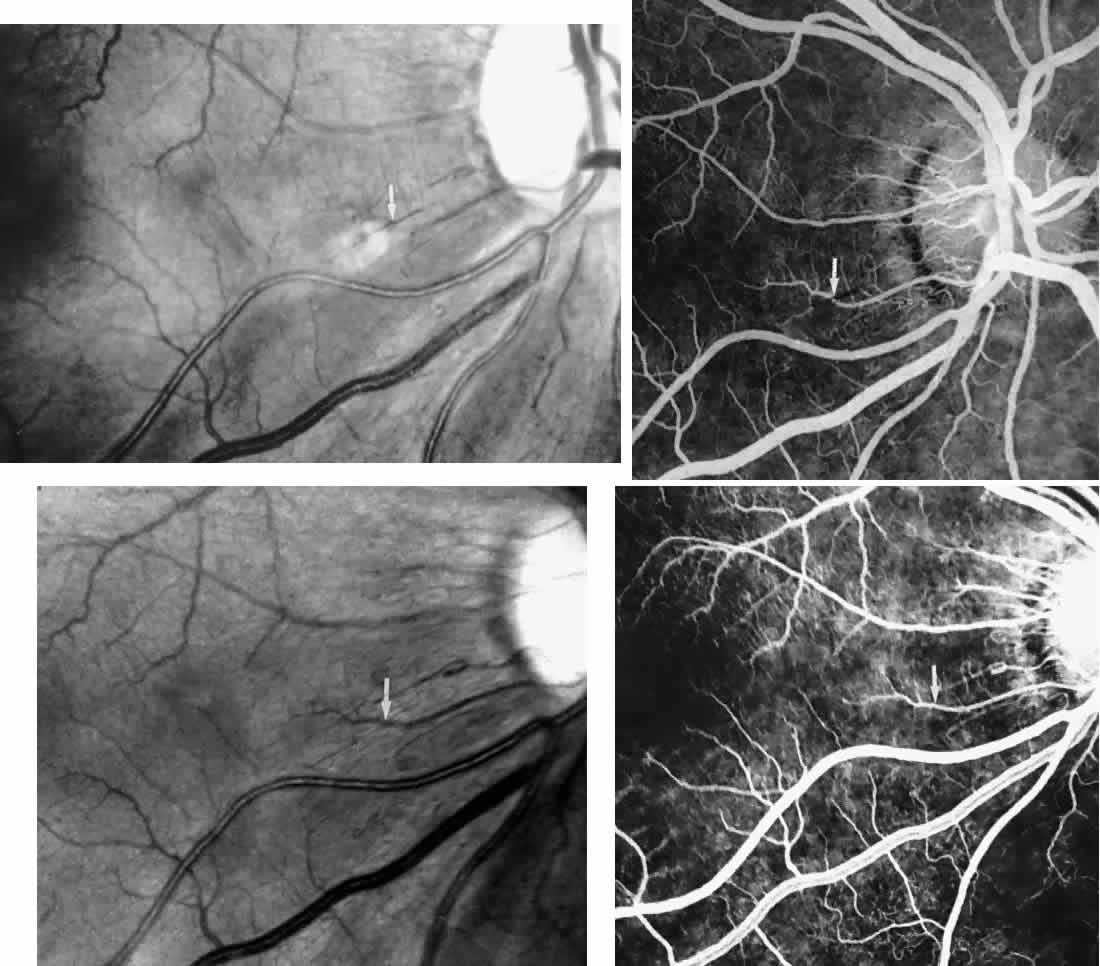
| Fig. 10. A 33-year-old woman with SC disease and stage III sickle cell retinopathy. A. Photograph of the right eye shows a cotton-wool spot with a dark segment identifying the occluded vessel (arrow). B. Fluorescein angiogram demonstrates nonfilling of the occluded vessel (arrow). C. Eighteen months later, the occluded vessel is still visible (arrow). D. Fluorescein angiogram demonstrates that there is still nonfilling of the vessel (arrow). |