|
|
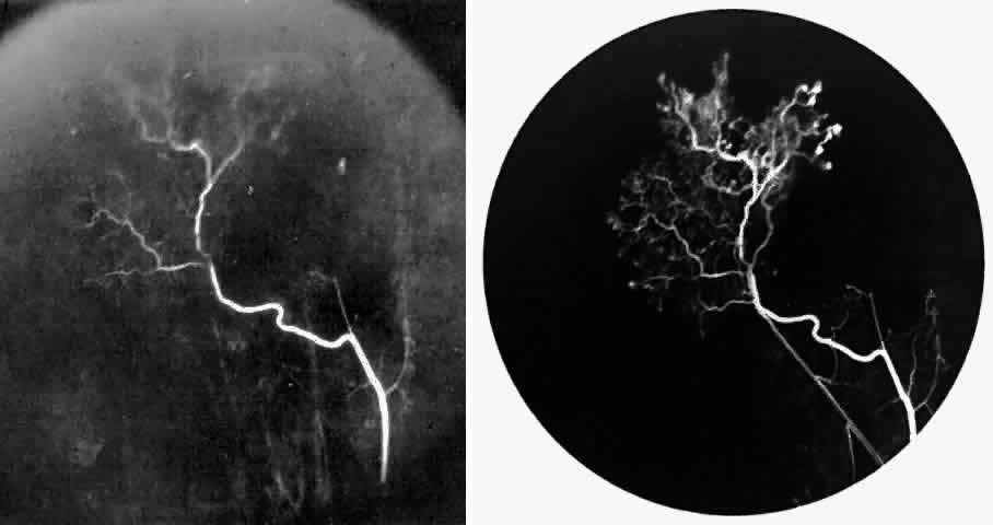
| Fig. 26. A. Arterial filling phase of the fluorescein angiogram of a sea fan demonstrates tortuosity of the feeding arteriole. B. Early arteriolar-venular filling phase demonstrates straightening of the draining venule. Note that this sea fan is adjacent to the qualitatively normal peripheral retinal vasculature demonstrated in Figure 23. |