|
|
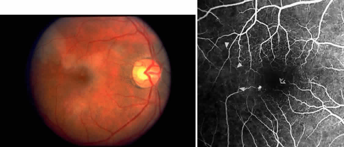
| Fig. 7. Transient perimacular arteriolar occlusions in a 32-year-old patient with SC disease, who presented with decreased vision in the right eye (20/40) after being tackled while playing football. A. Photograph of right macula showing a white, edematous retina and a cherry red spot due to multiple arteriolar occlusions. B. Fluorescein angiogram shows multiple avascular areas, particularly at the temporal raphe (arrowheads), and an irregular perifoveal capillary network (open arrows). |