|
|
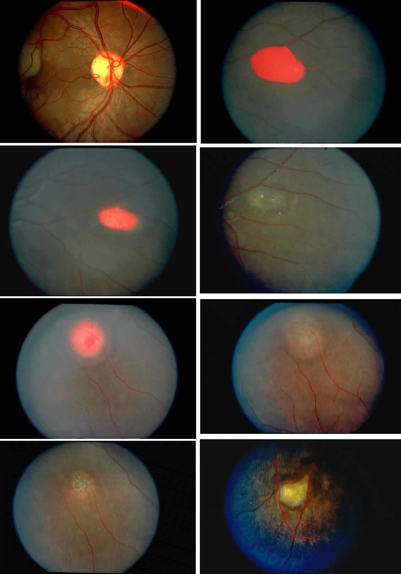
| Color Plate 1 .A. Photograph of the disc in the same patient as in Fig. 16A, demonstrating the disc sign with multiple dilated vascular segments. B. Salmon-patch hemorrhage with preretinal blood obscuring the retinal vasculature. (B through G; Gagliano DA, Goldberg MF: The evolution of salmon-patch hemorrhages in sickle cell retinopathy. Arch Ophthalmol 107:1814, 1989.) C. Two weeks later, the hemorrhage shown in B has a central grayish white color as it begins to resolve. D. Two years later, the hemorrhage shown in B and C has resolved, and an iridescent spot remains. E. Salmon-patch hemorrhage with pre-, intra-, and sub-retinal blood. F. Two months later, the hemorrhage shown in E has resolved, and a lightly pigmented area surrounded by a depigmented halo is seen. G. Four years later, a well-pigmented black sunburst adjacent to the arteriole remains from the salmon-patch hemorrhage shown in E. H. Iridescent spot with refractile copper-colored granules. |