|
|
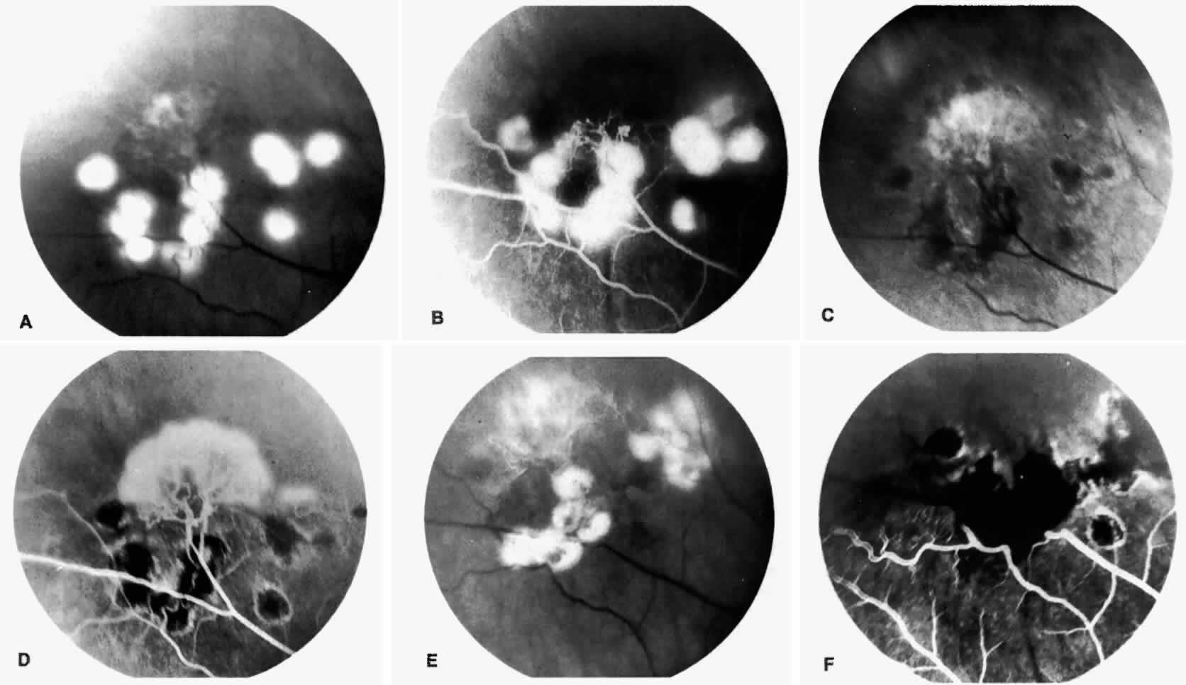
| Fig. 29. Feeder vessel photocoagulation. A. Photograph of feeder vessel photocoagulation applied to the feeding arterioles and draining venules of a sea fan. B. Fluorescein angiogram corresponding to A, showing nonperfusion of the sea fan. C. Photograph taken 2 weeks after treatment, demonstrating pigmented laser lesions and an increased fibrovascular appearance of the sea fan. D. Fluorescein angiogram corresponding to C, showing laser spots and perfusion of the previously closed sea fan. E. Photograph showing retreatment using the feeder vessel photocoagulation technique over the pigmented spots. F. Fluorescein angiogram corresponding to E, showing nonperfusion of the sea fan after retreatment. |