|
|
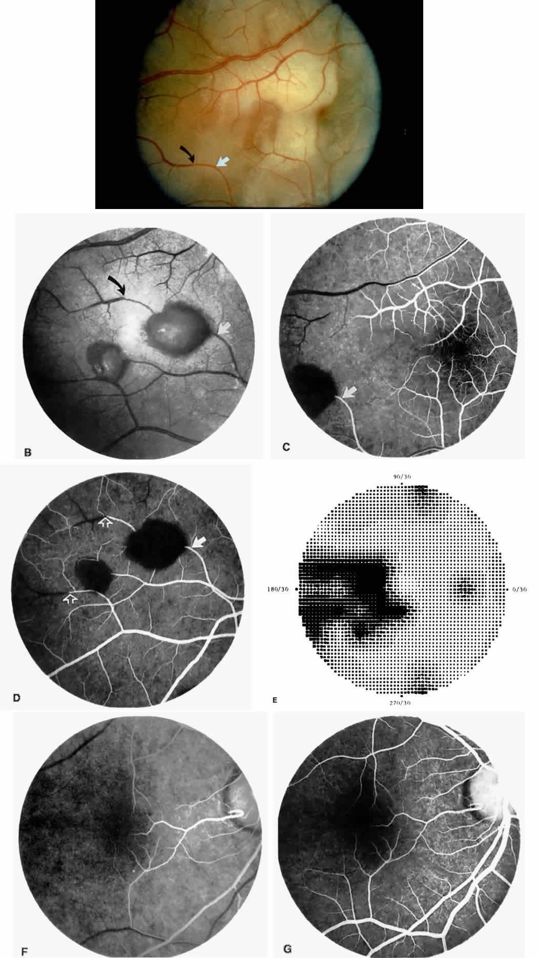
| Fig. 16. A 15-year-old boy with homozygous sickle cell anemia, stage II sickle cell retinopathy, and an ISC count of 23.8% noted an acute nasal visual field defect in the right eye. Visual acuity was 20/20 OU. A. Photograph of the right eye demonstrating arteriolar occlusions in the temporal macula, with white, edematous retina and cherry red spot in the fovea. Note the arteriolar occlusion inferiorly, with darkening of the vessel distal to the occlusion site (black arrow). (The closed white arrow identifies a corresponding arteriolar bifurcation in A through D.) B. Red-free photograph 6 days later showing two salmon-patch hemorrhages. The superior salmon-patch hemorrhage overlies the occlusion site noted in A. Note the distal movement of the occlusion site along the arteriole (black arrow). C. Early fluorescein angiogram, showing loss of capillary network in the temporal macula but preservation of the perifoveal network. D. Late phase of the fluorescein angiogram, showing salmon-patch hemorrhages temporally and the occlusions distal to the hemorrhages (open arrows).E. Visual field performed 9 months later shows persistent nasal field defect, but visual acuity has remained 20/20. F. Two years later, a fluorescein angiogram shows a cilioretinal artery perfusing the nasal macula. G. The late phase of the fluorescein angiogram demonstrates complete loss of the temporal macular capillary network. |