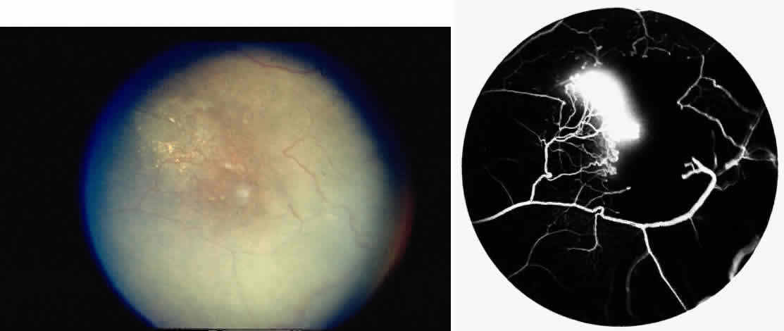
Fig. 20.
A.
Photograph of an iridescent spot with neovascularization within the schisis cavity.
B.
Fluorescein angiogram reveals a neovascular membrane within the iridescent spot.