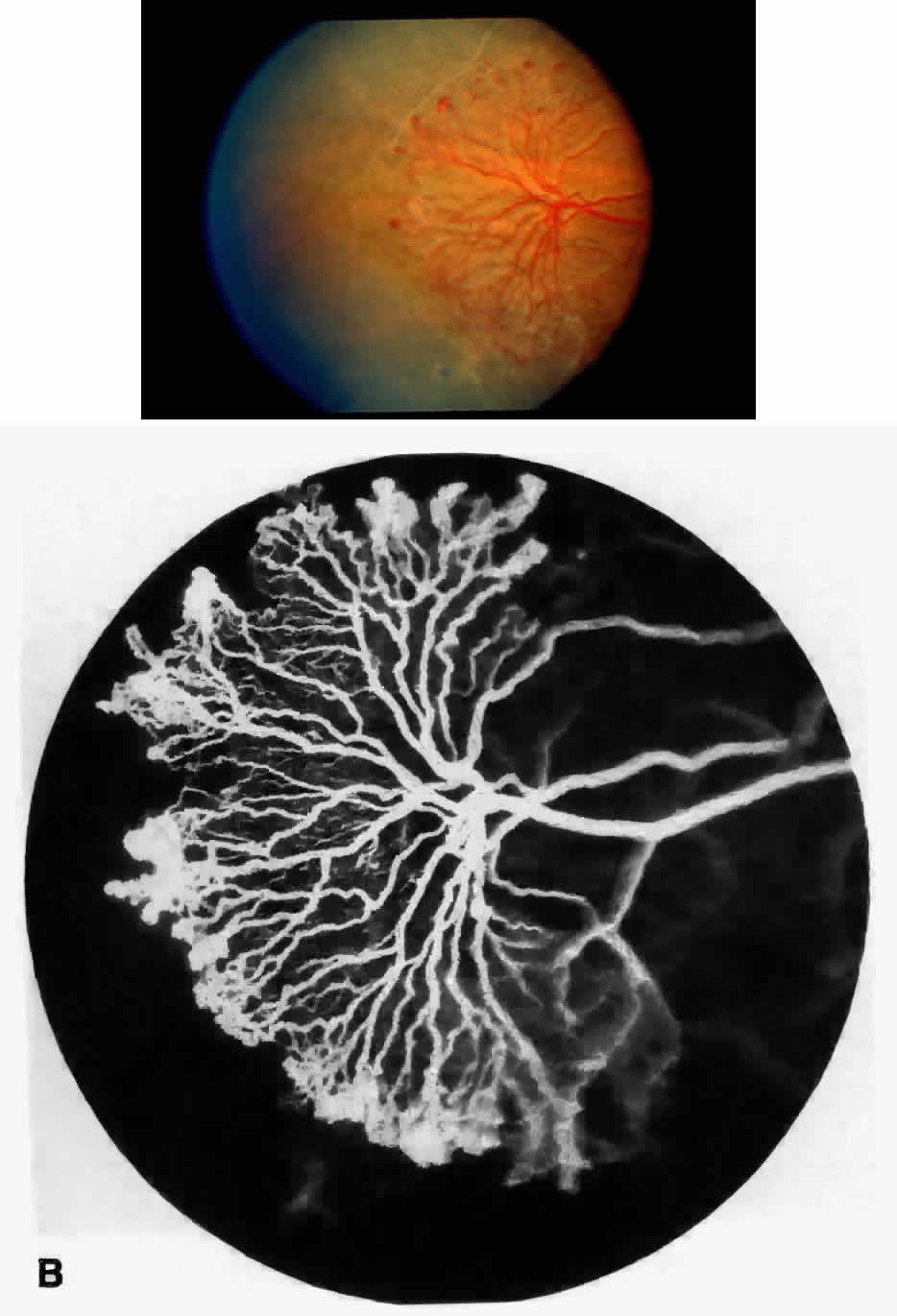
Fig. 27.
A.
Photograph of sea fan neovascularization with hemorrhages at the margins and a white line demarcating perfused and nonperfused retina.
B.
Fluorescein angiogram shows multiple feeding arterioles and draining venules.