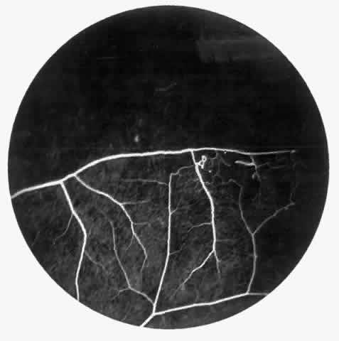
Fig. 23.
Fluorescein angiogram of continuous arteriolar-venular anastomosis demonstrating stage II retinopathy. (Note that this is the same eye demonstrating the qualitatively abnormal peripheral capillary border in
Figure 25
.)