|
|
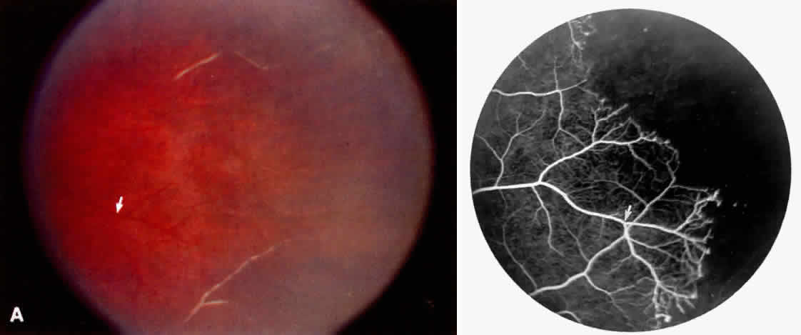
| Fig. 21. A. Photograph of the peripheral retinal vasculature shows sheathed vessels and absence of peripheral vascular perfusion. B. Fluorescein angiogram shows area of nonperfusion representing stage I sickle cell retinopathy. White arrow points to corresponding vascular bifurcation in A and B. |