|
|
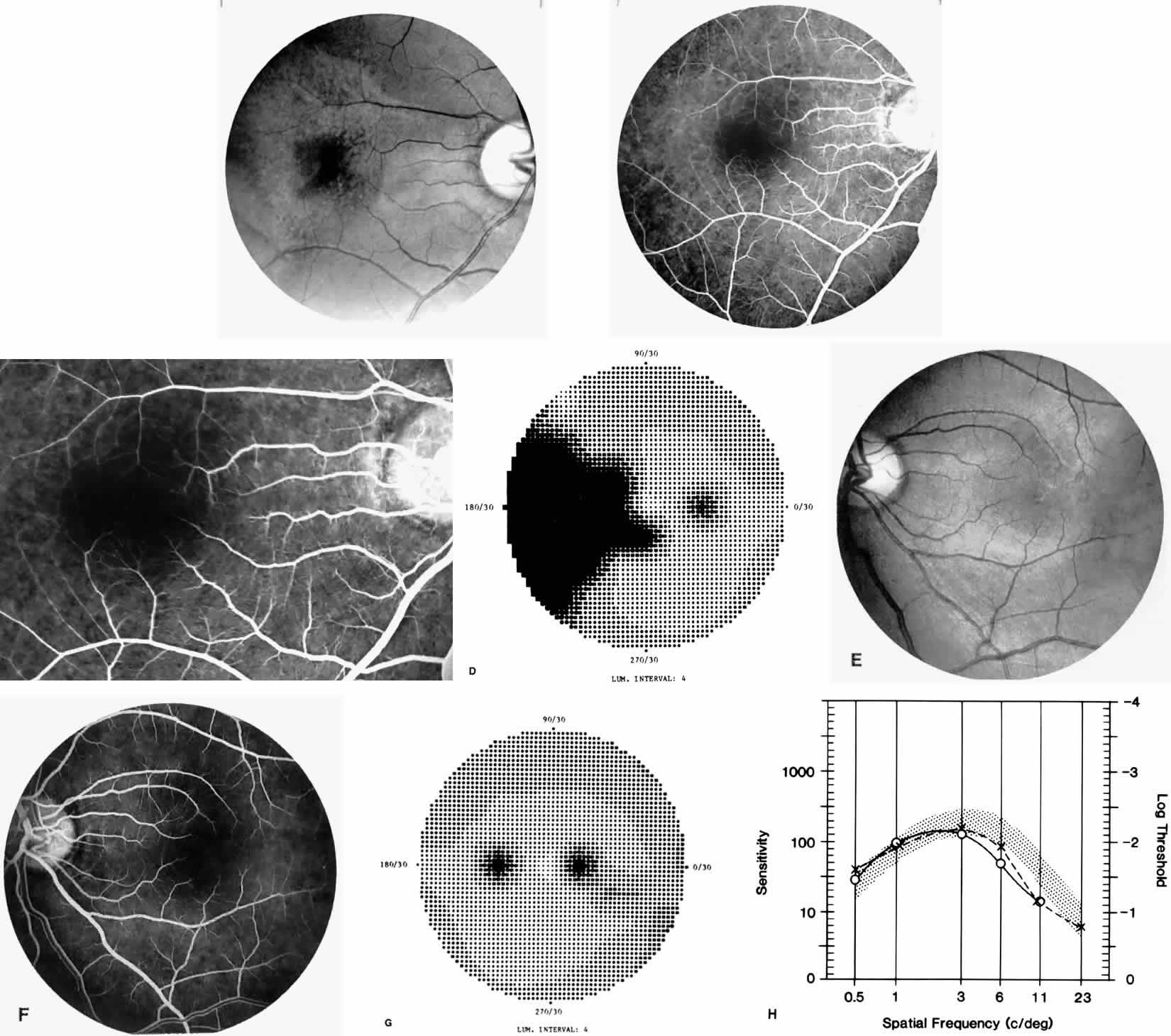
| Fig. 14. A 25-year-old woman with Hb S-β-thalassemia and bilateral proliferative sickle retinopathy (visual acuity 20/25 OD and 20/15 OS). A. Photograph of the right eye demonstrates an irregular macular reflex believed to represent a macular depression sign. B and C. Fluorescein angiogram shows loss of capillary filling corresponding to the area of irregular reflex. D. Octopus perimetry of the right eye shows a large nasal visual field defect corresponding to the area of retinal nonperfusion. E. Photograph of the left eye demonstrates an irregular retinal reflex from the temporal macula, believed to represent a retinal depression sign. F. Fluorescein angiogram shows loss of capillary filling corresponding to the area of irregular reflex. G. Octopus perimetry of the left eye shows a scotoma corresponding to the area of nonperfusion. H. Contrast sensitivity test demonstrates decreased high spatial frequency thresholds (O—O = OD, X- - -X = OS). |