Chapter 5
Topical Diagnosis: Prechiasmal Visual Pathways
JOEL S. GLASER
Main Menu   Table Of Contents

Search

Part I. The Retina
SYMPTOMATOLOGY
HEREDODEGENERATIONS AND ABIOTROPHIES
CHORIO-RETINAL INFLAMMATIONS
METABOLIC STORAGE DISORDERS
RETINAL ARTERIAL OCCLUSIONS
RETINAL VASCULITIS
UVEO-MENINGEAL SYNDROMES
HUMAN IMMUNODEFICIENCY VIRUS INFECTION AND AIDS
TOXIC RETINOPATHIES
CONGENITAL HAMARTOMA SYNDROMES
Part II. The Optic Nerve
CONGENITAL OPTIC DISC DYSPLASIAS AND ANOMALIES
HEREDODEGENERATIVE OPTIC ATROPHIES
ACQUIRED OPTIC NERVE DISEASE: AN OVERVIEW
THE “SWOLLEN DISC”: DIFFERENTIAL DIAGNOSIS
PAPILLEDEMA WITH RAISED INTRACRANIAL PRESSURE
INFLAMMATORY OPTIC NEUROPATHIES: OPTIC NEURITIS
ISCHEMIC OPTIC NEUROPATHIES
GLAUCOMA AND PSEUDOGLAUCOMA
NEOPLASMS AND RELATED CONDITIONS
NUTRITIONAL AND TOXIC OPTIC NEUROPATHIES
TRAUMATIC OPTIC NEUROPATHIES
REFERENCES

Part I. The Retina
Symptomatology

Heredodegenerations and Abiotrophies

Pigmentary Retinopathies

Cone and Cone-Rod Dystrophies

Chorio-retinal Inflammations

Metabolic Storage Disorders

Retinal Arterial Occlusions

Carotid Atheromatous Disease

Other Retinal Arterial Occlusions

Retinal Migraine

Retinal Vasculitis

Uveo-Meningeal Syndromes

Human Immunodeficiency Virus Infection and

AIDS

Toxic Retinopathies

Congenital Hamartoma Syndromes

Tuberous Sclerosis

Neurofibromatosis

To suppose that the eye, with all its inimitable contrivances for adjusting the focus to different distances, for admitting different amounts of light, and for the correction of spherical and chromatic aberration, could have been formed by natural selection, seems, I freely confess, absurd in the highest possible degree.

Charles Darwin

“Organs of extreme perfection and complication”

In The Origin of Species, 1859

Accurate diagnosis of disorders of the visual sensory system is dependent on knowledgeable history taking and competent evaluation of visual function, including acuity, visual fields, and color perception, of pupillary light reactions and of the fundi. There are no valid diagnostic shortcuts; rather, there are guiding principles and axioms that should at least determine a provisional impression and course of investigation, if not provide a conclusive diagnosis. Part I of this chapter is intended to provide a reasonably detailed overview of those retinal disorders and diseases that may confound or otherwise touch on neuro-ophthalmologic diagnosis.

Back to Top
SYMPTOMATOLOGY
Diseases and pathologic alterations involving the retina provoke the least clinical dilemma in that, for the most part, the ophthalmoscope resolves the question of the anatomic level at which the visual pathways are involved. The problem of complicated neurodiagnostic studies should not arise, although fluorescein angiography, electroretinography (ERG), and other retinal function tests (see Volume 2, Chapter 2) may prove valuable. There are two major pitfalls to be avoided. First, minimal retinal changes may be misconstrued as the cause of disproportionately perturbed visual function, or a normal macula may be misinterpreted as abnormal. For example, a patient with relentless monocular visual loss, a central field depression, and afferent-defect pupil, with a few drusen or minimal derangement of retinal pigment epithelium at the fovea, must not be dismissed with an inappropriate diagnosis of “macular degeneration.” In this instance, the afferent-defect pupil indicates a conductive lesion of the optic nerve and cannot be attributed to minimal retinochoroidal disease. Second, true macular disease, especially when bilateral and ophthalmoscopically subtle, should raise the question of macular dystrophies that masquerade as neurologic disease or for which no cause is apparent (Fig. 1). Indeed, there are increasing numbers of retinal disorders that produce subtle or even insignificant objective fundal changes that may escape conventional ophthalmoscopic detection.

Fig. 1. A 14-year-old boy referred for occult neurologic disease or malingering who had undergone magnetic resonance imaging and psychiatric counseling. Visual acuity was 20/100 in both eyes. Fundi (A and B) show thinned rounded macular reflexes and mild pigment changes at the foveae. Fluorescein angiography (C) disclosed marked macular pigment epithelial disturbance. Diagnosis: juvenile macular degeneration (Stargardt's type).

These “hidden” retinal disorders include the following: congenital and hereditary photoreceptor or pigment epithelial abiotrophies and dystrophies; immune-mediated retinopathy associated with distant carcinoma; and an enlarging list of acute zonal occult outer retinopathies, conveniently labeled AZOOR by Gass and associates.1,2 Previously lumped as the “big blind spot syndrome,” AZOOR now encompasses a variety of heterogeneous, presumably inflammatory, retinopathies to be discussed below. Other lesions such as serous detachment of the macula (central serous choroidopathy) or cone dystrophies may be quite subtle on funduscopic examination alone, even during biomicroscopy with the use of a corneal contact lens or Hruby lens. It is in such situations that auxiliary tests of visual function, including color function, Amsler grid, photostress, ERG, and fluorescein angiography are critical in distinguishing modest lesions involving the choroid and retina from early optic nerve compression and demyelination.

In the assessment of visual function, the role of retinal aging alone is noteworthy. Even as macular photoreceptors are incompletely developed at birth and do not reach maturity until at least 4 years post partum, so virtually every parameter of visual function declines from mid-adulthood onward. Age-related degradations in reading acuity, color perception, and feature recognition (contrast sensitivity) are caused by senescent changes of neuronal elements, and, in fact, over a 70-year life span there is a loss of almost 50% of retinal ganglion cells, some half of which serve the macula.3 After the age of 40 years, there is apparently a linear net loss of cone photoreceptors especially from the fovea, to which is added the non-neural factors of increasing pupillary miosis and decreasing lens transparency.4 Other studies dispute an age-related reduction in rod and cone counts.5

The temporal profile of monocular visual deficit is often an important clue in differential diagnosis. Abrupt, non-transient visual symptoms usually indicate retinal artery or vein occlusion, infarction of the optic disc, retinal detachment, or hemorrhage into the vitreous. Age-related macular degeneration may suddenly cause hemorrhage beneath the fovea. In addition, on occasion, optic neuritis may run a course that is interpreted by the patient as abrupt, rather than subacute or rapidly progressive. Transient visual events are considered below (see Table 3).


TABLE 3. Transient Obscurations of Vision

  Amaurosis fugax (retinal microembolization or hypoperfusion)
  Papilledema of raised cerebrospinal fluid pressure
  Migraine: retinal, monocular; cortical, hemianopic
  Hyaline bodies of optic nerve
  Hypotension (orthostatic, arrhythmia)
  Refractive errors (myopia)
  Congenital dysplasias of optic disc
  Anemia
  Arteritis
  Polycythemia/thrombocythemia
  Coagulopathies
  Angle-closure glaucoma
  Spontaneous anterior chamber micro-hyphema, especially after lens implantation


Back to Top
HEREDODEGENERATIONS AND ABIOTROPHIES

PIGMENTARY RETINOPATHIES

Heredodegenerative retinal disorders such as retinitis pigmentosa are almost always separable from “neurologic” causes of visual deficits on the basis of chronicity, bilaterality, and typical funduscopic appearance of pigmentary retinopathy, attenuated arteriolar tree, and waxy disc pallor. “Retinitis pigmentosa” is actually a misnomer twice over: the progressive photoreceptor degeneration is not inflammatory, as “-itis” would imply; and pigmentary changes evolve relatively late and may not be obvious. The term “retinitis pigmentosa sine pigmento” is not a separate entity, but it represents a stage of disorder when the retina appears relatively unaltered but photoreceptor function is defective.

A thorough account of retinal photoreceptor dystrophies is beyond the scope of neuro-ophthalmologic subjects, but A. C. Bird's Jackson Lecture6 provides an overview of current concepts of clinical classification, molecular biology, and therapeutic advances. Hereditary transmission may follow autosomal dominant or recessive, or X-linked, patterns, with a spectrum of responsible gene mutations. Indeed, molecular geneticists have now identified numerous mutations in literally dozens of genes that are associated with or cause photoreceptor degeneration, findings suggesting an extraordinary vulnerability of these cells, possible stemming from their high-oxygen-requiring metabolism and physiologic population culling. These detailed genetic elaborations are replacing imprecise, if more classic, clinical descriptions.6

In the fully developed disorder, there is usually profound constriction of the fields with relative sparing of the central fixational area, resulting in so-called “gun-barrel” or “tubular” field constriction. End-stage retinitis pigmentosa is actually one of the few organic causes of markedly narrowed visual fields. The causes of such “gun-barrel” field constrictions are listed in Table 1. The retained field function actually mimics a cone (see Volume 2, Chapter 2, Fig. 19), because the central remnant enlarges as the testing distance is increased between the patient and, for example, the tangent screen. The field diameter does not enlarge with increasing testing distances in hysteria or malingering, and, in fact, the diameter of the field may shrink further if this possibility is suggested to the naive patient.


TABLE 1. Constricted Field with Retained Acuity

  Glaucoma, late
  Retinitis pigmentosa
  Post-papilledema optic atrophy
  Hyaline bodies (drusen) of optic disc
  Bilateral occipital infarcts with “macular sparing”
  Malingering or hysteria


Atypical cases of selective pigmentary degeneration of the nasal retinal sectors (Fig. 2) produce field defects that may mimic bitemporal hemianopias. Such preferential involvement of the fundus nasal to the optic disc occurs in at least one-third of patients with sectoral retinitis pigmentosa. Nerve fiber bundle scotomas, somewhat mimicking glaucoma, are also recorded.7,8

Fig. 2. Retinitis pigmentosa. A. Advanced field loss showing dense annular defects. Deficits start in the 20° to 30° middle zone (as compared with Bjerrum's zone defects in glaucoma) and proceed toward fixation and outward toward the periphery. Central fixation is relatively spared, producing “gun-barrel fields.” B. The pseudobitemporal field defects of sector retinitis pigmentosa. Unlike chiasmal interference, the defects cross the vertical meridian. C. Left fundus of patient with nasal-sector retinitis pigmentosa.

Acuity is reduced when cystic or wrinkling changes occur at the fovea or as the central aperture of field finally darkens. Disc pallor and retinal arteriolar attenuation are not simply the result of ganglion cell death because the ganglion cell and nerve fiber layer of the retina are affected only late in the disease. Unilateral or bilateral disc hyaline bodies (drusen), at times most marked in peripapillary and juxtapapillary locations, occur in some 10% of all genetic subtypes.9

Central field defects with acuity loss occur with photoreceptor degeneration that especially affects the macula, so-called retinitis pigmentosa inversa, but this disorder likely represents a separate nosologic class, the cone-rod dystrophies (see below) (Fig. 3). Severe visual defect in early infancy is frequently enough caused by a primary outer segment retinal abiotrophy, Leber's congenital amaurosis, although this is probably not a single clinical entity. This disorder is characterized by the following: severe impairment of vision, present at birth or becoming evident during early to late infancy; a fundus that may initially approach normal, but within years, optic atrophy, diffuse fine pigmentary degeneration, and attenuation of the arteriolar tree are evident; and either an absent or a markedly reduced ERG response. Other variable features include nystagmus, photophobia, digito-ocular maneuver (forceful eye rubbing with sunken globes; “blindisms”), strabismus, cataracts, hyperopia, mental retardation, deafness, renal anomalies, seizures, hydrocephalus, and focal neurologic deficits (e.g., cerebral diplegia). Hereditary transmission is typically autosomal recessive10 (see Volume 2, Chapter 13).

Fig. 3. Cone-rod dystrophy or so-called “retinitis pigmentosa inversa” in a young man with progressive spinocerebellar degeneration and 20/200 acuity in each eye.

CONE AND CONE-ROD DYSTROPHIES

As noted above, the macular (“inversa”) form of abiotrophic pigmentary retinopathy is actually a selective loss of cone function, but rod function also is defective. Indeed, retinal dystrophies accompanying many systemic disorders are typically, but not exclusively, this macular form. Of special neuro-ophthalmologic interest is the association of this and other geographic pigmentary patterns with hereditary cerebellar ataxias,11,12 with olivopontocerebellar and spinocerebellar degenerations,12–14 and with Friedreich's ataxia.15 These disorders are usually dominantly inherited, with variable expression of early life onset of progressive spasticity, ataxia, slowed saccades or supranuclear ophthalmoplegia, and chorioretinal macular dystrophy. Trinucleotide gene expansion (point mutation) is incriminated, but not exclusively. In Friedreich's ataxia, the most common inherited, if clinically inhomogeneous, spinocerebellar ataxia, a mitochondrial location of frataxin (Friedreich's ataxia protein) has been identified; this locus on chromosome 9 reflects the relationship with vitamin E deficiency ataxia and certain neuropathies with mutations in nuclear genes.16

Other subsets of pigmentary retinopathies are due to mitochondrial DNA mutations and are associated with migraine, ataxia, dementia, and Leigh's disease17; the sporadic or maternally inherited MELAS syndrome of mitochondrial myopathy, encephalopathy, lactic acidosis, and stroke-like episodes, usually presents in the teens as cognitive regression, headaches, and cerebral lesions causing field defects.18–20 Even in family members, there is considerable variation of genotypic and phenotypic specificity in metabolic disorders mediated by mitochondrial DNA aberrations, including Kearns-Sayre progressive external ophthalmoplegia (see Volume 2, Chapter 12) and other conglomerations of pigmentary retinopathies. Further identification of gene point mutations will eventually provide a more precise classification.

Progressive cone degenerations present as bilaterally diminished acuity, defective color vision, and aversion to bright lights (photophobia) with “day blindness” (hemeralopia). Central field defects progress, at times showing a fenestrated central scotoma. This widespread loss of cone function usually begins in the first 2 decades of life, but severe cone disease may begin at any age. Heredity is autosomal dominant, but sporadic cases are common, and both severity and rate of progression are variable. The fundus may appear quite normal, a finding that, when coupled with photophobia, provokes an impression of hysteria, but defective pigment epithelium in the form of a “bullseye,” nonspecific mottling, and crystalline deposits are usually enhanced by fluorescein angiography (see also Fig. 1). Mild disc pallor is not unusual. Abnormal single-flash photopic and flicker (i.e., cone-mediated) responses define the disorder by ERG21; fluorescein angiography and full-field ERG may be inconclusive, yet focal ERG is abnormal.22 If considered sooner rather than later, full-field or focal ERG should obviate more exhaustive and inappropriate diagnostic studies for optic nerve disease.

Cones may be congenitally absent, as in the case of rod monochromatism, a rare form of “color blindness” with acuity of about 20/200 and reduced visual capacity under bright light situations; that is, these children show a preference for dim illumination. Pendular or mixed jerk-pendular nystagmus patterns are typical, and the fundus foveal reflex may be blunted. ERG shows absent or markedly subnormal photopic (cone) responses, but normal scotopic (rod) responses.

Pigmentary retinopathies, or more typically cone-rod dystrophies, keep company with a large number of other neurodegenerative and metabolic disorders, such as Bassen-Kornzweig syndrome, Refsum's disease, Kearn-Sayre progressive ophthalmoplegia (see Volume 2, Chapter 12), Laurence-Moon-Bardet-Biedl cerebellar ataxia, and sensorineural hearing loss. A complete description of the retinal abiotrophies, dystrophies and degenerations may be found in topical reviews, and pertinent texts. Age-related macular degenerations and other macular dystrophies and abiotrophies without neurologic implications are beyond the scope of this chapter.

Back to Top
CHORIO-RETINAL INFLAMMATIONS
As noted above, Gass1 and others have elaborated a class of acute and subacute, diffuse or focal, presumed inflammatory zonal disorders of the outer retinal layers, now termed AZOOR. Roughly described as “enlarged blind spots” and thus confounding neurologic diagnosis, this somewhat heterogeneous rubric includes MEWDS (multiple evanescent white dot syndrome), multifocal choroiditis, acute macular neuroretinopathy, pseudo-presumed histoplasmosis, and idiopathic blind spot enlargement. These entities share a constellation of signs and symptoms and so raise the question of a spectrum of common origin involving geographic zones of retinal photoreceptors and pigment epithelium. Characteristics include the following: predilection for female patients; acute onset in one or both eyes, associated with photopsias; minimal fundus findings at onset, but eventual minor pigment epithelial disturbances; ERG abnormalities; fluorescein angiographic evidence of geographic thinned pigment epithelium; vitreous cells; and permanent field depressions often close to the physiologic blind spot. Taken as a group, Jacobson and colleagues2 investigated the nature of retinal dysfunction and found patchy but dense scotomas and ERG abnormalities, but no evidence of autoantibodies to specific retinal antigens. Recurrent central nervous system (CNS) inflammation in association with AZOOR is reported,23 characterized by cerebrospinal fluid (CSF) lymphocytosis and multiple magnetic resonance imaging (MRI) signal abnormalities, followed in 6 years by an episode of cervical myelopathy.

Acute multifocal retinitis, another idiopathic inflammation in eyes of healthy young to middle-aged adults, may be associated with optic disc edema and may follow flulike prodromes.24 Mild vitrous reaction and macular exudative stars are characteristic, and a self-limited benign outcome is usual, with no evidence of specific infectious or autoimmune causes. Other relatively acute multifocal chorioretinal inflammations include presumed ocular histoplasmosis, coccidioidomycosis, Pneumocystis carinii infection, cryptococcosis, mycobacterial or syphilitic choroiditis, birdshot (vitiliginous) retinochoroidopathy, punctate inner choroidopathy, and acute multifocal posterior placoid pigment epitheliopathy (AMPPPE). Although specific infectious agents may also be incriminated in meningeal reactions, such as cryptococcosis, AMPPPE is associated with CSF pleocytosis and protein elevation, findings suggesting possible viremia.25 The foregoing discussion has focused on retinal pigmentary and macular disease in relation to neurologic disorders, not simple isolated macular dystrophies, which are considered at length elsewhere.23,26

Back to Top
METABOLIC STORAGE DISORDERS
Biochemical assays have considerably clarified the nosologic status of the group of storage diseases previously classified as the “cerebromacular degenerations,” of which Tay-Sachs disease is the eponymous prototype. Although these disorders share a superficial resemblance, showing a progressive neurodegenerative course with variable fundus findings, they are now best classified by abnormal storage products (e.g., sphingolipidoses, mucopolysaccharidoses, and mucolipidoses) and lysosomal enzyme deficiencies. Lysosomes contain hydrolytic enzymes that degrade proteins, polysaccharides, and nucleic acids; if undegraded, these materials accumulate in lysosomes and impair cell function. The complex lipids and saccharides indigenous to neural cells produce symptoms and signs related to eye and brain, including corneal clouding, macular “cherry-red spot,” pigment epithelial degenerations, optic atrophy, mental deterioration, seizures, motor incoordination, myoclonus, and death.

The ganglion cell layer of the retina is a principal site of abnormal accumulation of anomalous storage products, such that ophthalmoscopic changes are observable either in the form of retinal “graying” or the well-known cherry-red spot. The ganglion cell layer densely surrounds the thin fovea, which transmits the normal red color of underlying choroid (Fig. 4). The storage disorders with cherry-red spot or macular graying are listed in Table 2.27

Fig. 4. “Cherry-red spot” of advanced Tay-Sachs disease (gangliosidosis). Note the central foveal window surrounded by a ring of densely opaque retinal ganglion cell layer; also, optic atrophy.


TABLE 2. Storage Diseases with Macular Changes

  Sphingolipidoses

  Gm1 gangliosidosis (“Generalized” gangliosidosis)
  Tay-Sachs disease (Gm2 gangliosidosis type I)
  Sandhoff's disease (Gm2 gangliosidosis type II)
  Niemann-Pick disease
  Lactosyl ceramidosis
  Childhood metachromatic leukodystrophy


  Mucolipidoses

  Mucoliposaccharidosis (MLS) type I (lipomucopolysaccharidosis)
  Farber's disease
  Sea-blue histiocyte syndrome or chronic Niemann-Pick disease (yellow perifoveal granules)
  Mucolipidosis I (lipomucopolysaccharidosis) (Spranger)


  Unclassified


In addition to retinal changes, pallor of the optic disc occurs in numerous storage disorders, the nerve being affected by several mechanisms. Optic atrophy occurs when abnormal glycolipids are stored in retinal ganglion cells with subsequent neuronal death and nerve pallor, such as occurs in Tay-Sachs disease (Gm2 gangliosidosis), but ophthalmoscopic evidence of optic atrophy is not invariable. Tay-Sachs is an autosomal recessive inherited disease, resulting from mutations of the hexosaminidase (Hex) A gene coding for the alpha-subunit of beta-D-N-acetyl-hexosaminodase. Juvenile and adult Hex A deficiencies are rare, less severe variants. Both infantile and late forms are most prevalent among Ashkenazi (roughly, Middle and Eastern European) Jews, but are reported in non-Jews, in whom clinically benign mutations may occur.28,29 MRI findings include hyperintensities in basal ganglia and thalamus, with marked brain atrophy and diffuse white matter lesions; these findings likely reflect accumulation of Gm2 ganglioside.30 Similarly, in Gm1 gangliosidosis (generalized gangliosidosis; “Tay Sachs disease with visceral involvement”), there is also an abnormal deposition of ganglioside in the retina, with subsequent atrophy of the nerve, but it is a mucopolysaccharide that accumulates in the viscera.

Metachromatic leukodystrophy (sulfide lipidosis) is an autosomal recessive deficiency of arylsulfatase A, with ceramide-galactose sulfate in retinal ganglion and glial cells and macrophages in the optic nerve; a cherry-red spot occurs occasionally. The optic atrophy of Krabbe's disease (formerly considered one of the familial diffuse scleroses) is due to extensive demyelination associated with the accumulation of globoid bodies containing ceramide-galactose resulting from beta-galactosidase deficiency.

The ceroid lipofuscinoses are among the most commonly inherited childhood neurodegenerative disorders. These autosomal recessive syndromes are caused by accumulation of an insoluble, complex lipopigment that demonstrates autofluorescence and appears microscopically as characteristic curvilinear bodies. Histopathologic examination discloses degeneration of rods and cones beginning at the macula, narrowing of retinal vessels, atrophy of pigment epithelium with pigment migration, and curvilinear bodies in ganglion cells. These progressive disorders are not genetically homogeneous and are separable into infantile, late infantile, juvenile, and Finnish variants. These share seizures, motor disturbances, visual impairment, dementia, and autosomal inheritance.31

The late infantile type (Bielschowsky-Jansky) is a fatal recessively inherited disorder with genomic defect localized to chromosome 13q22, thus delineating this disease as a separate entity.32 Presentation is with developmental arrest and seizures at 2 to 5 years of age, with motor and visual symptoms thereafter. There is massive tissue accumulation of lysosomal hydrophobic subunit-c protein of the mitochondrial adenosine triphosphate synthase. Ganglion cells are decreased in number, with thinning of the nerve fiber layer and optic atrophy. In addition, there is involvement of the outer segments with degeneration of the rods and cones and pigmentary clumping in the outer retinal layers. On MRI, hyperintense periventricular signals correlate with severe loss of myelin.33

Juvenile amaurotic idiocy (Batten-Mayou, Vogt-Spielmeyer) presents as visual loss in early adolescence and is characterized by retinal pigmentary changes predominantly at the macula, with late and minimal pallor of the disc. Apoptosis of photoreceptors and brain neurons has been confirmed as the mechanism of cell death.34 At a later stage, motor and mental deficits evolve. The late or adult form of amaurotic idiocy ascribed to Kufs paradoxically is associated neither with amaurosis nor with retardation to the degree of idiocy, but rather, with dementia with cerebellar and extrapyramidal motor signs.

Optic nerve involvement may occur in the mucopolysaccharidoses (MPS), taking the form of optic atrophy or papilledema. Although true papilledema doubtlessly occurs in association with hydrocephalus seen in these disorders, there are other instances of “papilledema” in which fundus descriptions or photographs are not convincing. Goldberg and Duke35 reported the ocular histopathologic findings in a patient with Hunter's syndrome (MPS II), in which premortem examination included the observation of “bilateral chronic papilledema.” On microscopic examination, the optic nerve was normal, despite marked retinal pigmentary degeneration, showing neither consecutive atrophy nor changes compatible with chronic papilledema. Similarly, Kenyon and colleagues36 reviewed the systemic mucopolysaccharidoses and included an instance of a 26-year-old man with Hunter's syndrome (MPS II), whose ophthalmoscopic examination revealed “blurred disc margins in both fundi (without venous congestion, hemorrhages or exudates) compatible with mild chronic papilledema,” but no further comment was made in elucidation of this finding. Hunter's syndrome is an X-linked recessive disorder characterized by facial and skeletal dysmorphism, stiff joints, and mental slowing. Deafness and chronic disc edema occur, without raised intracranial pressure, and mucopolysaccharide deposition in the sclera and optic nerve septa, especially at the lamina cribrosa, is described.37 MRI may disclose large multifocal cystic areas of hypointense or hyperintense signals in white matter, including the corpus callosum, likely reflecting deposition of mucopolysaccharide and increased fluid content.38

Mailer39 reviewed 16 patients with optic atrophy in gargoylism and concluded that communicating hydrocephalus was the most frequent cause. Although hydrocephalus has long been recognized to occur in mucopolysaccharidosis, it is uncovered more frequently at autopsy than clinically. Goldberg and associates40 reported a case of Maroteaux-Lamy syndrome (MPS VI) with hydrocephalus and papilledema, treated with a ventriculojugular shunt. Those authors discussed the following possible pathogenetic mechanisms of visual dysfunction due to optic nerve involvement in mucopolysaccharidosis: optic atrophy secondary to glaucoma, retinal pigmentary degeneration, or mucopolysaccharide deposition in the retinal elements; infiltration of the nerve substance or meninges; narrowing of the optic canals; and hydrocephalus, with or without papilledema. Retinal pigmentary dystrophy occurs in MPS I-H (Hurler), MPS III (Sanfilippo), and MPS I-S (Scheie), all resulting from storage of heparan sulfate.

The cherry-red spot myoclonus syndrome is an autosomal recessive oligosaccharidosis due to deficiency of lysosomal acetylneuraminidase. The syndrome is comprised of typical cherry spot macula, resting and intention myoclonus, and preserved intellect. Sogg et al41 also described flutterlike ocular oscillations attributed to possible cerebellar involvement.

Back to Top
RETINAL ARTERIAL OCCLUSIONS
Retinal arterial occlusions of neuro-ophthalmologic pertinence occur in patients with carotid athero-occlusive disease, heart disease, arteritis (giant cell, collagen vasculitides), and, rarely, migraine. Cranial arteritis is considered in a subsequent discussion of ischemic optic neuropathies (see Part II of this chapter).

The onset of retinal arterial occlusion rarely goes unnoticed by the patient, but unlike venous thrombosis, arterial occlusions of a minor degree may be difficult to discern ophthalmoscopically, especially if days or weeks pass before the fundus is examined. Muci-Mendoza et al42 used fluorescein fundus angiography to demonstrate ophthalmoscopically occult emboli and post-embolic endothelial damage after episodes of amaurosis fugax (see below, Carotid Atheromatous Disease).

With acute infarction, the retina becomes opaque and takes on a creamy or gray appearance. Atheromatous material in the form of “bright plaques” of cholesterol or other microemboli may be seen, especially lodged at arterial bifurcations (Fig. 5). Segmental arteriolar mural opacification (see Fig. 5D) may follow retinal microembolization by weeks to months, and such sheathing may be as useful as the recognition of the cholesterol embolus itself.43 ERG may show diminished B-wave amplitude, a finding indicating inner retinal ischemia. Weeks following retinal infarction, the optic disc becomes pale, and the arterial tree becomes narrowed in the sector corresponding to the arterial occlusion.

Fig. 5. Retinal microembolic phenomena. A. Bright cholesterol plaque (arrow) impacted at an arterial bifurcation. Thin crystal does not obstruct flow. B. Cholesterol crystals in disc vessels (arrows). Often, the plaque appears larger than the vessel diameter. C. Infarcted opaque retina. The artery contains emboli (? fibrin platelets) that have obstructed flow. D. Reactive opacification of the arterial wall. Fluorescein angiography demonstrated flow through this formerly occluded vessel.

The visual field defects with arterial occlusion are variable, but they usually take the form of arcuate scotomas or “altitudinal hemianopias” of the superior or inferior half fields (Fig. 6). These altitudinal or pseudo-quadrantic defects are dense and are easily discovered by hand- or finger-counting confrontation techniques. The localizing value of the position of the “vertex” of quadrantic and wedge-shaped defects was pointed out by Alfred Kestenbaum. When the wedge originates at or points toward the blind spot, the defect is due to a retinal arterial occlusion or a lesion at the edge of the optic disc (including the arcuate defects seen in glaucoma). The differential diagnosis of arcuate scotomas, that is, with radial borders originating at the blind spot (see Fig. 6, right field), includes glaucoma, ischemic optic neuropathy, branch retinal artery occlusion, hyaline bodies of optic disc, congenital optic pit, juxtapapillary inflammation, and, rarely, chiasmal interference (see Volume 2, Chapter 6, Fig. 1F).

Fig. 6. Field defects of vascular origin. RE. Arcuate nerve fiber bundle defect of the right eye extending from the blind spot into the nasal quadrant. LE. Inferior altitudinal “hemianopia” of the left eye. These patterns are common to both retinal arterial occlusions and segmental infarction of nerve head (ischemic optic neuropathy).

CAROTID ATHEROMATOUS DISEASE

The pathogenesis of embolic retinal arterial occlusions is diverse, but most are associated with atheromatous degeneration of the cervical carotid arteries. Since the observations by Fisher44 of mobile intravascular material during episodes of transient retinal ischemic episodes (amaurosis fugax), an embolic source is commonly invoked, especially when any material is detected ophthalmoscopically within the retinal circulation. Such retinal emboli have been examined histopathologically and may consist of cholesterol crystals,45 platelet aggregates,46 fibrin and blood cells,47 and neutral fat.48 A study of the prevalence of asymptomatic retinal emboli among 3654 persons aged 49 years or older disclosed photographic evidence in 1.4%, the majority being judged as cholesterol; risk for subsequent stroke is not yet calculated.49 On the other hand, the incidence of visible emboli in retinal arterial occlusion is estimated at about 20%.50 Wilson and coworkers51 compared the incidence of carotid and cardiac disease in 103 patients with retinal artery occlusions and reported cardiac valve abnormalities in 28; of 62 carotid arteriograms, 35% were normal, 13% were occluded, and the remainder showed irregularity or significant stenosis. In a small group of 41 patients with transient monocular visual loss or retinal artery occlusion, no cardiac or carotid source was uncovered in two-thirds of subjects, ipsilateral carotid disease was seen in 11 (27%) cases, and a cardiac source was noted in a single patient.52

Even in those patients without ophthalmoscopically visible emboli, it is possible that the embolus either has lodged in the retro-laminar portion of the optic disc or has disintegrated and passed to the retinal periphery. Primary thrombosis in the retinal arterial circulation, beyond evidence of frank vasculitis or hypercoagulable states, is considered obsolete. Vasospasm, on the other hand, is an unlikely candidate to explain arterial occlusion, although rare instances of otherwise unexplained transient visual loss are reported.53 Other noncarotid sources of emboli, including cardiogenic, are discussed below.

Transient ischemic episodes involving the retinal arterial tree produce the well-known symptom of amaurosis fugax (“fleeting darkness, or blindness”; ocular transient ischemic episode). This phenomenon may be defined as a painless unilateral, transient loss of vision that usually progresses from the periphery toward the center of the field. Often, the visual deficit takes the pattern of a dark curtain descending from above or ascending from below. Complete or subtotal blindness follows in seconds and lasts from 1 to 5 minutes (rarely longer). Vision returns to normal within 10 to 20 minutes, at times by reversal of the pattern of progression. Incomplete variations produce less distinctive sensations such as “looking through a fog … through raindrops … through haze.” By convention, the term amaurosis fugax is reserved for the ocular symptoms as described, which are usually distinguishable from other episodic visual disturbances (see Volume 2, Chapter 1), and it implies transient ischemia in retinal arterial circulation. Those conditions with principally monocular transient visual loss are listed in Table 3.54

It is likely that microembolic material in the form of fibrin-platelet aggregates momentarily occludes retinal vessels, then fragments and passes into the retinal periphery. If disaggregation with reconstitution of blood flow does not take place within several minutes, ischemic damage to inner retinal layers may be irreversible, and permanent visual defects may ensue. Most evidence indicates that the major source of retinal microemboli is the extracranial internal carotid artery, specifically an ulcerating atheromatous lesion at the level of the bifurcation.55 The incidence of carotid atheromatous disease in patients with retinal strokes or amaurosis fugax, as noted above, ranges from a low of 27%52 to an estimated high from 57 to 67%.56 Moreover, retinal ischemic events may be more frequent when carotid stenosis is greater than 50% to 70% or in the presence of ulcerative atheromatous plaques.57,58 The diagnosis of amaurosis fugax and the implied underlying carotid atheromatous disease may usually be made on the basis of signs and symptoms (Table 4).


TABLE 4. Symptoms and Signs of Carotid Atheromatous Disease

  Transient symptoms

  Amaurosis fugax
  Photopsias
  Orbital and ocular pain


  Ophthalmoscopic signs

  Arteriosclerotic retinopathy
  Hypertensive retinopathy
  Bright plaques
  Occluded arteries
  Optic atrophy


  Chronic ocular hypoxia

  Dilated episcleral veins
  Edematous cornea
  Anterior chamber reaction
  Rubeosis and iris atrophy
  Synechiae
  Cataract
  Glaucoma (or hypotony)
  Retinal venous dilation
  Microaneurysms, hemorrhages
  Neovascularization
  Arterial occlusions


The presence of a carotid bruit is suggestive evidence of turbulent flow, but, however practical, auscultation and tender palpation of the carotid arteries and performance of ophthalmodynamometry or ocular plethysmography no longer suffice. At any rate, in the patient with amaurosis fugax, in whom carotid disease must be evaluated, further technical procedures are mandatory. Such tests of carotid morphology and hemodynamics assume two distinct forms: arteriographic, requiring intraluminal contrast opacification of arteries; and nonarteriographic (“noninvasive”), using ultrasound, Doppler imaging, and, most recently, MRI (magnetic resonance angiography or MRA). Noninvasive panels are commonly employed for preliminary assessment, with the advantages of risk-free, outpatient application. Carotid duplex scanning combines simultaneous real-time B-mode ultrasonography with gated, pulsed Doppler ultrasonography. These studies provide color-coded images of the degree of stenosis and plaque morphology, although not without limitations and artifactual data.59 MRA imaging of carotid vessels is currently an evolving field, but preliminary drawbacks include overestimation of stenosis and perturbation by signal voids related to turbulant flow. MRA images are useful in combination with duplex ultrasonography, especially when the carotid bifurcation is involved.58

The general consensus58 is that there is a sharp decline in the risk of stroke and in benefit from endarterectomy as the degree of angiographically defined stenosis diminishes from 99% to 70%, and this critical point underscores the importance of precise measurement of stenosis. Whereas modern techniques of angiography carry a 0.09% to 0.3% stroke rate risk, it is generally recognized that duplex ultrasonography be used as a screening tool to exclude patients with no detectable carotid disease from further testing, but patients suspected of harboring carotid disease, who are otherwise suitable candidates for endarterectomy, should undergo confirmatory contrast angiography. Ultrasonography may also be applied to monitor for progression of stenosis. Therefore, the elimination of conventional carotid angiography in favor of combined Doppler and MRA, to assess for stroke risk or determination of proper therapy, is premature, cost-containment strategies notwithstanding.

In contrast to transient symptoms and signs of retinal microembolic episodes, a condition of chronic ocular hypoxia (ocular ischemic syndrome) occurs less frequently, resulting from diffuse vascular occlusive disease of the aortic arch or common carotid artery. Acute or chronic occlusion with insufficient collateralization produces an ischemic pseudo-inflammatory uveitis, which variably includes an injected painful globe, corneal edema, aqueous flare and cells, a mid-dilated fixed pupil, rubeosis and iris atrophy, rapidly advancing cataract, either hypotony or elevated intraocular pressure (“neovascular glaucoma”), retinal microaneurysms and new vessel formation, posterior pole and mid-peripheral blot hemorrhages, macular edema, venous dilation and “sausaging,” cytoid infarcts (cotton-wool spots) of the nerve fiber layer, and arterial occlusions (Fig. 7; see Table 4). The hypoxemic fundus changes constitute a picture of venous stasis (low-pressure) retinopathy, perhaps the commonest ocular sign of chronic carotid obstruction.

Fig. 7. Ocular hypoxia with subacute carotid occlusion. The patient complained of a painful red eye. A. Anterior segment shows an irregular, fixed pupil and iris rubeosis (arrows). B. Fundus demonstrates combined retinochoroidal infarction with acute excavation of the optic disc. Arteriography revealed right internal carotid occlusion.

Ischemic photoreceptor metabolism accounts for subjective afterimages following exposure to bright light, including a positive photostress test60 (see Volume 2, Chapter 2). Low retinal arterial pressure may be detected by observing pulsation or collapse of the disc arterioles with even slight fingertip pressure exerted on the globe. Such borderline perfusion associated with carotid stenosis and retinopathy may be heralded by postprandial visual loss.61 In the situation of chronic, subacute, or rapidly progressive ischemic oculopathy, giant cell arteritis must be considered in the differential diagnosis, and chronic venous obstruction or diabetic retinopathy may produce similar fundus appearance.

From a series of 32 patients62 with ocular ischemic syndrome manifested by anterior segment neovascularization, the following data were accrued: mean age 68 ± 8 years; visual symptoms presented as amaurosis fugax, 15%, gradual, 28%, or sudden, 41% loss; initial visual acuity less than or equal to 20/400 in 64%, and in 77% at final follow-up; iris neovascularization, 87%; iridocorneal angle neovascularization, 59%; disc pallor, 40%, or cupped, 19%, or edematous, 8%; retinal circulatory stasis, 21%; retinal hemorrhages, 24%. Intraocular pressures ranged widely from 4 mmHg to 60 mmHg. Ipsilateral carotid occlusion or severe stenosis (80% to 99%) was found in 74%, but endarterectomy in 7 patients did not influence visual outcome, which was poor at onset. Other significantly associated systemic diseases included diabetes (56%), arterial hypertension (50%), coronary artery disease (38%), and previous stroke or transient ischemic attack (TIA) (30%).

Visual outcome in such ischemic globes is guarded, but endarterectomy may prevent progressive infarction of ocular tissues and may alleviate pain (ocular “angina”). Improvement or stabilization of vision and some resolution of retinopathy are reported, especially if carotid reconstruction is performed before irreversible neovascular glaucoma.63 A vasospastic form of ocular ischemia is reported, with improvement with the calcium channel blocker verapamil.64

It is exceedingly difficult to predict which patients with amaurosis fugax will develop permanent visual loss or suffer a hemispheral vascular accident, and it is not yet clear that patients with asymptomatic carotid stenosis benefit from arterial surgery. From the exacting North American Symptomatic Carotid Endarterectomy Trial (NASCET),65 in patients with 70% or greater stenosis and cerebral TIAs within the past 4 months, stroke risk is estimated at up to 25% per year; with first-time amaurosis fugax and high-grade stenosis, the risk is 8.5% per year, but apparently with less severe stoke deficits.66 These risks are compared with surgical complication rate of carotid endarterectomy, with a perioperative stroke or death rate of 5.8% in the NASCET study, but as much as 8.5% even in academic centers,67 and the surgical morbidity/mortality figures may be greater outside major vascular surgery facilities.

Regarding the Asymptomatic Carotid Atherosclerosis Study (ACAS),68 in 1662 patients with greater than 60% stenosis but no symptoms, after 4465 cumulative patient-years, aggregate risk of stroke or death was 4.8% among surgical patients and 10.6% among patients who did not have surgery; both groups received aspirin and attempts at reduction of risk factors; perioperative morbidity/mortality risk was 3%, but patients were selected to avoid those with confounding factors to avoid excessive surgical risk. Interestingly, patients with 80% stenosis or greater had a lower subsequent events rate than those with less stenosis.

To reiterate, carotid endarterectomy may be recommended for properly selected asymptomatic patients with stenosis of 80% to 90% by modern contrast angiography and when angiography and surgery can be performed with a combined stroke or death rate of less than 3%. In patients with asymptomatic carotid stenosis who are prepared to undergo coronary artery bypass grafting for symptomatic coronary artery disease, there are no data suggesting benefit from prophylactic endarterectomy.69

In a British study,70 retinal infarction was believed to have some prognostic value, the presence of retinal emboli being associated with increased mortality rate of 8% per year. It was emphasized that the most deaths in such patients were related to cardiac infarctions.

In recent years, aspirin, in daily low doses of 75 mg to 325 mg, has been used as a platelet antiaggregant. A collaborative overview71 summarizing data from 173 randomized trials of antiplatelet therapy in patients at high risk for occlusive vascular disease showed a definite protective value for myocardial infarction, nonfatal stroke, and vascular death, with respective risk reductions of one-third, one-third, and one-sixth. Other previous analyses showed stroke reduction rates as high as 42% in both men and women; dipyridamole (Persantine) has no established role in stroke prophylaxis.72 Most interestingly, an analysis of published randomized studies of the medical treatment (chronic anticoagulation or platelet inhibitors, versus no treatment or placebo) of TIAs showed that neither treatment modality significantly reduced mortality rates.73 Failure to prevent cardiac complications appears independent of therapeutic effectiveness in reducing the incidence of cerebral strokes in patients with TIAs.

Not without controversy, there is general agreement with the following management recommendations regarding indications for endarterectomy, as modified from Trobe.74 Patients for whom carotid endarterectomy is indicated: cerebral hemispheric ischemic symptoms within the carotid artery distribution occurring within the past 6 months; ipsilateral stenosis of 70% or greater, without intraluminal thrombosis or substantial syphon stenosis (i.e., distal intracranial segment stenosis does not exceed cervical segment stenosis); age less than 75 years; no evidence of significant disease (organ failure) of kidney, liver, lung; no severe disabling stroke or progressing neurologic dysfunction; no recent myocardial infarct, unstable angina pectoris, or a cardiac valvular or rhythm disorder that could be a source of embolic symptoms; no uncontrolled hypertension; life expectancy of at least 2 years and adequate quality of life. Patients with acute ocular ischemia (amaurosis fugax, recent retinal infarction, ischemic optic neuropathy) do not qualify. Again, it is imperative that a proficient vascular surgical team with a perioperative stroke and death rate less than 2% to 4% should be available. It also must be reiterarated that atherosclerotic disease is multifocal, and most patients succumb to myocardial infarcts, not stroke.

Other nonatheromatous carotid diseases may manifest as transient visual obscurations. Fibromuscular dysplasia, most common in younger women, is also a likely source of recurrent microembolization, requiring angiography for confirmation, and surgical intraluminal dilation may be required.75 Spontaneous dissection of the cervical segment of the internal carotid artery is increasingly recognized as a cause of stroke, with a mean age of 45 years (range, 16 to 76 years), and an estimated annual incidence of 2.5 to 3.5 per 100,000.76 Symptoms of dissection frequently include headache, neck and jaw pain, dysphagia, metallic dysgeusia, Horner's syndrome, and amaurosis fugax. According to Biousse and colleagues,76a nearly two-thirds of patients have ocular signs or symptoms, 5,270 at presentation; almost half have a painful Horner's syndrome, one-third with transient monocular vision loss, and rarely ischemic optic neuropathy occurs. Trivial trauma or exertion such as weight-lifting may be inapparent contributing factors, but predisposing conditions include fibromuscular dysplasia, Marfan's or Ehler-Danlos syndrome, and cystic medial necrosis.77 Diagnosis is made on angiography, which shows irregular narrowing, and MRI discloses hyperintense signal of mural hematoma. Duplex echography demonstrates arterial enlargement due to mural hematoma and stenosis distal to the point of dissection. Recanalization is the anticipated outcome, but antithrombotic therapy such as heparin is usually used to prevent subsequent embolization.

Pulseless disease (Takayasu's arteritis), suggested by the inability to obtain peripheral pulses in the arms and confirmed by aortic arch angiography,78 is also associated with transient obscurations of vision and chronic retinal ischemic changes. This disorder is an idiopathic chronic inflammation of the aorta and proximal segments of its major branches, producing progressive stenosis and end-organ hypoperfusion. Although there is a predilection for Asians and for women, pulseless disease has been described in all racial groups. Ophthalmologic signs and symptoms are considered late manifestations, and visual loss may be noted when the patient assumes an erect posture. Ischemic retinopathy, iris neovascularization, cataract, vitreous hemorrhage, and anterior segment ischemia are chief ophthalmic findings.

Color Doppler ultrasound imaging is a relatively recent noninvasive technique to assess blood flow dynamics in the eye and orbit, although the accuracy and applicabilities are not yet fully explored. Nonetheless, preliminary investigations suggest effective application in retinal arterial occlusions, cranial arteritis, ischemic neuropathies, and carotid artery disease.79 In a study of 24 persons with greater than 75% carotid stenosis,80 all patients showed lower mean peak systolic velocities in the central retinal, posterior ciliary, and ophthalmic arteries, with improvement after endarterectomy.

OTHER RETINAL ARTERIAL OCCLUSIONS

Although atherosclerotic disease of the extracranial carotid system is by far the most common source of emboli to the retina and brain, other sites should be considered. Embolic material may originate from damaged endocardium following myocardial infarction, a significant difference being noted between observed and expected probabilities of stroke at 1 and 2 months.81 Rheumatic or atherosclerotic aortic and mitral valvular disease may serve as a nidus for recurrent embolus formation. Patent foramen ovale is found in one-third of normal hearts at autopsy, as well as other septal defects (right-to-left shunts), and atrial fibrillation, are all potential sources of retinal and cerebral microemboli. In recent years, mucoid degeneration of the mitral valve and chordae tendineae (“prolapsing mitral valve”; Barlow's syndrome) has been recognized as a source for ocular and cerebral stroke and transient ischemic events.82,83 In 59 patients with mitral prolapse, Lesser and colleagues83 found an incidence of 22% with amaurosis fugax. This syndrome should come to mind especially in nonhypertensive patients younger than the sixth decade; both men and women may be affected, and most patients with native valve endocarditis have mitral valve prolapse.84 This condition is suggested by chest pain, dyspnea and cardiac arrhythmias, and mid-systolic click or murmur. The diagnosis is confirmed by echocardiography and angiocardiography. Otherwise, endocarditis of native or prosthetic valves is rarely unaccompanied by systemic manifestations of malaise, fever, petechiae, and heart murmur. Other ocular manifestations include conjunctival petechiae, Roth's spots, choroidal septic emboli with subretinal vascularization, embolic retinitis, and endophthalmitis.85

Retinal arterial obstructions in children and young adults are only rarely the result of embolization in the absence of detectable cardiac disease. Of 27 patients with retinal artery occlusions occurring before the age of 30 years, Brown and colleagues86 found a history of migraine headaches in 8 patients, but no instance of a previous history of “retinal migraine” attacks (see below), and emboli were detected in only 7%. In a similar series of retinal arterial occlusions in 27 eyes of 21 persons aged 22 to 33 years, 67% of whom were women, Greven and coworkers87 found identifiable emboli in 7 (33%) patients and cardiac valvular disease (atrial myxoma, bacterial endocarditis, mitral valve lesions) in only 4; hypercoagulable or embolic factors included various admixtures of oral contraceptives, cigarette use, pregnancy, obesity, and migraine; 2 patients with either anticardiolipin antibody elevation or protein S deficiency were both pregnant, and no patient in the series had typical migrainous symptoms at the time of retinal arterial occlusion. The role of transesophageal echocardiography is stressed, especially in young patients with unaccountable arterial occlusions, with or without visible emboli.88 In a retrospective review89 of 16 patients with idiopathic recurrent branch arterial occlusions and no particular common risk factor, the long-term visual, neurologic, and systemic prognosis remained favorable after a mean follow-up of 9 years, with no systemic thromboembolic events.

Coagulation studies (e.g., prothrombin and partial thromboplastin times, total platelet count) are generally unrevealing unless specific thrombophilic (coagulants and fibrinolytics) factors are evaluated, such as lupus anticoagulants, other immunoglobulins against phospholipids, proteins C and S (inhibit clotting cascade), antithrombin III, and homocysteine.90,91 The antiphospholipid antibody syndrome accounts for both venous and arterial occlusions. Antibodies to negatively charged phospholipid include anticardiolipin antibodies, lupus anticoagulant, and those responsible for biologic false-positive Venereal Disease Research Laboratory tests. Antiphospholipid antibodies are detectable in 2% of healthy persons, but in 35% to 50% of patients with lupus erythematosus and in 25% to 50% of young patients with stroke.92 However, in a prospective study93 of 75 patients with retinal vascular occlusions, mostly venous, no increased prevalence of antiphospholipid antibodies was found. With regard to homocysteine, there is unequivocal evidence that hyperhomocysteinemia is a risk factor in carotid artery stenosis,94 likely related to impaired production of endothelium-derived relaxing factor, to stimulated proliferation of smooth muscle cells that play a key role in atherogenesis and affecting the expression of thrombomodulin and activation of protein C.

Among young patients with stroke, 17% exhibit a deficiency of natural anticoagulants, with protein S deficiency in 12%, protein C deficiency in 2%, and antithrombin III deficiency in 2%. However, 5 to 10 times more common than these conditions is functional resistance to activated protein C (APC), especially in youthful patients with venous thrombosis, and it is prevalent in the general population in 2% to 5% of individuals. APC, also known as factor V Leiden, is due to a single point mutation altering coding for residue 506 from arginine to glycine and is dominantly inherited.92

Branch arterial occlusion is documented in association with Lyme disease,95 and arterial and venous retinal vascular disease is rarely associated with Crohn's ulcerative colitis.96 Other systemic (“collagen”) vasculitides such as lupus erythematosus, polyarteritis nodosa,97 and dermatomyositis are also infrequent causes of retinal arterial occlusions, but they must be suspected especially in young women. Serum complement (C3, C4) and antinuclear antibody (ANA) levels, erythrocyte sedimentation rate (ESR), and other rheumatologic evaluations are mandatory.

The single and multiple influences of some risk factors remain speculative. For example, the previously described mitral valve prolapse syndrome, with or without “sticky platelets,” occurs in some 20% of otherwise healthy women, migraine affects at least 10% (other estimates run as high a prevalence as 20% in men and 30% in women) of the population, and millions of women regularly use oral anovulatory agents.

It bears emphasizing that the origin of transient, monotonously stereotyped visual obscurations in healthy young persons often proves elusive, but fortunately most of these attacks are self-limited and benign, with neither identifiable risk factors nor need for therapy. Of course, echocardiography should be considered. Extrapolating from general stroke data,98 there is no consensus that low-dose oral contraceptives increase the risk of retinal vascular occlusions. However, other reviews99 suggest that the presence of complex or prolonged migraine aura, or of additional stroke risk factors (increased age, smoking, hypertension), likely increases the ischemic stroke risk further in patients with migraine when oral contraceptives are added. Otherwise, intra-ocular pressure, occult vasculitis, and hemoglobinopathies should be considered in patients with retinal arterial occlusions, as well as the multiple causes considered here, and judiciously selective laboratory assessments should be applied. That is not to say that definitive diagnosis is forthcoming, nor do abnormal laboratory data necessarily imply cause and effect; for example, antiphospholipid antibodies are found in healthy persons. Optimal, or even minimal, therapy remains controversial, and only basic regimens currently suffice: systemic corticosteroids to suppress antibody production; anticoagulants to block thrombosis; and antiplatelet agents. In the acute period, perhaps hours after occlusion, ocular massage may lower intraocular tension, and microcatheter infusion of urokinase or tissue plasminogen activator is advocated,100 but there is little evidence to support anterior chamber paracentesis or other medical therapies.85

Retinal vein occlusion has far fewer neuro-ophthalmologic implications than do arterial retinal infarctions. Significant associations are found with hypertension, diabetes, glaucoma, cardiovascular disease, and increased ESR in women, but not with estrogen use, alcohol consumption, or physical activity.101 Vein occlusion is reported in association with hyperhomocysteinemia,102 and with antiphospholipid antibody syndrome,103 although in larger series94 no direct relationship with anti-cardiolipin antibodies or lupus anticoagulant is found. However, in patients younger than 45 years, dominantly inherited APC resistance (see above) is a distinct risk factor.104 Venous stasis retinopathy may be found in instances of arteriovenous shunts or fistulas, as discussed in Volume 2, Chapter 17. The general problem of venous occlusions is reviewed elsewhere.105

RETINAL MIGRAINE

Retinal migraine implies stereotypical transient monocular loss of vision of rapid onset, usually followed by ipsilateral headache, in persons usually with other migrainous symptoms, including typical common migraine headaches. The following case history is exemplary:

A 22 year-old male medical student complained of “blackouts” in the left eye, lasting between 10 and 20 minutes, often followed by mild diffuse headache localized to the left side of the head. Cephalgia usually began as the visual deficit was clearing. These episodes began at age 16, at first occurring once or twice per year, but currently every 4 weeks for the past 6 months, especially at “exam time.” The patient had no other particular headache pattern, and the family history was negative. Standard cardiac echography was unremarkable.

The retinal variety may be admixed in a person who suffers the more conventional attacks of migraine. It is presumed that vasospasm in the retinal circulation determines transient hypoxia, perhaps somewhat similar to the visual cortical event. On rare occasions, the fundus has been examined during typical retinal migraine episodes, and arterial constriction has been described. Wolter and Burchfield106 photographically documented such an episode and demonstrated mild “retinal edema”; vessel narrowing is also evident (Fig. 8). Fortunately, permanent complications of retinal migraine are rare. These may take the form of central retinal artery occlusion or ischemic papillopathy (see Volume 2, Chapter 16); nerve fiber bundle visual field defects may be demonstrated (Fig. 9).

Fig. 8. Retinal migraine. A. During amaurotic episode. Note the dusky appearance of the fundus, increased retinal sheen (possibly edema), and dark narrowed veins (arrows). The disc is also hyperemic. B. Fundus after episode. Compare paired arrows. (Courtesy of Dr. J. Reimer Wolter)

Fig. 9. An 18-year-old student with recurrent episodes of left retinal migraine. After a typical attack, he noted an inferior field defect. A. Fundus shows a defect in the superior arcuate nerve fiber bundle (between arrows: compare fiber layer below disc). B. Visual field defect corresponds to a retinal nerve fiber layer defect.

Back to Top
RETINAL VASCULITIS
Mild monocular visual blurring and a fundus picture of disc swelling with dilated veins and multiple small nerve fiber layer hemorrhages constitute a problem in diagnosis and management. In young patients whose vision is retained at good levels and the abnormality spontaneously regresses, a diagnosis of central retinal vein occlusion (see above) is usually made, without disclosure of underlying inflammation, hyperviscosity, or coagulopathy.

When disc swelling seems disproportionate to other hemorrhagic features, the vague term “papillophlebitis” is clinically applied, admittedly with little or no evidence of actual inflammatory disease. Although the course may be protracted (up to 18 months), the outcome is benign and apparently is unaltered by corticosteroid therapy. In a series of 40 patients with this diagnostic rubric,107 some 7 cases of mitral valve prolapse were found, and 8 of 23 patients studied hematologically showed an increase in platelet coagulant activity concerned with the initiation of early stages of intrinsic coagulation. Nine patients had macular edema that contributed to lowered acuity, at times worse than 20/200, and “all patients had a fluorescein angiogram that was consistent with a central retinal vein occlusion of the nonischemic type.” So-called papillophlebitis may be associated with retinal arterial occlusions and may possibly represent a form of idiopathic vasculitis or a coagulation defect of pregnancy.108

In contrast to this benign form of papillary or retinal “vasculitis,” Cogan109 documented a “severe vasculitis” category, including cases of periarteritis nodosa, Behçet's disease, multiple sclerosis, and granulomatous vasculitis. The cause in patients without systemic disease is speculative, but Cogan demonstrated in his patients with “severe vasculitis” that there was round cell infiltration of the venular walls.

The question of retinal arteritis, vasculitis, and autoimmune mechanisms was admirably reviewed by M. D. Sanders,110 who presented clinical information on 150 cases examined at St. Thomas' Hospital, London. Retinal vasculitis should be considered when inflammatory changes (focal or extensive sheathing or occlusion of vessels; retinal infiltrates and hemorrhages; cellular debris in vitreous; vascular leakage of fluorescein) occur in relation to retinal vessels. Diagnostic categories include the following: infectious (tuberculosis, syphilis, herpes simplex, cytomegalovirus [CMV]); multiple sclerosis with phlebitis; polyarteritis nodosa, lupus erythematosus, Wegener's and Goodpasture's syndromes, sarcoidosis, Behçet's disease; autoimmune vasculitis without systemic disease, polymyositis, dermatomyositis, polyarteritis nodosa, Whipple's disease, and ulcerative colitis. Despite these purported associations, in well patients with primary retinal vasculitis and no medical history suggestive of underlying systemic disease, results of diagnostic batteries are meager, and follow-up data do not suggest subsequent manifestations of specific causes.111 Although it is admittedly unrewarding, minimal evaluation would include complete blood count, ESR, urinalysis, fluorescent treponemal antibody absorption test, rapid plasma reagent, and chest roentgenogram.111

Retinal vasculitis exceptionally is associated with CNS vasculitis (angiitis). In 1882, Eales described a variety of retinal periphlebitis characterized by “retinal hemorrhage associated with epistaxis and constipation” seen in young men in southern England. Patients present with recurrent, usually monocular, vitreous hemorrhages that may persist for years and ultimately involve the second eye. Neurologic complications must be rare, but subacute myelopathies, chronic lymphocytic meningitis, and middle cerebral artery stroke have been reported.112 Distinction from the retinal vasculitides discussed above is problematic, and “Eales' disease” remains a diagnosis of exclusion.

Microangiopathy of the brain, retina, and inner ear (Susac's syndrome) is a rare disorder predominantly affecting women of child-bearing age, but without a specific origin or systemic manifestations. An immune or coagulopathic background is unproved. Patients present with the following: vision loss due to branch retinal arteriolar occlusions with vessel hyperfluorescence on fluorescein angiography, and delayed leakage; hearing loss; multiple CNS infarctions.113 Efficacy of treatment with corticosteroids and immunosuppressive agents is uncertain, but hyperbaric oxygenation has been beneficial in a single case, with rapid visual improvement.114

Other infrequent causes of multifocal segmental retinal vasculitis, co-mingled with neuroretinitis and choroiditis, include Lyme disease,115 Rochalimaea infection (cat-scratch disease),116 and intraocular lymphoma.117 Without neurologic or systemic implications, a syndrome of retinal vasculitis, with aneurysmal dilatation of arterioles, capillary nonperfusion, and exudative neuroretinitis with marked decrease in acuity, has been described, with an age range of 9 to 49 years and a female preponderance; oral corticosteroids had no beneficial effects.118 Ten patients with isolated retinal vasculitis with family history of multiple sclerosis, or positive HLA-B7 typing, underwent MRI, which showed white matter lesions resembling those of multiple sclerosis in 3 instances, suggesting a causative relationship.119

Back to Top
UVEO-MENINGEAL SYNDROMES
As noted, surprising numbers of etiologically diverse diseases simultaneously or sequentially involve the retina, uvea, and brain. Of these, the Vogt-Koyanagi-Harada syndrome is best known, characterized by bilateral diffuse granulomatous panuveitis, whitening (poliosis) especially of eyebrows and lashes, skin depigmentation (vitiligo), alopecia, meningismus with headache and CSF pleocytosis, rarely focal CNS signs, tinnitus, hearing loss, and vertigo. Autoimmune inflammation directed against melanocytes seems the basic mechanism, with an epidemiologic predilection for pigmented racial groups, especially in Japan and other parts of Asia; it is uncommon in whites. Prolonged corticosteroid and other immunosuppressive therapy is effective.120

Ureitis, including periphlebitis (“venous sheathing”), and pars planitis, is infrequently discovered in association with multiple sclerosis. Delay between onset of neurologic and ocular symptoms occurs, and the possibility of a shared genetic factor is raised.120a

Zonal outer retinopathies (AZOOR, see above) can show choroidal changes and are reported with CSF pleocytosis, cervical myelopathy, and periventricular white matter lesions on MRI, abnormal ERG, perivenous sheathing, and retinal pigment migration.121 Posterior placoid pigment epitheliopathy (APMPPE, see above) also is reported to occur in association with headaches, CSF aseptic cellular reaction, TIAs, inner ear symptoms, and multiple strokes, requiring immunosuppressive agents for presumed cerebral vasculitis.122 The classification found in Table 5 highlights the difficult diagnostic dilemma posed by these diverse disorders.


TABLE 5. Uveo-meningeal Syndromes

  Infections

  Syphilis, Lyme borreliosis, tuberculosis, fungal infections
  Cytomegalovirus, herpes simplex, herpes zoster, rubeola, subacute sclerosing panencephalitis
  Toxoplasmosis, Whipple's disease


  Chronic inflammations

  Sarcoidosis
  Vogt-Koyanagi-Harada syndrome
   Behçet's syndrome
  Lupus erythematosus


  Multiple sclerosis
  Malignancies

  Reticulum cell sarcoma
  Lymphomas: B or T cell
  Metastatic carcinoma


  Miscellaneous disorders

  Acute posterior pigment epitheliopathy
  Acute zonal occult outer retinopathy
  Sympathetic ophthalmia
  Inflammatory bowel disease


Back to Top
HUMAN IMMUNODEFICIENCY VIRUS INFECTION AND AIDS
The spectrum of ocular, orbital, and CNS involvement with human immunodeficiency virus (HIV) infection, and subsequent acquired immune deficiency syndrome (AIDS), is vast and complicated, characterized by peculiar neoplasia and a host of opportunistic infectious agents that invade the retina, optic nerve, leptomeninges, and brain, at times mimicking other neurologic syndromes. Co-existing manifestations in the eye and brain present confounding factors for neuro-ophthalmologic localization. The usual parsimonious medical expectation that a single basic disorder encompasses all manifestations of an illness is inoperative in the patient with severely depressed cellular immunity, and, indeed, unrelated problems may be mislabeled. Ocular findings are detected in the majority of patients with AIDS at sometime in the course of the disease, the most common being relatively asymptomatic noninfectious microangiopathy consisting principally of microinfarcts (“cotton-wool spots”) admixed with small hemorrhages, occurring in at least 50% of patients with AIDS, 34% of those with AIDS-related complex, and 3% of persons with asymptomatic HIV infection.123 Interestingly, there is distinct evidence124 of abnormal visual function (contrast sensitivity function, automated perimetry, color sense) in HIV-positive patients without ophthalmoscopic evidence of retinopathy, with normal global neuropsychologic function, and unrelated to disease state as determined by markers including CD4 T-lymphocyte count. It is speculated that such visual function may be related to HIV infection at some level of the visual pathways, or it may be an effect of antiviral or other therapeutic agents. Morphometric techniques125 have demonstrated markedly lower mean axonal populations in AIDS-affected optic nerves, possibly reflecting a form of primary optic neuropathy (see Part II of this chapter for discussion of optic neuropathies in AIDS).

Opportunistic infections, particularly CMV retinitis, are major causes of severe visual loss and blindness; CMV retinitis is estimated to occur in 37% of patients with AIDS.126 CMV retinitis is a relatively late manifestation of the basic disease and is associated with CD4 T-cell counts of less than 0.10 × 10⁹/L. Other tissues are affected, including the brain, lungs, and gastrointestinal tract. CMV retinitis presents as patches of opaque retina with intraretinal hemorrhages and exudative borders of advancing necrosis, which may eventuate in retinal detachment. Intravenous or intraocular ganciclovir and intravenous foscarnet are effective in controlling (viro-static) CMV retinitis, but they do not eliminate the virus.126

Other opportunistic fundus infections include the following: toxoplasmosis (retinochoroiditis, at times also involving optic nerve127), frequently accompanied by toxoplasmic encephalitis, the most common cause of focal CNS dysfunction in AIDS; varicella-zoster virus, producing acute retinal necrosis128; herpes simplex; and Pneumocystis carinii choroidopathy. Central retinal vein occlusions are documented, with pathologic examination disclosing no histologic evidence of HIV, a finding suggesting other hemorheologic factors.129 The protean manifestations of AIDS are exemplified by a case of sudden blindness in a 50-year-old man who showed, at autopsy, simultaneous CMV infection of the retina, herpes simplex in the optic nerve, and nonHodgkin's lymphoma of the optic tract.130

Back to Top
TOXIC RETINOPATHIES
Given the vast array of pharmaceutical agents and their extensive usage, and ingenious “recreational” drug usage, toxic effects on the retina only infrequently are encountered (see Part II of this chapter for Toxic Optic Neuropathies). Aside from the well-known problems with observable pigmentary maculopathies secondary to long-term intake of the antipsychotic phenothiazines, and hydroxychloroquine131 (for rheumatoid arthritis, lupus erythematosus), and the acute systemic response to methanol poisoning, included here is retinal toxicity with peculiar symptoms or a clinical course that could be misconstrued in neuro-ophthalmologic context.

Ocular symptoms of the cardiac glycosides have been recognized since the time of Withering, who wrote on “foxglove” in 1785. Visual anomalies include blurring, peri-central scotoma, abnormal dark adaptation, xanthopsia (“yellow vision”), a peculiar sensation of whitish glare described at times as “frosting,” and other aberrations of color perception. Symptoms may be continuous or intermittent and are reversible with diminished dosage levels, although other systemic signs of digitalis toxicity may be absent, and, indeed, serum levels may be well within the normal therapeutic range.132 Although this condition is classically attributed to optic nerve dysfunction, evidence provided by ERG and color vision data implicate a cone dysfunction syndrome, likely related to inhibition of adenosine triphosphate in rod outer segments, or ganglion cell intoxication.132 Pain on eye movement is also reported.133 Fisher134 recorded visual disturbances in five elderly patients who were receiving quinidine therapy, disturbances that were at first attributed to transient ischemic episodes, consisting of temporary dark shadows or bright afterimages noted only on awakening, and lasting a few minutes to less than 1 hour. Four of these patients were also receiving digoxin, and a synergistic effect cannot be dismissed. Although the long-term effect of quinine on retinal ganglion cells is well known, causing severe visual field contraction, optic atrophy, and narrowing of retinal arteries, Fisher considered these morning scotomas to be symptoms of transient failure of retinal light adaptation.

The oral antiestrogen agent tamoxifen, used for breast cancer, even at high daily doses has infrequent ocular side effects, including the deposition of fine crystalline deposits in the retina and occasional macular edema,135 and intra-arterial cisplatin for glioblastoma may produce severe retinotoxic effects.136 Interferon produces retinopathy characterized by hemorrhages and cotton-wool spots.137 In patients undergoing renal transplantation, to prevent organ rejection, a murine monoclonal antibody, OKT3, is used and is associated with profound and irreversible visual loss at the retinal level.138

Most dramatically, temporary blindness occurs following transuretheral prostate resection (TURP), during which a nonelectrolyte, nonconducting irrigating fluid (glycine) is absorbed through prostatic venous sinuses into the systemic circulation.139 The TURP syndrome consists of confusion, bradycardia, nausea, hypertension, convulsions, and visual disturbance (even to no light perception) lasting minutes to several hours, and it may be related to hyponatremia, glycine retinal toxicity, or cortical edema. Retinol (vitamin A) is fat soluble, absorbed in the small intestine, and stored in the liver; deficiency occurs in malnutritional situations, liver dysfunction, and malabsorption states,140 including mastocytosis,141 and it is characterized by night blindness, visual loss, and abnormal ERG findings.

Along with other remote effects of carcinoma on the nervous system must be included an immune-mediated retinal photoreceptor degeneration, cancer-associated retinopathy (CAR). Described initially, and most commonly, with small cell carcinoma of the lung, visual disturbances include usually subacute loss of acuity and field depression, color anomalies, narrowed arterioles, pigment epithelial disturbances, and vitreous cells. The ERG is severely diminished, and pathologic examination discloses loss of rods and cones and thinning of the outer nuclear layer of the retina, but only mild disruption of inner retinal layers. Antiretina antibodies have been isolated in sera from patients with CAR syndrome, and rising titers of CAR antigen prove useful in identifying this disorder.142 Acute night blindness and sensations of shimmering lights have been reported as paraneoplastic effects of melanoma (MAR), with ERG and other psychophysical responses consistent with interruption of bipolar rod function and selective disruption of magnocelluler neurons.143 Thirkill144 provided a review of the CAR syndrome and noted the evidence of elaboration of autoantibodies reactive with the 23 kDa retinal CAR antigen, located within the photoreceptors. Although paraneoplasia antibodies can be reduced by plasmapheresis and immunosuppressants, only questionable results are reported, and controversy persists over consequences of reducing the immune competence of cancer patients. Intravenous immunoglobulin is an alternative treatment option.144a In the setting of cancer, especially with rapid visual loss and normal ERG, the likelihood of carcinomatous meningitis should be considered, as well as the possibility of complications of chemotherapy. Autoimmune retinopathy may occur without evidence of cancer, but with typical photopsias, field depression, ERG abnormality, and sera containing antiretinal antibodies directed against inner plexiform layer, as opposed to CAR.145

Back to Top
CONGENITAL HAMARTOMA SYNDROMES
The “neurophakomatoses” are a diverse group of disorders nosologically related by the presence of hamartomatous lesions, and, indeed, the term “hereditary hamartomatosis” is a more accurate description. However, whereas neurofibromatosis, tuberous sclerosis, and von Hippel-Lindau disease are transmitted with irregular dominance and considerable variation in penetrance, no hereditary basis of Sturge-Weber or angio-osteohypertrophy (Klippel-Trenaunay-Weber) syndrome has been established.

A hamartoma is a tumor of anomalous origin composed of elements normally present in the tissue in which it originates and with a limited capacity for proliferation. The following tumors may be classified as hamartomas: (1) in neurofibromatosis: optic gliomas (see Chapter 6), neurofibromas, and ganglioneuromas; (2) in tuberous sclerosis: retinal and cerebral astrocytomas, cutaneous angiofibromas (“adenoma sebaceum”), rhabdomyomas, and leiomyomas; (3) in von Hippel-Lindau disease: hemangioblastomas of the cerebellum and retina (including optic nerve head) and renal hypernephromas or cysts; (4) in Sturge-Weber disease: facial and choroidal cavernous hemangiomas and meningeal angiomatous malformations; and (5) in Klippel-Trenaunay-Weber syndrome: cutaneous nevi, visceral and limb hemangiomas, and orbitofacial venous varices.

If all disorders with neurocutaneous manifestations are considered, the term phakomatoses (Greek, phakos, “spot,” “birthmark”) is appropriate, and the catalog of “related” disorders becomes cumbersome. “The Phakomatoses,” Volume 14 of Vinken and Bruyn's Handbook of Clinical Neurology, is extraordinarily complete and serves as a source of detailed clinical descriptions of these diseases.146 Syndromes characterized by vascular hamartomas, that is, retinal-cerebellar angiomatosis (von Hippel-Lindau), and other angiomatous malformations, are discussed in Volume 2, Chapter 17.

TUBEROUS SCLEROSIS

Tuberous sclerosis, so-called because of cerebral tubers (potatoes), is a multiorgan complex that often shows the stigmata of retinal astrocytic hamartomas in epipapillary and parapapillary locations, as well as in the retinal periphery (Fig. 10; see Color Plate 5-1B). These characteristic lesions appear as elevated semitransparent domes in the nerve fiber layer of the retina and may undergo calcification as the patient ages. The calcified hamartomas, when on or near the optic disc, have been termed “giant drusen.” These tumors should not be confused with the much more common drusen (hyaline bodies; see Part II of this chapter, Optic Nerve) within the substance of the nerve head, which are nonhamartomatous lesions and are not characterized by astrocytic hyperplasia. There is no evidence to support the suggestion that hyaline bodies of the optic disc are minor manifestations of tuberous sclerosis.

Fig. 10. Tuberous sclerosis. Retinal astrocytic hamartomas in epipapillary, parapapillary, and peripheral sites. A. Superficial translucent lesions through which retinal vessels may be seen. B. Peripheral calcified “mulberry” lesion. C. Cutaneous stigmata include facial fibroma (“adenoma sebaceum”) and periungual fibroma of the toes and fingers.

Color Plate 5-1. A. Myelinated nerve fibers. Retina is white, opaque, with feathered edges. B. Calcified astrocytic hamartoma of retinal nerve fiber layer in tuberous sclerosis. C. Hypoplasia of the optic nerve. Disk is small and with pigment rim and surrounding paler ring. Disk vessels appear disproportionately large. D. Inferior crescent. Disk is small and horizontally oval with scleral crescent at lower border. Contiguous inferior fundus sector is hypopigmented and appears albinotic; foveal reflext is indistinct. E. Pseudopapilledema; congenital elevated disk (compare with true papilledema, F). Note absence of central cup, vessels arise at disk apex. Vascular anomalies include excessive number of major disk vessels and multiple bifurcations. Nerve fiber layer does not obscure vessels at disk margins. F. Chronic moderate papilledema (compare with pseudopapilledema in E.) Note retention of central cup, flame hemorrhage at superior border, absence of anomalous vessel pattern, small arterioles are obscured in nerve fiber layer.

The retinal hamartomas are said to occur in approximately half of patients with tuberous sclerosis. Though rarely symptomatic themselves, they are of great help in establishing diagnosis in the setting of seizures, facial angiofibromas and variable mental retardation. Seizures and EEG abnormalities are present in 80% to 90%, adenoma sebaceum in 80%, mental retardation in 60%, intracranial calcifications in 50%, as well as cardiac rhabdomyomas or hamartomas.147 Because of ventricular obstruction by giant cell astrocytomas, papilledema may develop and consequently visual loss.148 On rare occasions abnormal capillaries on optic disc astrocytomas may abruptly alter vision because of intravitreal hemorrhage.149 Familial occurrence in first-degree relatives should be sought, but some 60% of cases arise as new mutations.

Neuroimaging in tuberous sclerosis demonstrates subependymal (periventricular) nodules, calcifications, and parenchymal hamartomas (cortical tubers); although calcifications are difficult to discern, MRI is considered a better screening procedure.150

NEUROFIBROMATOSIS

Neurofibromatosis embraces at least two disease entities. General neurofibromatosis (NF-1; von Recklinghausen's disease) is characterized by café-au-lait skin lesions, neurofibromas, schwannomas, optic-chiasmatic gliomas, and Lisch's nodules on the iris. NF-1 is the most commonly inherited CNS disorder, with an estimated prevalence of 1 in 3000 in Western countries, with gene locus at chromosome region 17q11.2. Lesions of the fundus are rare in neurofibromatosis, although some authors believe that the astrocytic hamartomas typical of tuberous sclerosis also occur in patients with neurofibromatosis. Although there is said to be an increased incidence of medullated retinal nerve fibers, no convincing evidence exists. Multiple gray-brown to yellow, placoid choroidal nevi may be seen at the posterior pole,151 representing a proliferation of Schwann's cells and occurring in 50% of patients with neurofibromatosis.152 Other stigmata include neurofibroma and neurilemmoma of the orbit and lid, neuronal hamartomas of the iris (Lisch's nodules) and trabecular meshwork, congenital glaucoma, and sphenoid wing dysplasia with pulsating enophthalmos or exophthalmos.

Neurofibromatosis-2 (NF-2) is an autosomal dominant trait characterized by bilateral acoustic schwannomas associated with other neurofibromas, gliomas, and schwannomas. NF-2 is caused by a mutation in the chromosome region 22q12, with a prevalence rate of only 0.1 in 100,000.147 Cutaneous lesions such as café-au-lait spots and neurofibromas are also found in NF-2. Various cataracts, posterior subcapsular or cortical, are described, as well as small tuberous sclerosis-type retinal nerve fiber and pigment epithelial hamartomas and macular epiretinal membranes, but not Lisch's iris nodules, as found in NF-1.153 A limited study154 of the behavior of optic nerve gliomas in NF-1 versus those in NF-2 suggests that there is better survival rate with less likelihood of tumor progression in NF-1. In addition, in NF-1 tumors are located more anteriorly, that is, in the orbit and chiasm, than in the optic tract or hypothalamus. Optic gliomas are discussed in Part II of this chapter.

Back to Top
Part II. The Optic Nerve
Congenital Optic Disc Dysplasias and Anomalies

Nerve Hypoplasia

Colobomas and Pits

Dysversions and Crescents

Anomalous Disc Elevations: Pseudo-papilledema and Hyaline Bodies

Heredodegenerative Optic Atrophies

Recessive Optic Atrophy

Simple

Complicated

Wolfram's Syndrome and Juvenile Diabetes

Dominant (Juvenile) Optic Atrophy

Leber's Disease

Neurodegenerative Syndromes

Acquired Optic Nerve Disease: An Overview

Clinical Characteristics

Neuroimaging Techniques

The “Swollen Disc”: Differential Diagnosis

Papilledema with Raised Intracranial Pressure

Pseudotumor Cerebri Syndrome

Primary: Idiopathic Intracranial Hypertension

Secondary Pseudotumor Cerebri Syndrome

Inflammatory Optic Neuropathies: Optic Neuritis

Demyelinative Disease

Immunemediated and Atypical Optic Neuritides

Infective Neuropathies

Slowly Progressive and “Chronic” Optic Neuritis

Contiguous Inflammations

Ischemic Optic Neuropathies

Common (Arteriosclerotic, Non-arteritic) Ischemic Optic Neuropathy

Cranial (Giant Cell) Arteritis

Diabetes Mellitus Papillopathy

Infrequently Associated Disorders

Glaucoma and Pseudoglaucoma

Neoplasms and Related Conditions

Masses of the Optic Disc

Optic Gliomas

Perioptic Meningiomas

Secondary Neoplasms: Carcinoma and Lymphomas

Paranasal Sinus Disease

Orbitopathies: Graves' Disease

Vascular Compression: Aneurysms

Nutritional and Toxic Optic Neuropathies

Vitamin Deficiencies and “Tobacco-Alcohol Amblyopia”

Drugs and Toxins

Central Scotoma Syndromes

Traumatic Optic Neuropathies

Orbito-cranial Injuries

Radiation and Thermal Burns

My lad asked me to look up a word in the French Dictionary and I could not make out the letters because of the bouncing grey dots. The next day I could see peripheral colours but the central zone of vision was covered with what seemed like a grey asbestos mat that occluded all light. The next day saw a further deterioration, loss of colour and general darkening…. In three days all visual response had been lost.

High on the right of my circle of vision, through the murk, I could make out my fingers fluttering after two weeks of blackness. “Hello, Cobalt Blue!” Response to the ophthalmoscope. Strong white light was blue….

It was a thrill to see the cloud and identify its shape at such distance even though that mid-grey was as near as I could get to white…. A friend brought me a plant with red flowers…. “You may have difficulty with reds.” … Another pot of flowers but what fascinated me was the pointilist-like disintegration of each detail … At home. The grey flicks are still there but … I add this ironic homage … “You won't notice any difference but a doctor could tell.”

Artist Peter MacKarell went blind in one eye and then recovered, no doubt due to optic neuritis.

New Scientist, February, 1982

The optic nerve, roughly the diameter of thick spaghetti, is subject to a staggering variety of congenital anomalies and acquired disorders. Or perhaps it is even more remarkable that most persons are born without and remain free of optic nerve disease over a long lifetime. From simple myopia, optic neuritis, glaucoma, infarcts, and even its unfavorable location in the crowded skull base, the second numbered cranial nerve is victim to the widest spectrum of developmental malformations and disease processes. Consider the gross anatomic relationships (Fig. 11): at the nerve head, the peculiarities of vascular supply and ocular tissue tension (intraocular pressure) are unique; in the orbit, the nerve may be compromised by muscle enlargement or soft tissue tumors; the optic canal is flimsy protection from fractures or sinus inflammations; and the neighborhood relationship with the pituitary gland and arterial circle is clearly an evolutionary disadvantage. Indeed, malfunctions of the optic nerves and chiasm, taken together as the anterior visual pathways, constitute a large portion of all neuro-ophthalmologic diagnostic and management challenges.

Fig. 11. Axial anatomic section of the course of the optic nerves. G, globes; small black arrows just behind globes show the subarachnoid space within the optic nerve sheaths; S, sphenoid and E, ethmoid air cells; AC, anterior clinoids with dark bone marrow; open black arrows on the canalicular segments of the optic nerves indicate the thin bony medial optic canal wall shared with the sphenoid sinus; white arrow indicates the tuberculum of the planum sphenoidale; asterisk indicates chiasm bordered by carotid arteries, C; P, pituitary stalk; oculomotor nerves, 3 with open arrows. (Courtesy of Dr. Renate Unsold)

Back to Top
CONGENITAL OPTIC DISC DYSPLASIAS AND ANOMALIES
Considerable variation in optic disc size exists, influenced at least in part by refractive error. At times, it is difficult to determine ophthalmoscopically when the size of the disc falls outside the limits of normal. Megalopapilla, other than those disc variants occurring in myopia or with frank colobomatous malformations, must be exceedingly rare, but micropapilla (disc hypoplasia) is not and warrants inclusion in the diagnostic dilemma of the child with poor vision in one or both eyes. Other congenital disc malformations are accompanied by field defects mistaken for acquired disease, or they are regularly misinterpreted as nerve head swelling.

Among those benign conditions mistaken for new disease is aberrant myelination of the retinal nerve fiber layer on and around the disc, which presents a funduscopic picture of intensely white patches with feathered edges (see Color Plate 5-1A). Said to occur in less than 1% of ophthalmologic patients, slightly more common in men, and bilateral in 20%, such myelination is anecdotally linked to numerous conditions, but it may be associated with the triad of amblyopia, strabismus, and myopia.1

NERVE HYPOPLASIA

Hypoplasia of the optic disc may be unilateral or bilateral, marked or minimal (see Color Plate 1C), and associated with good or poor visual function. The condition may occur in isolation or may accompany other ocular or forebrain malformations. This anomaly has alternately been termed micropapilla, partial aplasia, or, incorrectly, aplasia. Literature related to optic disc hypoplasia has been for years almost exclusively composed of isolated case reports, but recognition of associated endocrine and central nervous system (CNS) anomalies and the relative ease of magnetic resonance imaging (MRI) have provoked considerable rewakened interest. This is especially true when optic hypoplasia is not simply part of gross ocular maldevelopment, but rather is discovered as a cause of diminished vision, strabismus, nystagmus, or growth retardation, in various combinations. Some authors2,3 contend that increased abuse of alcohol and drugs has contributed to an increased prevalence of optic nerve hypoplasia (ONH), but surely also there is heightened awareness of this funduscopic anomaly and its systemic ramifications.

When hyperplasia is bilateral and is accompanied by poor vision and nystagmus, most patients harbor other developmental abnormalities, but such defects occur in only about 20% of unilateral or segmental cases of hypoplasia.4 When the nerve head is slightly or segmentally reduced, especially in the presence of normal acuity, fundus diagnosis may be supported by careful side-by-side comparison of disc photographs (Fig. 12), or calculation from photographs of the ratio of the disc center-to-fovea distance (DM), to disc diameter (DD); this ratio (DM/DD) is significantly higher in hypoplasia than in normal fundi, a ratio greater than 3.0 being considered diagnostic.5 Otherwise, optic disc dimensions may be assessed using the Goldmann three-mirror contact lens and adjustable slit-lamp beam to measure vertical and horizontal disc diameters and applying the formula for area of an ellipse, according to the method of Jonas and colleagues.6 In eyes with minimal spherical refractive error, Zeki and associates7 found that a ratio of disc-to-macula to DD of 2.94 provides an 95% population upper limit, whereas disc hypoplasia has a mean ratio of 3.57.

Fig. 12. Relative optic nerve hypoplasia. Top. R, right optic disc; L, left optic disc. Note smaller left disc, with otherwise normal morphology and no peripapillary pigment disturbance. Bottom. Left visual field shows a dense nasal wedge defect. A left afferent pupil defect was present, and the case misdiagnosed as optic neuritis due to multiple sclerosis.

Field defects with ONH are variable and include central depressions, nasal and temporal wedges and hemianopias, inferior “altitudinal” loss, and generalized constrictions.8,9 Disc hypoplasia need not be accompanied by severe diminution of visual acuity, but, as a rule, the smaller the disc, the worse is the vision. The size of the optic disc may also be diminished in albinism, aniridia, and in other disc anomalies such as inferior scleral crescents and in the “crowded” elevated discs of pseudopapilledema (see below).

Maternal diabetes has been implicated in simple ONH, but especially in segmental hypoplasia of the superior portion of the nerve head.10 Otherwise incriminated as teratogenic are a variety of toxic agents, including phenytoin, quinine, alcohol, lysergic acid,2 and cocaine.11

ONH may be the result of a spectrum of malformations occurring at several sites along the developing visual system, from the retinal ganglion cells and nerve head through the chiasm to occipital cortex.12 Hoyt et al13 described “bow-tie” hypoplasia (homonymous hemioptic hypoplasia) in eyes opposite congenital cerebral hemisphere lesions, with simple ONH in the homolateral fundus, resulting from transsynaptic retrograde axonal degeneration.

The congenital syndrome of septo-optic dysplasia (de Morsier's syndrome) may be recognized by the clinical triad of short stature, nystagmus, and optic disc hypoplasia. Neuroimaging demonstrates a single anterior ventricle (holoprosencephaly) without a midline partition; that is, the septum pellucidum is absent (Fig. 13). Other important but variable aspects include the following: neonatal hypotonia, seizures, and prolonged bilirubinemia; growth hormone, corticotropin, and antidiuretic hormone deficiencies; and mental retardation. Brodsky and colleagues14 reported sudden death in patients with corticotropin deficiency, diabetes insipidus, and thermoregulatory disturbances, aggravated by fever and dehydration. The forebrain dysplasia is important to recognize because accompanying growth hormone deficiency (and often diabetes insipidus) may be corrected, resulting in resumption of normal skeletal growth patterns.15 Septo-optic dysplasia is not necessarily a strictly separate entity, and various radiologic findings and endocrine deficiencies carry specific prognostic clinical implications. MRI16 is essential in identifying several clinical subgroups of ONH, as follows: isolated ONH; absence of septum pellucidum; posterior pituitary ectopia (posterior pituitary bright spot absent or ectopic hyperintense focus in tuber cinereum, absence of infundibulum, and especially growth hormone deficiency and diabetes insipidus); hemispheric migration anomalies (schizencephaly, cortical heterotopia); and intrauterine or perinatal hemispheric injuries (e.g., periventricular leukomalacia).

Fig. 13. Septo-optic dysplasia: magnetic resonance coronal images. Left. Absent septum pellucidum with a single midline ventricle; arrows indicate prechiasmal optic nerves: L, left nerve is hypoplastic (compare with right, R). Right. At chiasm (open arrow) note the hypoplastic left nerve contribution (arrow).

Dysplastic disc development may accompany congenital tumors that involve the anterior visual pathways, such as optic glioma and craniopharyngioma, such nerve heads being described as hypoplastic, truncated, or irregularly oval.17 Other investigators18 have reported enlarged discs with optic glioma.

It seems rational that patients with bilateral ONH should undergo neuroradiologic imaging, preferably MRI, and endocrinologic assessment, and that patients with unilateral ONH, or with segmental disc hypoplasia, in whom growth and development are normal, should undergo regular ophthalmologic and pediatric examinations. Children with unilateral ONH and reduced vision may undergo a trial of occlusion therapy to discover the additional role of amblyopia.

True aplasia of the optic nerves is an exceedingly rare condition, only some 30 cases reported in the world literature, with no evidence of a hereditary tendency or consistent environmental factors.19 In an otherwise healthy infant, bilateral aplasia of optic nerves, chiasm, and tracts is documented.20

COLOBOMAS AND PITS

Optic nerve colobomas (Greek, meaning mutilation or curtailment) or pits are congenital malformations that enlarge or distort the nerve head circumference and assume several forms (Fig. 14; see also Volume 2, Chapter 13, Fig. 4): enlarged discs with deep excavation; enlarged, relatively round disc filled with retained embryonic glial and vascular remnants, at times projecting forward as a funnel (“morning glory syndrome” of Kindler); discs posteriorly displaced within excavated peripapillary staphylomas; dysplastic, excavated, vertically oblong discs contiguous with retinochoroidal colobomas, located especially inferiorly; and slightly enlarged, irregular discs containing pits within the borders of the nerve head (see below). In most instances, the expanded peripapillary area is irregularly pigmented and is crossed by numerous anomalous vessels (Fig. 14A). These conditions probably represent different degrees of dysplasia in the spectrum of optic nerve malformations possibly related to faulty closure of the embryonic ventral (fetal) fissure of the optic stalk and cup.21 Rare instances of coloboma show momentary dynamic changes in the surrounding peripapillary staphyloma, described as “contractile” or “pulsatile,” and possibly related to mesodermal fat or smooth muscle replacing the optic meninges.22,23

Fig. 14. Congenital anomalies of the optic disc. A. Large disc coloboma is surrounded by staphylomatous sclera. The disc is partially filled with dysplastic glial tissue. The patient had a transsphenoidal encephalocele that presented as a nasopharyngeal mass. B. Moderate-sized disc coloboma with a central cavity. C. Typical optic pit (arrows) in the inferotemporal aspect of an enlarged disc. D. Unilateral anomalous disc. Nerve substance (outlined by arrows) is truncated nasally and surrounded by a scleral ring. The patient complained of transient visual loss lasting minutes. Vision was 20/20 (6/6) with a mild superotemporal field defect (see also Fig. 15). E. Inferior crescent (Fuchs' coloboma; tilted disc). The actual disc substance (arrows) is hypoplastic, with a large inferonasal scleral crescent. Note hypopigmentation in the inferonasal retinal pigment epithelium and choroid (see field defect in Fig. 16) and anomalous trifurcation of the inferior retinal artery. F. Myelinated nerve fibers on an anomalously elevated disc with no central cup. Note anomalous venous trifurcation (arrow) (see Color Plate 5-1A).

Savell and Cook24 recorded a family with 15 members affected by bilateral colobomas and a pattern of autosomal dominant inheritance, but most colobomatous dysplasias are unilateral and sporadic, especially the morning glory type.3,22 Gopal and coworkers25 categorized several variants of disc position and morphology in the spectrum of coloboma, the majority of discs being included in the retinochoroidal defect itself. Simple disc colobomas may be accompanied by a variety of systemic disorders,3 including Aicardi's syndrome, CHARGE association (congenital heart disease, choanal atresia), oculo-auricular dysplasia (Goldenhar), linear sebaceous nevus, and orbital cyst. Bilateral disc coloboma is reported in association with bilateral retro-bulbar arachnoid cysts,26 and with Dandy-Walker cyst27; unilateral coloboma is recorded with basal vascular system lesions that include carotid occlusions, moya-moya collateralization with dolichoectasia, and absent ophthalmic artery.28

Of great clinical importance is the association of disc malformations, especially morning glory,29 and congenital forebrain anomalies, including basal encephaloceles. Herniated brain tissue may present as pulsating exophthalmos (spheno-orbital encephalocele, most commonly in neurofibromatosis), hypertelorism with a pulsatile nasopharyngeal mass (transsphenoidal encephalocele), or a frontonasal mass, with or without hypertelorism (fronto-ethmoidal encephalocele) or other mid-facial malformation. The physical findings of transsphenoidal or transethmoidal basal encephalocele are listed below:

  Midline facial anomalies

  Broad nasal root
  Hypertelorism
  Midline lip defect
  Wide bitemporal skull diameter
  Cleft palate


  Nasopharyngeal mass

  Midline pharyngeal space
  Pulsatile
  Symptoms of nasal obstruction
  “Nasal polyp” (true polyp rare in infancy)
  Hypopituitarism/dwarfism
  Ocular


  Congenital disc anomalies (colobomatous dysplasias)

  Chiasmal field defects, poor vision
  Exotropia


Neuroimaging of the skull base, the chiasm, and the inferoanterior brain structures affirms the diagnosis. Biopsy or attempted resection of posterior nasopharyngeal masses should be vigorously deprecated because these “masses” invariably are encephalomeningoceles, and surgical manipulation may result in meningitis with tragic outcome. Patients with congenital disc malformations may complain of transient obscurations of vision lasting seconds to minutes, but the mechanism of visual disturbance is unknown (Figs. 14D and 15).

Fig. 15. A young woman complained of transient obscurations of right vision lasting 15 seconds. A. Right optic disc is large and surrounded by staphylomatous sclera. The retinal vessels are somewhat attenuated. The remainer of ocular examination is unremarkable. B. Normal left disc. (Courtesy of Dr. Peter Rosen)

Pits of the optic disc are usually definable ophthalmoscopically as intrapapillary pearly gray dimples or slits containing filmy pale glial material, located typically just within the scleral rim of the disc margin, extending about 2 clock hours or one-third of the disc diameter. The disc border is frequently distorted and may be highlighted by contiguous mild pigment epithelial changes (see Fig. 14C), and cilioretinal vessels may traverse the depression.30 Pits usually occur singly in a temporal location, less frequently centrally, or in an inferior, superior, or nasal quadrant, but they may be multiple and bilateral in perhaps some 15% of patients. They are rarely familial, but an autosomal dominant inheritance pattern is possible. Indeed, Ragge and colleagues31 suggested that there is evidence that cavitary anomalies of the disc form a spectrum ranging from pits to colobomas, and the existence of persons with pits in one eye and contralateral coloboma implies that these anomalies are variations of the same genetic or environmental insult; moreover, pedigrees are reported that contain the various phenotypic expressions of the cavitary anomalies, some related to mutations of the PAX2 gene.

The association of temporal pits with serous detachment of the macula, and consequent diminished acuity, is well known, with an incidence perhaps in some 50%32 of patients and presenting in young adulthood. Otherwise, stable visual field changes take the form of dense nerve fiber bundle defects that extend from the blind spot, especially toward fixation in the papillomacular zone.33 In contrast to congenital disc pits, acquired pits are part of the spectrum of glaucomatous excavation, perhaps more frequent with normal ocular tension, typically with dense field depressions in the central portion of the visual field.34 Pulsatile communication of fluid between the vitreous cavity and a retro-bulbar cyst via an optic pit has been demonstrated.35

DYSVERSIONS AND CRESCENTS

Field defects associated with congenital dysversions of the optic disc (“tilted discs,” situs inversus) and accompanying depigmented peripapillary crescents may be confused with the bitemporal hemianopia of acquired chiasmal lesions. The most common variety of crescent is that located inferiorly (inferior conus, Fuchs' coloboma), first described by Fuchs in 1882 (see Fig. 14E). Not only is the disc hypoplastic, ovoid, and vertically truncated, but also the fundus in the sector contiguous to the crescent takes on a semialbinotic or tigroid appearance because of hypopigmentation of pigment epithelium and choroid. Because the inferonasal retinal quadrant is involved most frequently, relative superior temporal field defects are found (Fig. 16), and they may simulate bitemporal hemianopia.36 As a rule, inferior crescents are associated with moderate myopia with astigmatism (usually manifest in the same axis as the dysversion of the disc, that is, between 90° and 110° in right eyes and between 90° and 70° in left eyes), slightly reduced corrected visual acuity, and abnormal foveal reflex. During field testing, failure to properly correct for optical anomalies enhances the refractive scotoma that is most profound at and above the area of the blind spot. Riise37 provided an excellent monograph on this entity. Disc abnormalities in craniofacial disorders, including hypertelorism, Crouzon's and Apert's syndromes, show a spectrum of forms: simple pallor, tilt and inferior conus, and coloboma, some with widespread fundus hypopigmentation.38

Fig. 16. Pseudobitemporal field defects with inferior crescents of the disc. A. Note that defects have vertices at or near the blind spots and the vertical meridian is not a limiting border. (RE: -7.00 + 1.50 cx 155°; LE: -8.00 + 3.00 cx 87°). B. Superior temporal defects slope across vertical meridian. Defects are slight (2, 3/1000 w) and relative.

ANOMALOUS DISC ELEVATIONS: PSEUDOPAPILLEDEMA AND HYALINE BODIES

Anomalous elevation of the optic nerve head, with or without ophthalmoscopically detectable hyaline bodies, is a major cause of unnecessary alarm and misdirected diagnostic procedures. Because this funduscopic appearance somewhat resembles acquired disc swelling, including papilledema of raised intracranial pressure, in previous decades patients were subjected to cerebral arteriography, pneumoencephalography, and even craniotomy for innocent headaches, vertigo, or more trivial symptoms. Nowhere in neuro-ophthalmology is funduscopic differentiation more critical, for once a pronouncement of “papilledema” is made, a course of neurodiagnostic procedures becomes inevitable, and the patient, usually a child or young adult, as well as family members, endure the suspicion of brain tumor.

Congenitally elevated discs have been termed pseudopapilledema or pseudoneuritis, but, when possible, more specific funduscopic characteristics should be described (e.g., intrapapillary hyaline bodies, simple hyperopia, persistent hyaloid tissue). It is likely that most cases of anomalous elevation are associated with hyaline bodies of the nerve head (alternately, drusen; because the term “drusen” is more frequently applied to the common multiple punctate pigment epithelial, subretinal lesions at the posterior pole, perhaps the term “hyaline bodies” is less confusing, histopathologic tinctorial niceties aside).

Disc hyaline bodies are acellular laminated concretions of unknown source, often partially calcified, and possibly related to accumulation of axoplasmic derivatives of degenerating retinal nerve fibers, in which orthograde axoplasmic flow is obstipated at an abnormally narrow scleral canal.39,40 Hyaline bodies slowly become (more) visible as they enlarge toward the disc surface and margins, but the overlying retinal nerve fibers also becomes progressively thinner. Anomalously elevated discs in children usually do not show ophthalmoscopically detectable hyaline bodies (said to be “buried”), which insidiously emerge by the early teens. Hoover et al41 reported first evidence in one or both eyes at a mean age of 12.1 years in 40 children, and other series included a low age range of 6 years.42

The occurrence of overt hyaline bodies in parents of children with anomalously elevated discs, but without apparent hyaline bodies, attests to both the progressive and the heredofamilial nature of this disorder. Indeed, some family members have visible hyaline bodies, whereas others have only elevated discs (see Color Plate 5-1E). Examination of family members is ideal when the distinction between true papilledema and pseudopapilledema is in doubt.

According to the genetic analysis of Lorentzen,43 disc hyaline bodies are inherited as an autosomal irregular dominant trait. Additionally, there appears to be a distinct tendency for occurrence in fair Caucasians.42,44

Although one disc may be more elevated than the other, both with or without apparent hyaline bodies, there is a tendency toward some degree of bilaterality. Lorentzen's43 figure for bilaterality in ophthalmoscopically visible hyaline bodies is 73%, and that of Rosenberg and colleagues44 is 69%. Mustonen45 studied 184 patients and found hyaline bodies to occur bilaterally in 66.9%, strictly unilateral in 25.5%, and with the contralateral eye showing disc elevation without hyaline bodies in 7.6%. There is no significant relationship between hyaline bodies and refractive error,44,45 a factor further minimized in light of unilateral occurrence. With the infrequent exception of retinitis pigmentosa,46 and angioid streaks (with and without pseudoxanthoma elasticum),47 there appears to be no statistically significant association of hyaline bodies with the numerous and diverse ocular and neurologic disorders (including tuberous sclerosis) with which they have been described.44,45

Hyaline bodies may become symptomatic by virtue of either insidious field loss or spontaneous hemorrhage, at times associated with choroidal neovascular membrane formation, even in children.41,48 Field defects usually take the form of blind spot enlargement, arcuate or other nerve fiber bundle patterns, or irregular peripheral contraction49,50 (Fig. 17). These deficits typically progress exceedingly slowly, with a predilection to involve the inferior nasal quadrant. Because enlarged blind spots occur in both pseudopapilledema and true papilledema, this finding is of no differential diagnostic significance. Loss of central field, that is, diminished acuity, should not usually be attributed to disc drusen unless it is due to hemorrhagic complications such as bleeding from submacular fibrovascular membranes (Color Plate 5-2D), arterial occlusions, or ischemic disc infarction51,52 or is associated with profound loss of peripheral field, which may occur rarely without obvious additional fundus findings.53

Fig. 17. Defects with hyaline bodies (drusen) of the nerve head. Irregular enlargement of blind spots is frequent and may be accentuated with derangement of peripapillary pigment epithelium (see Color Plates 2A and B). Nerve fiber bundle defects commonly course inferonasally; irregular, general peripheral contraction and wedge defects are also seen.

Color Plate 5-2. A. Hyaline bodies (drusen) of optic nerve. Note crystalline “rock candy” appearance. B. Hyaline bodies. Note anomalous arterial branching and marked reaction of pigment epithelium. C. Hyaline bodies in hypoplastic disk associated with inferior scleral crescent syndrome. D. Anomalous elevated disk (? Buried hyaline bodies) with spontaneous sub-retinal hemorrhage, in 5 year old child. Father had visible hyaline bodies. E. Resolution of hemorrhage (D), with proliferation of pigment epithelium and permanent visual loss. F. Leber hereditary optic neuropathy with typical tortuous vessels and nerve fiber layer thickening.

It is not uncommon to elicit a history of transient obscurations of vision in patients with hyaline bodies,43 and this symptom may further serve to confuse the clinical differentiation from true papilledema. These episodes may last seconds to hours, and vision may be profoundly affected during the episode.54 Sarkies and Sanders55 have documented the extraordinary history of 26 years of recurrent episodic visual loss associated with ischemic disc swelling, perhaps related to anomalous vasculature.

Pseudopapilledema, with or without visible intrapapillary hyaline bodies, is not an uncommon condition. Friedman et al56 cited the following figures for incidence of hyaline bodies: in Lorentzen's clinical study, 3.4/1000; in histologic series, 10 and 20.4/1000 (the latter figure indicating a frequency of 6 times that of the clinical diagnosis). Therefore, it should not be surprising that other ophthalmoscopic changes occur, but they need not be statistically related. I have seen a 54-year-old woman with long-standing bilateral hyaline bodies who developed mild but distinct disc swelling associated with a massive right hemispheral glioblastoma. In addition, a 15-year-old girl presented with bilateral pseudopapilledema and typical unilateral papillitis that recovered spontaneously.

Ophthalmoscopic criteria that distinguish between true and pseudopapilledema are listed below and are elaborated in Color Plates 5-1E and F and Color Plates 5-2A, B, and C.

  1. The central cup is absent; the disc diameter tends toward small.
  2. Vessels arise from the central apex of the disc.
  3. Anomalous branching of vessels occurs; an increased number of major disc vessels is noted; venous pulsation is present.
  4. The disc may be transilluminated with a focal light source of the ophthalmoscope or slit beam, with a glow of hyaline bodies when present.
  5. The disc margins are irregular with derangement of peripapillary retinal pigment epithelium.
  6. Superficial capillary telangiectasia is absent.
  7. No hemorrhages (rare exceptions) are seen.
  8. No exudates or cotton-wool spots are present.

If difficulty in fundus diagnosis persists, the following rules may prove valuable: a spontaneous venous pulsation militates strongly against papilledema of increased cerebrospinal fluid (CSF) pressure; if the patient is otherwise thriving, it is probably not papilledema; computed tomography (CT) scan57 and ultrasonography can reveal buried hyaline bodies (Fig. 18).

Fig. 18. Hyaline bodies (arrows) of optic discs on computed tomography. Note the incidental arachnoid cyst (C) of the temporal fossa, in middle and bottom.

Back to Top
HEREDODEGENERATIVE OPTIC ATROPHIES
Among the causes of insidious, bilateral, and symmetric loss of central vision must be considered the heredodegenerative optic atrophies. Although it is seemingly a simple task to uncover familial incidence, in many cases such patterns cannot easily be established or are confounded by variations in phenotypic expression. Optic abiotrophies may occur as monosymptomatic isolated bilateral central visual defects, or they may accompany other nervous system degenerations involving motor, sensory, and auditory function. Optic atrophy also evolves secondarily in heritable storage disorders, in which accumulation of abnormal material in retinal ganglion cells results in consecutive disc pallor (e.g., Tay-Sachs disease; see Volume 2, Chapter 5, Part I). Retinitis pigmentosa and other retinal dystrophies, including Leber's congenital amaurosis, show variable degrees of optic atrophy, but the primary disorder is the retinal degeneration.

Simple or complicated optic atrophies occur in various patterns of transmission and with graded symptomatology, such that a vast and heterogenous literature has accrued. Table 6 is an attempt at pragmatic clinical classification.


TABLE 6. Heredofamilial Optic Atrophies


 DominantRecessiveCytoplasmic
 Juvenile (Infantile)Early Infantile (Congenital); SimpleBehr's Type; Complicated*With Diabetes Mellitus; ± deafnessLeber's disease
Age at onsetChildhood (4–8 hr)Early childhood† (3–4 yr)Childhood (1–9 hr)Childhood (6–14 yr)Early adulthood (18–30 yr; up to sixth decade)
Visual impairmentMild/moderate (20/40–20/200)Severe (20/200-HM)Moderate (20/200)Severe (20/400-FC)Moderate/severe (20/200-FC)
NystagmusRare‡UsualIn 50%AbsentAbsent
Optic discMild temporaral pallor; ± temporal excavationMarked diffuse pallor (± arteriolar attenuation)§Mild temporal pallorMarked diffuse pallorModerate diffuse pallor; nerve fiber prominent especially in acute phase
Color visionBlue-yellow dyschromatopsiaSevere dyschromatopsia/achromatopsiaModerate to severe dyschromatopsiaSevere dyschromatopsiaDense central scrotoma for colors
CourseVariable slight progressionStableStableProgressiveAcute visual loss, then usually stable; may improve/worsen

FC, finger counting; HM, hand motions.
* See discussion of heredodegenerative neurologic syndromes.
† Difficult to assess in infancy, but visual impairment usually manifests by age 4 years.
‡ Presence of nystagmus with poor vision and earlier onset suggests separate form or associated vestibulopathy.
§ Distinguished from tapetoretinal degenerations by normal electroretinogram.


RECESSIVE OPTIC ATROPHY

Simple

Isolated optic atrophy of recessive inheritance must represent a relatively rare entity, the very existence of which has been called into question.58 In older literature citations, most such instances were not clinically well documented, where consanguineous parentage was roughly coupled with congenital or early childhood optic atrophy, and most cases were reported without electroretinography (ERG) recording, much less modern techniques that assess mitochondrial genome mutations. Those cases originally described by Behr in 1909 as complicated optic atrophy (see below) seem too heterogeneous for analysis, and citation search of the Index Medicus and Medline since 1968 reveals no clearly documented cases.58

Complicated

The previous reservations notwithstanding, instances are described of a form of complicated optic atrophy, also called infantile recessive atrophy, or Behr's syndrome. In 1909, Behr described six boys in whom optic atrophy was associated with mild mental deficiency, spasticity, hypertonia, and ataxia. Subsequent reports have indicated no sex predilection, although all of Behr's original patients were male. The disorder purportedly has its onset in childhood (1 to 9 years) and stabilizes after a variable period of progression. Pallor of the disc tends to be temporal; nystagmus is present in one-half the patients and strabismus in two-thirds, according to Francois.59 It is generally thought that Behr's infantile complicated optic atrophy may represent a transitional form between simple hereditary optic atrophy and the hereditary cerebellar ataxias of the Marie type. Two sisters with Behr's syndrome have been reported,60 including the autopsy findings of atrophy of the optic nerves, tracts and lateral geniculate, and mild changes in visual radiations and cortex. Also consistent with Behr's syndrome, a constellation of recessively inherited infantile optic atrophy, ataxia, extrapyramidal dysfunction (choreiform movements), variable cognitive function, and juvenile spastic paresis is reported among Iraqi Jews with elevated urinary excretion of 3-methylglutaconic acid.61

Axonal motor and sensory neuropathy is also documented in association with optic atrophy, with presumed autosomal inheritance.62 Occurring almost exclusively in the Finnish population is the PEHO syndrome (progressive encephalopathy, subcutaneous limb edema, hypsarrhythmia, and optic atrophy), presenting as intractable seizures, and infantile hypotonia, attributed to an autosomal recessive infantile cerebello-optic atrophy.63 Optic atrophy is also reported in autosomal recessive familial dysautonomia.64

WOLFRAM'S SYNDROME AND JUVENILE DIABETES

The association of early childhood-onset optic atrophy with diminished vision usually in the 20/200 range and juvenile diabetes is known as Wolfram's syndrome, and is recalled by the mnemonic DIDMOAD (diabetes insipidus, diabetes mellitus, optic atrophy, and deafness).65 The disc pallor is not directly related to degree of diabetic retinopathy and, indeed, is found even without background retinopathy. Lessell and Rosman66 reported an experience of 9 cases that included other associated manifestations, including ptosis, ataxia, nystagmus, seizures, mental retardation, abnormal ERG, elevated spinal fluid protein and cells, and small stature. Other systemic anomalies include ureterohydronephosis and neurogenic bladder. In the United Kingdom, the prevalence rate is 1/770,000, with median age at presentation of diabetes at 6 years and optic atrophy at 11 years; cerebellar ataxia and myoclonus tend to evolve in the fourth decade, and median age at death is 30 years (range, 25 to 49 years).67

Neuropathologic examination68 has disclosed the following: atrophy of olfactory bulbs and tracts, optic nerves, and chiasm; loss of neurons in small cell layers of the lateral geniculate; atrophy of the superior colliculus; fiber loss in the cochlear nerve and cochlear nuclei; olivopontocerebellar atrophy; and pyramidal tract demyelination. Other neurologic features include central apnea and respiratory failure, startle myoclonus, axial rigidity, and Parinaud's syndrome.69 MRI may show striking brain stem atrophy, especially of the pons and midbrain (Fig. 19).

Fig. 19. Wolfram's syndrome. A 23-year-old woman with diabetes mellitus, visual acuity 20/400 OU, and severe ataxia. Top. R, right and L, left optic discs with profound atrophy. Bottom. Magnetic resonance imaging (T1-weighted, sagittal section) shows severe atrophy of cerebellum, brain stem, and spinal cord.

The possibility of a mitochondrial defect has been raised, with rare patients that present with Leber-type visual loss (see below) and 11778 DNA mutation,70 and other studies demonstrate heteroplasmic 8.5-kb deletion in mitochondrial DNA71 and clusters of nucleotide exchanges at positions 4216 (similar to Leber-type optic atrophy), but with other distinct haplotype exchange variants.72 For these reasons, the Wolfram genotype may not be homogeneous.

To reiterate, the simple autosomal recessive optic atrophies are not common, and patients previously categorized as having recessive congenital or infantile optic atrophy may indeed have suffered from retinal dysplasias or other unrecognized hereditary patterns. Of the more complicated situations in which optic atrophy is associated with clinical manifestations in organs unrelated functionally or embryologically, the possibility of mitochondrial defects is now strongly implicated.

DOMINANT (JUVENILE) OPTIC ATROPHY

The monograph on dominant optic atrophy by Poul Kjer73 published in 1959 was an important milestone in nosologic analysis of the heritable optic atrophies, defining the clinical parameters and providing further evidence for distinction from Leber's hereditary optic atrophy, with which it had previously been confused. Kjer distinguished two dominant forms, separated primarily by the presence of nystagmus. Damien Smith74 provided an additional admirable review of dominant optic atrophy and defined diagnostic criteria and clinical variants, emended here with data from a study of 21 families from the Genetic Clinic of Moorfields Hospital75: (1) dominant autosomal inheritance; (2) insidious onset between the ages of 4 and 8 years; (3) moderately reduced visual acuity, from 20/30 to 20/70, but rarely so poor as 20/200, with some considerable asymmetry of acuities possible, and modest deterioration with age; (4) temporal pallor of optic discs, at times with temporal sectoral excavation, and striking thinning in the papillomacular nerve fiber layer (Fig. 20); other discs show diffuse atrophy; (5) centrocecal enlargement of the blind spot or mid-zonal temporal depressions, at times mimicking temporal central hemianopia (Figs. 21 and 22); (6) full peripheral fields but elevated threshold for motion detection; (7) acquired blue-yellow dyschromatopsia, which is pathognomonic when present (about 8%), but also with nonspecific or mixed color-confusion axes; and (8) reduced amplitude and delayed evoked potentials and reduced N95 component of pattern ERG.

Fig. 20. Dominant optic atrophy. White arrows indicate circumscribed temporal atrophy. A. Right eye with focal temporal atrophy and excavation (see vessel depression, black arrows). B. Right eye of the son of the patient in (A). C and D. Patient with right eye acuity of 20/80 and left eye acuity of 20/100; see D-15 color test and Humphrey visual fields (see Fig. 21).

Fig. 21. Dominant optic atrophy. For patient fundi, see Figure 20C and D. Top. Humphrey 30-2 visual field with cecocentral depressions mimicking chiasmal disease. Bottom. Plot of D-15 color vision score. Note the error pattern aligned at the tritan axis.

Fig. 22. Dominant optic atrophy. A. Family H pedigree. Proposita (P): 20/70 (6/21) and 20/100 (6/30) acuity and tritanopia. Father (f): 20/40 (6/12) and 20/50 (6/15) acuity and mild unclassified dyschromatopsia. Grandmother (g): 20/30 (6/9) and 20/40 (6/12) acuity and tritanopia. Cousin (c): 20/70 and 20/100 acuity and undetermined color defect. Arrows indicate persons examined. Other family members (stippling) have reportedly poor vision. B. Visual field in dominant optic atrophy. Note cecocentral scotomas and superotemporal depression of internal isopters I-2,1, which simulates the bitemporal pattern of chiasmal interference.

Kjer73 and others75 have noted that many patients are ignorant of the familial nature of their disease and, indeed, had themselves not been aware of visual defects. Frequently, no special schooling is required for these patients, nor are vocations regularly limited. These phenomena attest to the insidious onset in childhood, mildly progressive course, and usual mild degree of visual dysfunction. From Kjer's data (and those of the Moorfields Hospital study), there is some evidence of disease progression, because patients less than 15 years of age did not show vision worse than 20/200, whereas 10% of patients 15 to 44 years old, and 25% of patients 45 years and older, had visual function less than 20/200; none of Kjer's patients had vision reduced to hand-motion or light-perception levels, although the Moorfields data do include such low levels. Other familial studies76,77 also suggest a less sanguine visual outcome, with many patients experiencing insidious visual decline even to levels of legal blindness by middle age. In a three-generation Danish analysis,78 over a mean follow-up of 14 years (range, 1 to 38 years), one-third of patients showed no progression, whereas in two-thirds, visual deterioration occurred, at times quickly; presenting visual acuities in offspring were not worse than in parents, nor was there a gender difference. Genomic DNA linkage mapping has identified a genetic homogeneity at chromosome region 3q27–3q28.76–78

Regarding nystagmus, interestingly, none of Kjer's patients was unequivocally demonstrated to have the combination of optic atrophy and nystagmus. However, C.S. Hoyt79 reported nystagmus with sensorineural hearing loss in 2 families, and Grehn and colleagues80 carefully documented autosomal dominant optic atrophy of severe degree, hearing loss with mutism, deuteranomalous color defects, no nystagmus, and normal ERG when this test was conducted. Twenty-three members of a large kindred81 demonstrated progressive optic atrophy, acuity of 20/30 to 20/400, variable a- and b-wave ERG amplitude reductions (without pigmentary retinopathy), ptosis and moderate gaze palsies, hearing loss beginning in the second decade, and ataxia; CT scan and CSF results were normal. Other associated syndromes include a dominant form of relatively asymptomatic hereditary motor and sensory neuropathy (HMSN type IV) with optic atrophy.82 Thus, there is an obvious spectrum of dominantly inherited optic atrophies not encompassed by the relatively benign Kjer type.

Johnston and associates83 described a pathologic study, including diffuse atrophy of the retinal ganglion cell layer, normal inner and outer nuclear layers, and thinning of the papillomacular nerve fiber bundle; the optic nerves showed noninflammatory demyelination and loss of temporal disc substance. In addition, in a pathologic study of one of Kjer's original patients,84 there was ganglion cell loss in retina and geniculate body, normal cortex, and some axonal loss in both vestibulo-auditory nerves. These authors concluded that evidence pointed to primary retinal ganglion cell degeneration.

Sufficient case material has accrued to indicate that dominant (juvenile-onset) optic atrophy is the most common heredofamilial simple (monosymptomatic) optic atrophy. Visual dysfunction in this disorder is considerably milder than in either Leber's hereditary optic neuropathy (LHON) or any form of recessive optic atrophy. As a rule, progression is minimal, and prognosis good, but, in some instances, relatively rapid deterioration may occur, even after years of stable visual function.

LEBER'S DISEASE

In 1871, Leber described a nosologically distinct hereditary form of optic neuropathy that now bears his name. Leber's hereditary optic neuropathy (LHON) is characterized by sudden and severe loss of visual acuity associated with large, dense central scotomas, sequentially bilateral, occurring mostly in the second and third decades of life, affecting young males primarily. The disorder is inherited strictly in the maternal line (Fig. 23), but with incomplete penetrance, and affected males do not transmit the trait. Although most patients with LHON are otherwise healthy, some show cardiac conduction defects, and also well documented85,86 are major and minor neurologic abnormalities including dystonia, spasticity, ataxia, encephalopathic episodes and psychiatric disturbances, and a syndrome mimicking multiple sclerosis (MS). The degree of symmetry, high rate of failure to remit, mostly painless onset, and typical disc appearance serve to distinguish most cases of Leber's optic neuropathy from the retro-bulbar neuritis of MS. Nonetheless, MRI should be considered to rule out demyelinative disease, for which therapeutic trials are now available (see below). T2-weighted fast spin echo MRI does not show signal changes in the optic nerves in the acute stage of LHON.87

Fig. 23. Leber's optic atrophy. A. Family B pedigree. Three young sons with useful vision in one of six eyes. Mother, age 28 years, suffered acute bilateral loss of vision at age 14 years, with slowly progressive recovery over 6 years. Maternal uncle, age 74, suffered acute bilateral loss of vision at age 18. Note oblique transmission in the female line. Arrows indicate persons examined. B. Typical dense central field defects.

During the acute phase of visual loss, the nerve head usually appears hyperemic and swollen, as do the dense arcuate retinal nerve fiber bundles above and below the disc, accompanied by tortuousity of large and small peripapillary vessels (see Color Plate 2F). Variable arteriolo-venular shunting is best demonstrated by fundus fluorescein angiography (Fig. 24). Nikoskelainen et al88,89 demonstrated that this characteristic peripapillary microangiopathy occurs also in the presymptomatic phase of involved eyes and in a high number of asymptomatic offspring in the female line. Increased peripapillary capillary shunting and disc hyperemia herald the acute phase of visual loss; slowly, marked disc pallor evolves. Peripapillary and subhyaloid hemorrhage is quite rare. Thus, telangiectatic microangiopathy is a hereditary “marker” and signifies increased risk of acute optic neuropathy, with a phase of visual loss triggered by as yet unclear environmental or metabolic factors.

Fig. 24. (continued) C. Left optic disc of a 17-year-old boy with Leber's optic neuropathy. D through F. Fluorescein angiography demonstrates peripapillary microangiopathy, with mildly dilated retinal arterioles (D), capillary shunts in arteriovenous phase (E). The late phase (F) shows no fluorescein leakage from disc tissue.

The strict maternal transmission is determined by inheritance of defective cytoplasmic mitochondrial DNA from the mother, that is, a nucleotide mutation that substitutes adenine for guanine, affecting the organelle's capacity to manufacture adenosine triphosphate.90 Mitochondrial mutations at nucleotide position 11778 (Wallace mutation) account for an estimated 50% to 76% of LHON probands, the 3460 point mutation for 7% to 30%, and at point 14484 for about 10% to 31%, with some ethnic variability.88 In the series of Newman et al,91 82% of patients with the 11778 mutation were males, but in a large British series,92 male-to-female ratios were as follows: for mtDNA mutation 11778, 2.5:1; for 3460, 2:1; and for 14484, 5.7:1. Apparently, patients with the 14484 mutation may show a greater tendency for spontaneous recuperation of vision,92,93 which correlates also with younger age onset, especially less than age 15 years88; other analyses suggest that final acuity may be most favorable with the 3460 mutation.88 Although the disc remains diffusely pale, remarkable visual recovery includes recuperation from finger-counting levels to eventual 20/20 acuity. It is suggested that women with the 11778 mutation may suffer particularly severe visual loss, even to levels of only light perception, and that an MS-like illness may be observed in a large proportion in this group.92

Instances of apparently nonfamilial cases (“singleton”) are not rare, and the relative ease and accessibility of mitochondrial testing may disclose occult cases of LHON among otherwise undiagnosed chronic or acute, but bilateral, optic neuropathies. False-positive results for the 11778 mutation may occur and can be verified by concomitant loss of restriction endonuclease SfNI and MaeIII marker sites.94 Otherwise, point mutations at 11778, 3460, and 14480 have not been detected in normal control subjects.

Combined data indicate age at onset to be usually in the second or third decade, typically in the late teens to middle twenties, but ranging from 5 years well into the sixth and even seventh decades. In the analysis by Riordan-Eva and coworkers,92 visual loss developed between ages 11 and 30 years in 69%, with no significant differences between point mutation groups or gender. There are data88 that marginally support a slightly later age-onset incidence in females, but variable age of onset in some families contrasts with remarkably constant age of onset for other sibships. Asymmetry of onset is difficult to assess because of patient subjectivity, but some interval of weeks to a few months would appear to be the rule. According to Nikoskelainen and colleagues,88 40% of patients are unsure of the interval between eyes, but second eye onset was less than 2 months in 23%, from 2 to 6 months in 32%, and more than 6 months in only 6%. Newman et al91 reported simultaneous onset in 55% of patients with 11778 mutation and an inter-eye interval of 1.8 months, whereas in the British study,92 visual loss was simultaneous in 22% and sequential in 78%, with a median inter-eye delay of 8 weeks. Individual variations in severity and tempo may be confusing, but entirely unilateral cases are distinctly rare88,91,92; one case with the 11778 mutation was considered unilateral for 16 years.88 Of interest, monozygotic (“identical”) twins homoplastic for mitochondrial mutations 4216, 13708, and 11778 are reported, one with typical visual loss at age 34 years, the second with normal visual function and unremarkable optic discs after more than 6 years of observation.95

There are no known effective therapeutic measures, medical or surgical, although succinate and co-enzyme Q, co-factors for normal mitochondrial function, may be easily used. The role of endogenous and external epigenetic “trigger” factors has been questioned. It is reasonable to counsel individuals at risk to at least guard against potential toxins such as tobacco and alcohol. An entire issue of Clinical Neuroscience (Volume 2, 1994) is given over to LHON and is a rich source of additional specific information.

NEURODEGENERATIVE SYNDROMES

There is considerable overlapping of syndromes variously combining progressive degeneration of cerebellar and pyramidal systems, deafness, and optic atrophy. To add to the nosologic confusion, familial progressive polyneuropathies of the Charcot-Marie-Tooth type have occasionally been associated with optic atrophy96 or abnormal visual-evoked potentials.97 The association of optic atrophy with spinocerebellar degenerations, familial bulbospinal neuronopathy,98 cerebellar ataxia with sensorineural deafness (CAPOS syndrome),99 and familial dysautonomia64,100 is also well established. Optic nerve dysfunction, though not necessarily symptomatic, is documented in Friedreich-type and hereditary spastic ataxia.101 As previously noted, optic atrophy associated with neurologic signs may take the form of an acute optic neuritis, at least superficially resembling Leber's disease, or may occur as a recessive optic atrophy in childhood, associated with ataxia (Behr's syndrome). Other ocular findings associated with cerebellar ataxia include ophthalmoplegia with slow saccades, retinal pigmentary degenerations with primary or consecutive optic atrophy.

This frequent intermingling of spinocerebellar degenerations, heredoataxias, motor and sensory neuropathies, deafness, and optic atrophy strongly suggests a complex genetic continuum in which finite distinctions are not yet possible (Fig. 25). Transmission may be dominant, as in Charcot-Marie-Tooth polyneuropathy, some cases of Friedreich's ataxia, and in CAPOS syndrome, recessive, as in most cases of Marie's disease, or may not conform to strict mendelian rules, or as yet disclosed mitochondrial mutations.

Fig. 25. Continuum of heredodegenerative syndromes associated with optic atrophy. Behr's syndrome may represent a transitional form between simple (monosymptomatic) hereditary optic atrophy and the heredoataxias. Acoustic nerve degeneration is another frequent concomitant.

Back to Top
ACQUIRED OPTIC NERVE DISEASE: AN OVERVIEW
In neuro-ophthalmologic practice, many patients are victims of lesions of the prechiasmal visual pathways. These disorders are both varied and numerous and therefore constitute a common diagnostic challenge. The clinical distinction of optic neuropathies from maculopathies and other retinal disorders is elaborated in Volume 2, Chapter 2 and in Part I of this chapter.

CLINICAL CHARACTERISTICS

The typical and characteristic symptomatic deficits of visual function with optic nerve disease may be summarized as follows:

  1. Monocular deficits are the rule unless, of course, both optic nerves are involved. Hereditary atrophies and toxic-nutritional neuropathies are bilateral, but they may be asymmetric. Optic neuritis is occasionally bilateral and simultaneous, most frequently in childhood.
  2. Defects in central field function include diminished acuity, desaturation of color perception, a sense of reduced brightness, and sluggish direct pupillary light reaction.
  3. Field defects include central depression and nerve fiber bundle defects. Altitudinal defects are more usually vascular in origin. Optic disc disease (e.g., glaucoma, ischemic neuropathy, hyaline bodies) typically shows inferior nasal predilection.
  4. The appearance of disc pallor depends on the nature of the offending lesion, the time interval, the degree of axonal attrition, and, to some extent, the distance of the lesion from the optic nerve head. Disc swelling is discussed below.

Acquired optic nerve disease is usually heralded by acute, or subacute progressive, dimming of central vision. Abrupt onset of monocular visual dysfunction in the age group up to the fifth decade, with a normal-appearing optic disc, is highly suggestive of retro-bulbar neuritis, especially if accompanied by dull orbital pain or discomfort of the globe itself. Otherwise, pain is not generally a symptom of optic neuropathies. In the older age group, the single most common optic nerve disease that presents as apoplectic loss of vision is ischemic infarct of the disc, almost always with disc swelling during the acute phase, for which reason the term anterior ischemic optic neuropathy (anterior ION) has been popularly applied. Retro-bulbar disease producing abrupt to subacute loss in the elderly includes cranial arteritis (CA) and meningeal metastases. Slowly progressive monocular visual loss over many months, in neuro-ophthalmologic context, typifies chronic tumoral compression of the optic nerve in its prechiasmal portion.

Insidious bilateral, but not necessarily symmetric, central, or cecocentral scotomas are hallmarks of intrinsic optic nerve disease, resulting from nutritional deficiencies, intake of toxins, or heredofamilial atrophies. Rarely, demyelinative disease runs such a slowly progressive course.

When a central field defect is found in one eye, careful search of the temporal field of the contralateral eye is mandatory to rule out the possibility of junctional (nerve and chiasm) compression (see Volume 2, Chapter 6, Fig. 1A and C). Most prechiasmal optic neuropathies are due to inflammatory or vascular disease, whereas practically all chiasmal syndromes are due to pituitary adenomas, other neoplasms, or aneurysm compression.

Visual loss may be subjectively misinterpreted as sudden under circumstances in which the better eye is momentarily closed or obstructed, or when the involved eye is used unaccustomarily for monocular viewing (e.g., microscope, telescope, gunsight).

Optic atrophy, with few exceptions, is generally a nonspecific clinical observation, and ophthalmoscopic criteria that permit a retrospective etiologic diagnosis, without other clinical clues, may include arterial attenuation in vascular causes. However, Frisen and Claesson102 quantitatively demonstrated a reduction in central retinal artery caliber of 17% to 24% in nonischemic descending (retro-bulbar nerve, chiasm lesions) optic atrophy. Likewise, focal narrowing of retinal vessels correlates best with age and may be seen with a wide variety of optic nerve diseases, including glaucoma.103

NEUROIMAGING TECHNIQUES

With the exception of lesions or fractures of skull bones, MRI has supplanted CT in the elucidation of occult optic nerve disease. By clinical criteria, suspicion may fall on a particular segment of the nerve, such as intraorbital, intracanalicular, or prechiasmal (intracranial), and special anatomic attention should be addressed accordingly. Standard “brain” studies may provide few sections of orbital or basal skull structures. Therefore, the clinician should provide precise and specific instructions to the radiologist or technician. Relatively thin-section techniques (e.g., 3 mm) are required especially for adequate visualization of the optic canal, paraclinoidal and prechiasmatic portions of optic nerves, with and without gadolinium enhancement. Ideally, high-resolution (1 to 1.5 Tesla unit magnet systems) T1- and T2-weighted views should include axial, coronal, and sagittal sections; oblique views aligned with the long axis of the optic nerve (oblique sagittal) are of dubious value. The fat tissue of the orbit permits excellent contrast because fat appears bright (hyperintense) on T1-weighted images, whereas muscles, vessels, and nerves are darker (hypointense). Moreover, the optic nerve shares MRI characteristics with myelinated white matter of the brain. Blood vessels appear dark because of proton “flow voids.”104 Protocols that delineate optic nerves from orbital fat and minimize eye movement artifacts, such as fat-suppression fast spin-echo,105 are more effective than conventional T1- or T2-weighted images (see Volume 2, Chapter 14, Figs. 19 and 20). Subsequent illustrations of specific studies follow; a more exhaustive discussion of technical details is beyond the scope of this work, and the reader is referred to available comprehensive reviews.106

Ultrasonography (echography) is a practical, noninvasive adjunct to evaluate anatomic characteristics of optic nerve morphology. Standardized echography is useful in disclosing the presence of perineural fluid, highly suggestive of inflammatory optic neuritis. In addition, perineural fluid accumulation in papilledema may be confirmed and monitored.107

Back to Top
THE “SWOLLEN DISC”: DIFFERENTIAL DIAGNOSIS
Active or passive edematous swelling of the optic disc provides compelling objective evidence of perturbed distal optic nerve function, but by appearance alone it is rarely specific. The causes of optic disc “swelling” are legion, as outlined in Table 7. It is imperative to separate papilledema, that is, disc swelling due to increased intracranial pressure as defined in the following section, from all other causes of acquired disc edema. Disc swelling is usually interpreted as such a compelling sign of intracranial mass lesion that diagnostic studies often take an inappropriate, if no longer uncomfortable, course.


TABLE 7. Etiology of the “Swollen” Disc

  Congenital

  Anomalous elevation
  Hyaline bodies (drusen)
  Gliotic dysplasia


  Ocular disease

  Uveitis
  Hypotony
  Vein occlusion


  Inflammatory

  Papillitis
  Neuroretinitis
  ? Papillophlebitis


  Infiltrative

  Lymphoma
  Reticuoendothelial


  Systemic disease

  Anemia
  Hypoxemia
  Hypertension
  Uremia


  Disc tumors

  Hemangioma
  Glioma
  Metastatic


  Vascular

  Ischemic neuropathy
  Arteritis, cranial
  Arteritis, collagen
  Juvenile diabetes
  Proliferative retinopathies


  Orbital tumors

  Perioptic meningioma
  Glioma
  Sheath “cysts”
  Retrobulbar mass


  Graves' disease

  Elevated intracranial pressure
  Mass lesion
  Pseudotumor cerebri


  Hypertension


The distinction of congenitally elevated discs, that is, pseudopapilledema, from papilledema has already been elaborated. It should be recalled that true papilledema, even in the fully developed form, does not reduce acuity (unless the macula is encroached), nor does it present with field defects other than enlarged blind spots. Therefore, confusion should not arise in distinguishing papilledema from inflammatory papillitis or from ION (Table 8), two common causes of disc edema that regularly are associated with acute loss of acuity or field and diminished direct light reaction of the ipsilateral pupil.


TABLE 8. Clinical Characteristics of Optic Neuritis, Papilledema, and Ischemic Optic Neuropathy


 Optic NeuritisPapilledemaIschemic Neuropathy
Symptoms   
VisualRapidly progressive loss of central vision; acuity rarely sparedNo visual loss; ± transient obscurationsAcute field defect, commonly altitudinal; acuity variable
OtherTender globe, pain on motion; orbit or brow acheHeadache, nausea, vomiting; other focal neurologic signsUsually none; cranial arteritis to be ruled out
BilateralRarely in adults; may alternate in MS; frequent in children, especially papillitisAlways bilateral, with extremely rare exceptions; may be asymmetricTypically unilateral in acute stage; second eye involved subsequently with picture of “Foster-Kennedy” syndrome
Signs   
PupilNo anisocoria; diminished light reaction on side of neuritisNo anisocoria; normal reactions unless asymmetric atrophyNo anisocoria; diminished light reaction on side of disc infarct
AcuityUsually diminishedNormal acuityAcuity variable; severe loss (including NLP) common in arteritis
FundusRetrobulbar: normal; Papillitis: variable degree of disc swelling, with few flame hemorrhages; cells in vitreous variableVariable degrees of disc swelling, hemorrhages, cytoid infarctsUsually pallid segmental disc edema with few flame hemorrhages
Visual prognosisVision usually returns to normal or functional levelsGood, with relief of cause of increased intracranial pressurePoor prognosis for return; second eye ultimately involved in one third of idiopathic cases

MS, multiple sclerosis; NLP, no light perception.


Disc edema accompanying local ocular diseases, including uveitis, central retinal vein occlusion, or postoperative hypotony, should represent no diagnostic problem. Even simple posterior vitreous detachment108 may be associated with disc and peripapillary hemorrhage and edema.109 Primary nerve head tumors (melanocytoma, glioma, astrocytic hamartoma, hemangioma) are rare and usually are definable by ophthalmoscopy. Metastatic disc tumors, other than those arising in the adjacent choroid and retina, are extremely infrequent and are characterized by massive hemorrhagic elevation of the disc and peripapillary retina and drastic reduction of vision. Occasionally, the optic nerve head may be infiltrated by leukemia or similar hematologic process, usually with rapidly progressive visual loss.

Orbital mass lesions characteristically produce proptosis, but they may present as chronic, unilateral disc edema with insidiously advancing field loss. As a general rule, unilateral disc swelling should be considered a local vascular or inflammatory disorder of the nerve head or the result of a chronic perioptic mass lesion in the orbit (e.g., nerve sheath meningioma). On rare occasions, papilledema from increased intracranial pressure, including pseudotumor cerebri,110 may be remarkably asymmetric or even strictly unilateral. When any diagnostic dilemma arises, enhanced CT scan or MRI of brain and orbital optic nerves is mandatory and should be performed sooner rather than later, but obviously after thorough history-taking and meticulous examination.

As noted, thin-section (ideally 1.5 mm) contrast-enhanced CT scan or especially fat-suppression MRI displays fine details of optic nerve anatomy on both axial and coronal views, and increase or distortion of nerve diameter is regularly detected. Standardized A-scan ultrasonography107 also defines the morphologic characteristics (normal vs. enlarged, solid vs. sheath fluid) of various optic neuropathies; this technique can effectively distinguish among causes of chronic optic neuropathy (perioptic tumor mass vs. remote neuropathy) and disc edema (tumor vs. inflammatory neuritis vs. ischemic neuropathy). Conditions that enlarge the optic nerves include optic neuritis, papilledema of raised CSF pressure, Graves' orbitopathy, direct or indirect trauma, perioptic inflammatory pseudotumor and sarcoid, optic glioma, perioptic meningioma, and infrequent infiltrations.

Back to Top
PAPILLEDEMA WITH RAISED INTRACRANIAL PRESSURE
Although it is admittedly an arbitrary decision, in this discussion the term “papilledema” is reserved for the following situation: passive disc swelling associated with increased intracranial pressure, almost always bilateral, and without visual deficit (at least in those stages of development that precede atrophy). It is intended that this rather circumscribed usage will prevent confusion when other forms of disc swelling are considered or when ophthalmologists offer opinions to neurologists and neurosurgeons. Furthermore, this definition should take into account the possibility of brain tumor and must also include the urgent need for neurodiagnostic procedures (contrast-enhanced CT scan, MRI, lumbar puncture) to exclude or discover mass lesions.

The pathogenesis of papilledema is a confused and controversial issue. Elevation of intracranial pressure in acute and chronic experiments have provided variable results and conclusions, but it seems reasonable that raised pressure is transmitted in the vaginal sheaths of the optic nerves111 with resultant (or attendant?) stagnation of the venous return from the retina and nerve head. That nerve sheath pressure is critical has been demonstrated by Hayreh, who showed reversal of disc swelling by opening the nerve sheath, and subsequently Tso and Hayreh112 established that there is stasis of both fast and slow axoplasm flow at the lamina cribrosa of the nerve head. In essence, optic nerve fibers are compressed in the subarachnoid space of the intraorbital portion of the optic nerve because of an elevation of CSF pressure. The subsequent obstipation of intra-axonal fluid mechanics results in leakage of water, protein, and other axoplasmic contents into the extracellular space of the prelaminar region of the optic disc. This protein-rich fluid adds to the osmotic pressure of the extracellular space of the disc substance. Venous obstruction and dilation, and nerve fiber hypoxia and vascular telangiectasis of the disc, are secondary events. Therefore, it is likely that papilledema is primarily a mechanical rather than a vascular phenomenon. It has been suggested113 that the distal retro-bulbar portion of the optic nerve sheath is bulbous and distensible, and that movement of the globe in the orbit normally milks sheath fluid posteriorly, thus reversing flow and completing the circulation of CSF into the intracranial subarachnoid space. Therefore, perturbation of this pumping mechanism may also play a role in the evolution of papilledema.

No simple mechanistic explanation serves to include other circumstances in which papilledema develops. Patients with cyanotic congenital heart disease may show papilledema with markedly tortuous retinal vessels, in the absence of elevated CSF pressure.114 Decreased arterial oxygen saturation and polycythemia are believed to be etiologic factors, which similarly may play a role in the production of papilledema in sleep apnea.115 Other conditions include spinal cord tumors (with and without elevated CSF protein),116 thoracic disc herniation,117 syringomyelia without hydocephalus,118 inflammatory polyneuritis,119 thyrotoxicosis,120 and neurocysticercosis.121

The various underlying mechanisms that raise CSF pressure may be summarized as follows: intracranial mass lesions; increased CSF production (e.g., choroid plexus papilloma); decreased CSF absorption, the presumed cause of pseudotumor cerebri; high protein content or cellular debris that obstructs CSF absorption at arachnoidal granulations; obstructive hydrocephalus; increased cerebral blood volume (arteriovenous shunts and malformations122,123); and obstruction of cranial venous outflow (venous sinus thrombosis,124 neck surgery,125 and jugular vein compression126).

The clinical picture of chronic unilateral disc swelling most commonly results from obstruction of the sub-arachnoid space of the ipsilateral nerve by an intraorbital process such as sheath meningioma (with modest visual loss in the early stage and normal CSF pressure). As noted, on rare occasions, true unilateral papilledema does evolve from increased CSF pressure, including in the pseudotumor cerebri syndrome127; with respect to intracranial mass lesions, unilateral papilledema has no consistent lateralizing value. Previous optic atrophy may prevent disc swelling of one side, with papilledema developing on the other (the so-called Foster Kennedy syndrome, attributed to subfrontal masses). It is also speculated that a congenital nerve sheath anomaly may obstruct transmission of pressure such that the disc remains flat despite elevated intracranial pressure. However, Muci-Mendoza and colleagues128 demonstrated by CT scan bilateral optic nerve sheath distention in two cases of pseudotumor cerebri, with unilateral papilledema, thus raising the question of mechanisms at the distal portion of the optic nerve.

Although usually associated with slow-growing or subacute mass lesions, papilledema may develop within hours from subarachnoid or intracerebral hemorrhage,129 but curiously, papilledema seemingly is relatively rare in acute elevation of pressure that is due to spontaneous intracranial hemorrhage or craniocerebral trauma.130 Once the intracranial space is decompressed, venous congestion of the disc diminishes rapidly, but disc edema, hemorrhages, and exudates resolve more slowly. Well-developed papilledema resulting from mass lesions takes 6 to 10 weeks to regress after lowering of intracranial pressure.

Early, and even well-developed, papilledema may not be symptomatic. Neither visual field nor acuity is affected unless retinal hemorrhage, edema, or exudate involves the macular area. Enlargement of the blind spot is of no help in early diagnosis because ophthalmoscopically overt disc swelling precedes, and actually accounts for, this typical field change. The major clinical concept that separates papilledema of intracranial origin from other forms of acquired disc swelling is that visual acuity, field, and pupillary reactions are typically normal, whereas visual function (acuity, field, pupillary reaction) is almost always defective with papillitis (neuritis) or ION. When papilledema has existed for many weeks or months, nerve fiber attrition results in progressive field loss in the form of irregular peripheral contraction and nerve fiber bundle defects (as in Fig. 17). This is the atrophic stage of chronic papilledema, which can ultimately lead to severe visual loss and even blindness (Fig. 26).

Fig. 26. Visual defects with papilledema. A. Long-standing papilledema in pseudotumor cerebri. Blind spots may be of sufficient size to mimic bitemporal hemianopia. Field is generally constricted with preferential involvement of inferior nasal areas. B. Extreme loss of peripheral field with long-standing frontal glioma. Note retention of the central field in the left eye (LE). RE, right eye.

Patients with well-developed papilledema experience brief transient obscurations of vision. “Gray-outs,” “black-outs,” or other momentary dimming of vision may involve one or both eyes at a time, last seconds, and clear completely. Sudden changes in posture may precipitate such obscurations, or they can occur spontaneously. The cause of these visual disturbances is unknown, but it is probably related to transient fluctuations in nerve head perfusion as determined by the influence of increased intracranial pressure on cerebral blood flow mechanisms. Obscurations are unrelated to the location or nature of space-occupying lesions and occur with great regularity in the pseudotumor cerebri syndrome. The frequency of obscurations appears to be most closely correlated with high intracranial pressure (at least at the moment of the obscurations) and advanced degree of disc swelling. Transient obscurations without papilledema, but resolved by relief of intracranial pressure, are reported.131 It is problematic that a prognosis of ultimate visual function is related to the frequency or intensity of these episodes.

Other signs and symptoms associated with papilledema are related to the underlying pathologic processes that produce the increased intracranial pressure. Headache, nausea and vomiting, and diplopia resulting from lateral recti weakness are typical but nonspecific symptoms of raised CSF pressure, whereas hemiparesis, hemianopias or other field defects, seizures, or specific ocular motility disturbances all have localizing value.

It is helpful ophthalmoscopically to “stage” papilledema, which on occasion has considerable clinical value. As suggested by Jackson in 1871, papilledema may be classified into four temporal types: early, fully developed, chronic, and atrophic. The early phase of papilledema refers to the incipient disc changes that occur before the development of obvious disc swelling. Blurring of the nerve fiber layer and obscuration of the superior and inferior disc margins are early changes that may actually precede venous engorgement (see Color Plate 5-1F and Color Plates 5-4A and B). The use of red-free light (green filter) in the ophthalmoscopic may further delineate early nerve fiber layer changes in incipient papilledema. The veins of the retina ultimately become engorged, tortuous, and dusky, but usually not until disc swelling is well under way.

Color Plate 5-4. A. Papilledema of raised intracranial pressure. In patient with frontal astrocytoma, right disk shows early edema of superior pole. B. Left disk of same patient shows more advanced edema, yet absence of hemorrahges, exudates, or engorgment. C. Fully developed papilledema in a case of pseudotumor cerebri. Multiple superficial infarcts of nerve fiber layer (“cotton-wool spots”). Veins are dilated and tortuous. The disk diameter appears enlarged by edema that spreads laterally into, and elevates, the retinal nerve fiber layer. Center of disk relatively spared. D. Severe papilledema associated with dural venous sinus thrombosis in young boy. Note exudative partial “star” figure at fovea. E. Chronic papilledema of many months duration. “Champagne cork” appearance after resolution of hemorrhages. F. Chronic papilledema after detumescence of edema, revealing pallor and formation of retinochoroidal venous shunts.

Spontaneous pulsation of major disc veins is anticipated in approximately 80% of eyes, and its presence militates against the possibility of raised CSF pressure. It is estimated that spontaneous venous pulsation ceases when intracranial pressure exceeds 200 ± 25 mm H2O, but the absence or presence of pulsation is not a consistently useful sign.132

The occurrence of splinter hemorrhages in the nerve fiber layer at or just beyond the disc margin is a major confirmatory finding, especially in the course of repeated fundus observations. However, as noted previously, hemorrhages may infrequently occur with intrapapillary hyaline bodies or in chronic glaucoma, posterior vitreous separation, or for no apparent reason in the elderly.

In early papilledema, as well as in the more fully developed stage, the optic cup is retained. In fact, absence of the central cup is much more likely to be seen in pseudopapilledema than in incipient disc swelling. In the more chronic stage of papilledema, the central cup is likely to be slowly obliterated.

As edema progresses, the surface of the disc becomes elevated above the plane of the retina. Nerve fiber layer opacification obscures the scleral disc margins, and minor or major vessels are buried as they course off the disc. At this stage, that is, fully developed papilledema, disc elevation is consistently accompanied by multiple flame hemorrhages, nerve fiber layer infarcts (“cotton-wool” spots), serpentine tortuosity of veins, and marked disc hyperemia and hypervascularity attributable to telangiectatic dilatation of the superficial capillary bed of the disc surface (see Color Plates 5-4C and D). Swelling of the nerve fiber layer extends laterally into the retina, so that the area of the nerve head appears enlarged. The retina is raised up from pigment epithelium, and circumferential retinal folds (Paton's lines) may be seen around the swollen disc, representing the concentric lateral displacement of retina; these may extend even to the macula. Rarely, retinal exudates radiate spoke-like from the fovea in the form of a star (or half-star between the disc and fovea), with the apex toward the fovea (see Color Plate 5-1D). If intracranial pressure remains elevated, the acute hemorrhagic and exudative components resolve, and the disc progressively takes on the appearance of the dome of a champagne cork (see Color Plates 5-1F and 4E). The central cup remains obliterated, but peripapillary retinal edema resorbs. Small, round glistening “hard exudates” (axoplasm or protein remnants?) on the surface of the disc may simulate hyaline bodies. This stage of chronic papilledema indicates that disc swelling has been present for months. Nerve fiber attrition is predictable, leading to progressive field loss (see Fig. 26). As the disc detumesces, pallor slowly emerges, with apparent “sheathing” of vessels but no real loss of disc substance. Although the disc usually has a milky gray appearance (“secondary optic atrophy”) (see Color Plate 5-4E), at times it appears remarkably crisp and white. Even with fairly rapid detumescence of pre-atrophic papilledema, retinal exudates, changes in the foveal pigment epithelium secondary to edema or subretinal hemorrhage may permanently reduce central acuity.133

Choroidal folds, with and without acquired hyperopia, are described in association with papilledema, with neuroimaging that strongly suggests that these striae are likely related to distention of the most distal patulous portion of the optic nerve sheath, which flattens and foreshortens the posterior wall of the globe. It is reported134 that such folds may precede actually disc swelling.

The appearance of the optic discs, sequence of funduscopic changes, and ultimate visual outcome are dependent on variations of intracranial pathology, surgical interventions, and obscure hemodynamic events involving both the disc and the retro-bulbar visual pathways. Acute and drastic visual loss in the form of ischemic infarction of the disc is documented,135 as well as central retinal artery occlusion.136 In the presence of well-developed, usually chronic, papilledema, cranial decompression, including shunt procedures or ventriculography, is followed by visual loss that is abrupt or progressive over weeks. Such tragic outcome is neither understood nor predictable, and it may not be prevented even if shunting procedures precede decompression of the primary mass. This event may be related to arterial hypotension provoked by intracranial decompression, especially with the patient in a seated position (e.g., for posterior craniectomy), or to an increase in CSF pressure precipitated by anesthesia. A question of perfusion insufficiency of the distal optic nerve has been raised,137 but the precise mechanism(s) remain elusive.

Although much has been written regarding the use of fluorescein angiography in the diagnosis of papilledema, in instances of ophthalmoscopically definable papilledema, fluorescein fundus angiography is usually superfluous. In eyes in which disc changes are truly debatable, fluorescein angiography may be inconclusive and should not be considered the single indication for further complicated diagnostic studies. Thin-section CT views and fast spin-echo MRI,138 as well as standardized A-scan ultrasonography,107 are useful in confirming distended perioptic meninges, and the latter provides a convenient technique to monitor the effects of therapy on nerve sheath size.

For the ophthalmologist or neurologist, the finding of bilateral papilledema is, of course, an indication for immediate action. Following adequate history-taking, including queries regarding the use of pharmaceuticals associated with pseudotumor cerebri (see below), and otherwise competent neuro-ophthalmologic assessment including blood pressure evaluation, CT scan or MRI is mandatory to determine the state of the ventricular system and the potential presence of a mass lesion.

PSEUDOTUMOR CEREBRI SYNDROME

Pragmatically, ophthalmologists may distinguish perhaps three major clinical types of pseudotumor cerebri syndrome: purely idiopathic intracranial hypertension (IIH); a condition in which venous sinus thrombosis is demonstrable; and a condition in which other identifiable toxic or mechanical mechanisms secondarily cause raised intracranial pressure.

Primary: Idiopathic Intracranial Hypertension

IIH without discernable origin, perhaps somewhat inaccurately equated with a more general syndrome of “pseudotumor cerebri,” is strictly a diagnosis of exclusion, although in obese female patients it is a frequently anticipated cause of well-developed, often florid papilledema (see Color Plate 5-4C) and headaches. Other neurologic deficits, with the exception of nonspecific signs of raised CSF pressure (see below), are inconsistent with this diagnosis or, at least, occur so infrequently that a thorough investigation beyond simple contrast-enhanced neuroimaging is essential.

Although various factors (Table 9) are implicated in the production IIH, most cases reveal no clearly identifiable underlying cause, although there is a distinct female preponderance from the teens through the fifth decade, that is, the hormonally active, child-bearing years. In fact, there is no apparent gender difference for IIH in children or a tendency to obesity.139,140 Otherwise, these indisputable characterizations strongly imply an “endocrine connection,” although studies of hormonal function in IIH, including patients with radiologic empty sella or with long-standing raised CSF pressure, do not show significant abnormalities of the anterior or posterior pituitary or of the peripheral target glands.141


TABLE 9. Conditions Associated with Papilledema and Increased Intracranial Pressure (Excluding Space-Occupying Lesions)

  Renal diseases

  Chronic uremia


  Developmental diseases

  Syringomyelia
  Craniostenosis
  Aquaductal stenosis (adult type)


  Toxic conditions

  Heavy-metal poisoning:

  Lead, arsenic


  Hypervitaminosis A
  Tetracycline therapy
  Nalidixic acid therapy
  Prolonged steroid therapy
  Steroid withdrawal
  Lithium


  Allergic diseases

  Serum sickness
  Allergies


  Infectious diseases

  Bacterial

  Subacute bacterial endocarditis
  Meningitis
  Chronic mastoiditis (lateral-sinus thrombosis)
  Brucellosis


  Radical neck surgery

  Viral diseases
  Poliomyelitis
  Acute lymphocyte meningitis
  Coxsackie B virus encephalitis
  Inclusion-body encephalitis
  Recurrent polyneuritis
  Guillain-Barré syndrome


  Parasitic diseases

  Sandfly fever
  Trypanosomiasis
  Torulosis
  Neurocysticercosis


  Metabolic endocrine conditions

  Eclampsia
  Hypoparathyroidism
  Addison's disease
  Scurvy
  Oral progestational agents
  Diabetic ketoacidosis
  Menarche
  Obesity
  Menstrual abnormalities
  Pregnancy
  Thyrotoxicosis


  Degenerative diseases

  Schilder's disease
  Muscular dystrophy


  Head trauma
  Miscellaneous diseases

  Gastrointestinal hemorrhage
  Lupus erythematosus
  Sarcoidosis
  Syphilis
  Subarachnoid hemorrhage
  Status epilepticus
  Paget's disease
  Opticochiasmatic arachnoiditis


  Neoplastic diseases

  Carcinomatous “meningitis”
  Leukemia
  Spinal-cord tumors


  Hematologic diseases

  Infectious mononucleosis
  Idiopathic thrombocytopenic purpura
  Pernicious anemia
  Polycythemia
  Iron-deficiency anemia
  Hemophilia


  Circulatory diseases

  Congestive heart failure
  Mediastinal neoplasm
  Congenital cardiac cyanosis
  Hypertensive encephalopathy
  Pulmonary emphysema
  Dural-sinus thrombosis
  Chronic pulmonary hypoventilation
  Sleep apnea


(Adapted from Buchheit WA, Burton C, Haag B et al: Papilledema and idiopathic intracranial hypertension: report of a familial occurrence. N Engl J Med 280:938, 1969)


IIH affects about 1 to 2/100,000 in the general population, but 19 to 21/100,000 of obese women of reproductive age.142 The relative percentage of men with pseudotumor ranges from 17% to 35% in large series, with the Iowa study citing 16% in male patients older than 16 years of age,143 and including obesity and hypertension as risk factors, but discounting the roles of tetracyclines, steroids, vitamin A, or head trauma. More recently, the antiarrhythmic drug amiodarone144 and the androgen danazol145 were reported to induce pseudotumor cerebri, joining the classic list of steroid usage or withdrawal, nalidixic acid, hypervitaminosis A, tetracyclines, the insecticide chlordecone (Kepone),146 and lithium.147 The association with anovulatory agents (especially Norplant) may be fortuitous, these being so commonly used in the population at risk. Ironically, IIH has been reported without elevated CSF pressure,148 but in a typical obese, hypersomnolent young woman, with papilledema and headaches, normal MRI, and responding to standard therapy.

General symptoms and signs of increased CSF pressure include the following: daily diffuse or frontal headache; transient visual obscurations (“black-outs”) associated with usually florid bilateral papilledema (rarely unilateral127); neck stiffness; shoulder, arm, or leg pain, likely representing transmission of CSF pressure to dural sleeves of radicular spinal nerve roots; pulsatile tinnitus, related to transmitted increased pressure in the scala tympani of the inner ear; and diplopia due to sixth nerve palsy. Unusual ocular motor disturbances are infrequently encountered, including internuclear ophthalmoplegia, external ophthalmoplegia, vertical gaze palsies, and supranuclear palsies.149

Neuroimages of the brain are categorically normal, their role being to exclude other causes of raised CSF pressure: CT or MRI shows undisplaced normal or small ventricles and no signs of mass lesions. Dural venous sinuses may show evidence of thrombosis or slowed flow in an unknown proportion of otherwise idiopathic cases, but otherwise neither CT nor MRI has revealed much evidence in regard to pathogenesis; rarely, increase in brain white matter signal, representing a prolongation of the T2 relaxation time, indicates a general increased water content,150 consistent with previous theories of intracellular and extracellular edema.

Authoritative consensus dictates that lumbar puncture is vital in affirming a diagnosis of IIH, the only supportive finding being raised CSF pressure, but without abnormalities in protein level or cell count. Curiously, low-pressure pseudotumor is reported,148 as is IIH without papilledema.151 CSF pleocytosis or increased protein content suggests chronic inflammatory meningitis or encephalitis, or lupus arteritis. Corbett151a provided the figure 200 to 250 mm H2O for the upper limit of normal for CSF opening pressure, especially in the obese patient (Fig. 27).

Fig. 27. Cerebrospinal fluid pressures in normal obese, normal nonobese, and acute or chronic pseudotumor cerebri. (Corbett JJ: Problems in the diagnosis and treatment of pseudotumor cerebri. Can J Neurol Sci 10:221, 1983)

Although headaches may easily be controlled, the possibility of visual loss is real and demands regular monitoring, in children140 as in adults.152 Visual field defects relate to progressive nerve fiber atrophy consecutive to chronic papilledema, with fundus evidence of special predilection for thinning in the superior temporal arcuate nerve fiber bundles.153 Field defects take the form of blind spot enlargement, generalized field depression, arcuate nerve fiber bundle defects, and nasal contraction (see Fig. 26A). There is histopathologic evidence of axonal loss, especially in the peripheral rather than axial portions of the optic nerves, which may be as extensive as 80% to 90% attrition.154 Automated field strategies152 within the central 30° and along the nasal horizontal meridian disclose visual loss in more than 75% of involved eyes. Ultimately, the potential for visual loss is the single most serious complication of IIH, and proper management demands meticulous ophthalmologic surveillance.

That IIH is a self-limited disorder, with brief and transient manifestations, must be in considerable doubt. The precise pathophysiologic mechanism is not yet clarified, but various theoretical considerations include the following142: increased resistance to CSF outflow; vasogenic brain edema; and increased intracranial blood volume. Noteworthy is the relative rarity of IIH beyond middle age, suggesting either an endocrine transition or the mitigating effect of age-related brain atrophy. Whereas pathogenesis is confused, the efficacy of medical management is well established. Therapy commonly begins with the carbonic anhydrase inhibitor acetazolamide (Diamox), 500 mg twice daily; side effects include perioral and hand paresthesias, anorexia, metallic taste, and rarely renal stones or aplastic anemia and transient myopia with choroidal detachments.155 Furosemide (Lasix), 2 mg/kg three times daily, or thiazide diuretics are alternatives. It is not clear whether diuretics are effective by decreasing central plasma volume or by suppressing CSF production. Corticosteroids may be less useful considering the side effects of fluid retention, hypertension, and elevation of intraocular tension. However, in instances of relatively acute visual loss associated with florid papilledema, high-dose intravenous methyprednisolone may quickly reduce edema and may rapidly improve vision.156 The importance of a weight-reduction program bears emphasis; Johnson et al157 have suggested that as little as 6% weight loss may be critical to the success of standard acetazolamide therapy. Multiple lumber punctures may be effective but painful, and patient compliance is understandably tenuous. Therapeutic success is determined by relief of headaches, diminished frequency of transient visual obscurations, regression of papilledema, stability or improvement of field defects, and weight reduction.

When various optimum medical therapies fail, and disc swelling with field loss is progressing, operative decompressions are necessary. Lumbar-peritoneal shunts are undertaken frequently and are successful in acutely lowering intracranial pressure, but accumulated data158 indicate a high rate of shunt failure requiring multiple revisions. Shunting usually relieves headaches, but it may paradoxically produce headaches related to low CSF pressure. More importantly, delayed shunt failure may go undetected with recrudescence of intracranial hypertension and insidious papilledema with slow or sudden visual loss.159 Peculiarly, shunt failure may present with visual loss without papilledema.160

Direct fenestration of the optic nerve sheaths via medial or lateral orbitotomy has evolved as an effective and relatively simple procedure for relief of papilledema and stabilization of visual function.161,162 Nonetheless, such surgical manipulation may worsen vision through vascular events163 or neural damage.164 Interestingly, bilateral disc detumescence may follow unilateral optic sheath opening, and headache may be relieved, but to what extent the intracranial subarachnoid compartment is decompressed is debatable (see Volume 2, Chapter 14, Part II).

The co-existence of IIH with pregnancy is likely fortuitous, but perinatal hypercoagulable states are known to play a role in cerebral, pelvic, and deep vein thromboses. Although teratogenicity is debatable, acetazolamide may be safely used after 20 weeks, that is, beyond the critical teratogenic period. Peculiarly, some unknown proportion of patients with chronic daily headaches, without papilledema and with normal neuroimaging, may show elevated CSF pressures (range, 230 mm to 450 mm H2O); when treated with acetazolamide or furosemide, headache control improves.151,165 Of extraordinary interest is familial idiopathic pseudotumor, of which more than a dozen cases have been reported, including in siblings, but also in successive generations, strongly suggesting a hereditary disposition.166

Secondary Pseudotumor Cerebri Syndromes

With the advent of MRI, cerebral dural venous sinus thrombosis or venous outflow obstruction is demonstrated with increasing frequency. Such cases of dural sinus hypertension include surgical neck dissection, ligation of sigmoid sinus, thrombosed central intravenous catheters, tumoral compression or invasion of dural sinuses, and arteriovenous malformations.167 Indeed, it may be that elevated intracranial venous pressure is a universal mechanism in pseudotumor cerebri of various causes.124 The importance of otitis media, mastoiditis, and lateral sinus thrombosis in childhood pseudotumor is, of course, well known.139 Purvin et al168 have characterized the neuro-ophthalmic manifestations of cerebral venous obstruction, listing cases of noncompressive ( Behçet's disease, mastoiditis, thrombogenic factors), compressive (dural sinus meningioma, glomus tumor, cervical masses), and iatrogenic surgical ligation of dural sinuses or during neck surgery. Other underlying risks for hypercoagulable states include paroxysmal nocturnal hemoglobinuria.169

Various toxic or mechanical factors are incriminated in the pseudotumor syndrome (see Table 9), some of which are unsubstantiated; that is, association does not necessarily imply cause. Oral contraceptives are reported as a likely risk factor for migraine and vascular disease in the child-bearing population, and purportedly for venous thrombosis and pseudotumor cerebri, but in the absence of other risk factors (smoking, diabetes, hypertension obesity, hyperlipidemia), no substantial evidence exits.170,171 In children or young adults, growth hormone replacement,172 Addison's disease (primary hypoadrenalism),173 long-term corticosteroid use or withdrawal,139,174 vitamin A intoxication,175 nalidixic acid, minocycline,175a and thyroid replacement139 all seem to be genuine determinants. Chronic pulmonary insufficiency with pickwickian syndrome (obesity, hypoventilation, and somnolence) produces hypercapnia, increased cerebral blood flow, and increased CSF pressure.176

Skull growth anomalies such as achondroplasia177 or with craniosynostosis178 may be associated with insidious and severe visual failure related to chronic raised intracranial pressure and papilledema. In addition, many different abnormalities in CSF composition, as in the infections cryptococcosis, cysticercosis, and neurosarcoidosis, or in residual subarachnoid blood, may serve to obstruct absorption of CSF by arachnoidal villi, producing a secondary form of pseudotumor cerebri.179

Back to Top
INFLAMMATORY OPTIC NEUROPATHIES: OPTIC NEURITIS
The medical rubric “optic neuritis” is perhaps more a clinical syndrome than a topical disease. By the suffix “-itis” is understood inflammation of the nerve, but this modest definition fails to convey the complex nosologic spectrum that embraces the following conditions: demyelinative, other immune-mediated, infective, and idiopathic optic neuritides; inflammatory diseases of the adjacent paranasal sinuses, brain and meninges, cranial base, and orbit, which contiguously involve the optic nerves; granulomatous infiltrations such as sarcoidosis; and infections shared with the retina.

When the optic nerve is damaged by vascular, compressive, or unknown mechanisms, the more general term “optic neuropathy” is preferable. “Papillitis” refers to the intraocular form of optic neuritis in which disc swelling of variable degree is observed. The clinical distinction among “papilledema,” that is, passive disc edema associated with increased intracranial pressure, papillitis, and ischemic neuropathy with disc edema is summarized in Table 8.

The general clinical characteristics of noninfective optic neuritis may be summarized as follows:

  1. Relatively acute impairment of vision, progressing rapidly for hours or days; visual function usually reaches its lowest level by 1 to 2 weeks after onset, but it may actually be improving by that time.
  2. The typical episode involves one eye only, although in children especially it is not unusual for bilateral neuritis (with disc swelling) to follow viral illnesses, including measles, mumps, and chickenpox.
  3. Tenderness of the globe and deep orbital or brow pain, especially with eye movements, may precede or coincide with visual impairment.
  4. Visual function is depressed over the entire field, but most markedly involves the central 20°, with variable diminution of acuity, color sense, and contrast sensitivity; normal or near-normal acuity may be preserved.
  5. Perimetric findings include admixtures of central and cecocentral scotomas, nerve fiber bundle and altitudinal defects, and general constrictions (Fig. 28).

    Fig. 28. Optic neuritis. A. Central scotoma pattern with finger-counting vision. B. Inferior altitudinal (nerve fiber bundle) pattern, sparing the fixational area with acuity of 20/20. C. Complete loss of temporal field, 4/200 acuity. Peripheral field returned completely within 2 months, but central function remained diminished to 20/40.

  6. In the majority of cases, especially in demyelinating disease, visual function begins to improve in the second or third week, and many patients enjoy normal or near-normal vision by the fourth to fifth week; in others, following a fairly rapid improvement to modest levels of acuity (20/60 to 20/40), vision slowly but steadily improves over several months.
  7. In a small percentage of cases, vision does not improve to functional levels, and, even more rarely, vision does not improve at all after the initial precipitous loss.

Visual symptomatology in optic neuritis is related to the nature of neural conduction defects, which may be subjectively approximated by viewing through a neutral density filter or dark lens before the eye. In addition to diminished central acuity and field loss, the following symptoms are typical: drabness (desaturation) of colored objects, although specialized color vision testing180 suggests mixed types of dyschromatopsia without correlation with acuity, and fluctuations over time; apparent dimness of light intensities (e.g., room lighting appears reduced when viewed with affected eye); impairment of binocular depth perception (stereo-illusion), especially with moving objects (Pulfrich's phenomenon181), attributed to inter-eye disparity of light sense or retinal illumination; and increase in visual deficit with exercise (Uhthoff's symptom182) or other elevations of body temperature, typically noticed in the chronic or recovered phase. These visual defects may persist after return of reading acuity to normal levels, and thus patients continue to be visually symptomatic in spite of good acuity and field.

From the Optic Neuritis Treatment Trial (ONTT),183 visual field defects included the following: diffuse depression occurred in about 48%, and especially vertical altitudinal half and quadrant localized defects; strictly central or cecocentral scotomas constituted less than 10%; various single or double arcuate defects were reported; and unilateral nasal or temporal hemianopias were recorded, as well as chiasmal and retro-chiasmal patterns. At ONTT entry, field defects were found in two-thirds of fellow (non-acute) eyes.

In the common retro-bulbar form of optic neuritis, the fundus is unchanged during the acute episode, and subsequent pallor may range from profound to imperceptible. Papillitis, that is, disc swelling caused by local inflammatory processes of the nerve head, may be thought of as an intraocular form of optic neuritis, although etiologic considerations are not parallel. In children, papillitis is the common presentation of optic neuritis. In the Kennedy-Carroll series,184 22 of 26 children less than 15 years of age showed acute disc swelling. In addition, simultaneous bilateral neuritis is by far more common in children than adults. This latter point may be attributable to two factors: children with unilateral visual loss are less likely to complain than those with bilateral visual loss; the incidence of viral diseases (mumps, chickenpox, nonspecific fevers, and upper respiratory infections) is high in childhood, and these systemic disorders may be more prone to provoke symmetric optic neuritis than other demyelinative or inflammatory causes.

The role of vaccination and infections preceding optic neuritis in childhood was noted in a Scandinavian study,185 in which 8 of 11 children had bilateral nerve involvement; in this series, 10 patients eventually developed definite MS, implying that associated immune mechanisms may be risk factors for MS. From the Mayo Clinic study186 of 79 children less than 16 years old with isolated optic neuritis (39% unilateral, 57% bilateral, 3% recurrent), 13% had clinical MS by 10 years of follow-up, and 26% by 40 years; gender, age of onset, fundus findings, or acuity level had no predictive value, but the presence of bilateral sequential or recurrent optic neuritis increased the risk of MS, whereas the presence of infection within the 2 preceding weeks decreased the risk. It is likely that some degree of parainfectious encephalomyelitis (acute disseminated encephalomyelitis) exists in a subset of children with optic neuritis, as evidenced by the frequency of headache, nausea and vomiting, spinal fluid lymphocytosis, and some MRI abnormalities are indeed reported.187

Papillitis is frequently accompanied by cells in the vitreous especially just anterior to the disc, and deep retinal exudates may form a star figure at the macula, or half-star between the disc and fovea, termed Leber's stellate maculopathy (Fig. 29). When edema spreads to the peripapillary nerve fiber layer, the term neuro-retinitis is applied. This fundus appearance is not likely to be associated with subsequent disseminated sclerosis (see below). In certain patients, especially following febrile illnesses, a viral agent may be suspected. We have seen three instances of children with unilateral papillitis, with temporally related mumps in siblings. Otherwise, even recurrent neuroretinitis with mixed visual outcome, and no discoverable systemic cause, is not a harbinger of MS.188

Fig. 29. Papillitis; neuroretinitis. A. Fundus of an 8-year-old boy who complained of visual loss 2 weeks after his 4-year-old sister developed mumps. Note mild disc swelling and deep retinal exudates in the form of a macular star. Vision of 20/80 (6/24) ultimately cleared to 20/15 (6/5) with no therapy. B. Acute loss of vision in a 27-year-old healthy woman. Prepapillary haze is due to cells in the vitreous. Arteries are narrowed, and the peripapillary retina is thick and elevated. Retinal exudates surround the disc and form a macular hemistar. Vision did not recover (20/200) (6/60) despite multiple retro-bulbar steroid injections.

Visual prognosis with papillitis or uncomplicated neuroretinitis is surprisingly good, even in the presence of massive disc edema and hemorrhages or with initial severe loss of visual function. However, progressive atrophy may ensue regardless of therapeutic intervention, and good visual outcome is not guaranteed. In hopes of favorably influencing visual outcome, corticosteroids are used orally, but, as with retro-bulbar neuritis, there is no substantive evidence that eventual visual function is affected by therapy. In patients with neuroretinal edema or cellular debris in the vitreous, a short-term course of steroids seems reasonable.

Discussed previously (Part I of this chapter), but noted in passing here, optic neuritis, expressed in variable degrees of disc swelling, may accompany inflammation primarily of the uvea, retina, or sclera. The ocular signs and symptoms, cellular debris in the vitreous, and fundus characteristics are sufficient to establish a local, if nonspecific, cause of the neuritis. In such cases, reduced central vision may be due to cystoid macular edema rather than the papillitis, or a combination of both.

In clinical practice, the largest proportions of cases of optic neuritis present as a monosymptomatic event without clinically obvious cause. By history or physical examination alone, only rarely is a specific cause deduced (Table 10). History-taking should include the following points: symptoms of a preceding viral illness (e.g., upper respiratory or gastrointestinal infection, febrile illness); subjective sinus disease; previous or co-existing neurologic signs and symptoms (e.g., paresthesias, clumsiness of limbs, ataxia, diplopia, urinary incontinence); and concurrent viral illness in the family (especially children) or other close contacts. The presence of painful eye movement is an especially useful symptom, occurring in more than 90% of patients with optic neuritis.189 The minute details of disc swelling in some instances may be helpful in the distinction of optic neuritis from anterior ION (see below), the presence of altitudinal or pallid edema, hemorrhages, or arterial attenuation suggesting the latter diagnosis,190 but of course pain and younger age of onset favor neuritis.


TABLE 10. Causes of Optic Neuritis

  Unknown origin
  Multiple sclerosis
  Viral infections of childhood (measles, mumps, chicken pox) with or without encephalitis
  Postviral, paraviral infections
  Infectious mononucleosis
  Herpes zoster
  Contiguous inflammation of meninges, orbit, sinuses
  Granulomatous inflammations (syphilis, tuberculosis, cryptococcosis, sarcoidosis)
  Intraocular inflammations


The standard workup of patients with monosymptomatic optic neuritis, who are otherwise in good health and with an unremarkable medical history, is controversial. Unfortunately, many instances of isolated optic neuritis represent the clinical onset, or forme fruste, of disseminated demyelinating disease, that is, MS (see below). Even in a seemingly typical case of optic neuritis, neuroimaging studies, specifically MRI, are no longer considered superfluous, not only to rule out potential occult structural defects, but to detect brain white matter lesions. These most commonly take the form of discrete or confluent lesions contiguous with the ventricles (periventricular) (Fig. 30), but also in the anterior and posterior forceps, subcortical white matter, internal capsule, temporal lobes, and pons. On entry in the ONTT, fully 49% of patients had abnormal brain MRIs. With fat-suppression MRI techniques (Fig. 31), increased signal intensities in optic nerves may be found in 80% to 100% of patients with optic neuritis.191 Dunker and Wiegand,192 using short-time inversion recovery MRI technique suggested that optic nerve lesions greater than 17.5 mm in length, or lesions involving the intracanalicular segment, are more likely associated with incomplete or partial visual recovery.

Fig. 30. Magnetic resonance imaging (TR 2000, TE 30) in multiple sclerosis. Left. Periventricular and subcortical hyperintense white matter lesions. Right. White matter lesions in cortical gyri.

Fig. 31. Optic neuritis. A 45-year-old woman with left eye acuity 20/100, eye movement pain, and normal optic disc: magnetic resonance imaging. A. Fat-suppression T1-weighted scan, with contrast, high-intensity signal of the left optic nerve (arrow). B. T2-weighted, FLAIR sequence shows a hyperintense left nerve (arrow); compare with the right nerve, with central dark nerve surrounded by hyperintense cerebrospinal fluid. C. Fat-saturated T1-weighted image with gadolinium shows a hyperintense signal of nerve (between arrows).

Fig. 32. Left visual field in optic neuritis; cecocentral scotoma pattern and nasal depression (see case description in Fig. 31).

Vision rapidly regains acuity levels of 20/20 to 20/40 often within a few weeks and in 75% of cases by 6 months; recovery is only marginally influenced by corticosteroid treatment during the first 2 weeks, but without significant therapeutic effect on all parameters of visual function at 1 year.189 Paradoxically, in the ONNT, the regimen of oral steroids alone not only proved without benefit, but it was associated with an increased rate of new attacks (30%, compared with 14% in the group treated with intravenous methylprednisolone, and 16% recurrence rate in the group receiving placebo). The only predictor of poor visual outcome was very low vision at ONTT study entry, with 8 of 160 patients with acuity of 20/200 or worse still at that level at 6 months; remarkably, of 30 patients with initial vision of only light perception or worse, 20 (67%) nonetheless recovered to 20/40 or better.

Of great interest are the results of applying the same visual testing procedures to clinically unaffected eyes contralateral to acute optic neuritis193: 14% show diminished acuity, 15% abnormalities of contrast sensitivity, 22% dyschromatopsia, and 48% field defects. Intuitively, these phenomena infer the presence of bilateral optic neuropathy and the likelihood of disseminated demyelinative lesions, although in patients without subjective history of previous optic neuritis, this conclusion is debatable. (The details of tests of visual function, including color sense, contrast sensitivity, and evoked potentials are described in Volume 2, Chapter 2).

In spite of recovery to good levels of reading acuity, and failure to uncover specific defects by standardized techniques,194 loss of contrast at medium spatial frequencies and disordered depth and motion perception (see above, Pulfrich's stereo-illusion phenomenon) best correlate with subjective symptoms. Extraordinary complaints include phosphenes, photopsias, and subjectively better vision under dim (scotopic) levels of illumination.195,196

DEMYELINATIVE DISEASE

As noted above, the association of optic neuritis, usually of the retro-bulbar type, with demyelinating disease is well recognized. In fact, optic neuritis, internuclear ophthalmoplegia, and various nystagmus patterns are the most common ocular complications of MS. In the individual patient with a first episode of monosymptomatic optic neuritis and a normal MRI study, it is not yet possible to predict with precision the future development of MS. According to the ONTT,197 of 388 patients with acute optic neuritis, but without probable or definite MS, 5-year cumulative probability of definite MS was 30%, and it did not differ by treatment group. Neurologic impairment was generally mild. In 89 patients with 3 or more MRI abnormal white matter signals, 51% developed definite MS; 35% of patients with one or 2 lesions developed MS, as did 16% of 202 patients even with normal baseline MRI. Another study analysis198 showed 42 of 74 patients (57%) with isolated monosymptomatic optic neuritis to have multiple white matter changes on MRI, but all clinically asymptomatic lesions; during 5.6 years mean follow-up, 28% developed MS (of which 76% had initially abnormal MRIs); of 53 patients who did not develop clinically symptomatic MS, 26 (49%) had initially abnormal MRIs; this study found that abnormal CSF immunoglobulin G levels correlated more strongly than did abnormal MRIs with subsequent clinically definite MS. According to the ONTT,189 additional tests, including laboratory studies for lupus or syphilis, chest radiography, and lumbar puncture, proved of no diagnostic or prognostic value; white patients predominated, 77% of patients were women, and mean age was 32 years.

From the British experience at Moorfields Eye Hospital,199 after a mean follow-up of nearly 12 years, it was found that 57% of 101 patients presenting with optic neuritis had developed MS, almost all with clinically “definite” disease. With life-table analysis, the probability of developing MS was 75%, 15 years after initial optic neuritis, and the presence of HLA-DR2 or DR3 increased the overall risk. In a population-based study200 of 116 patients with monosymptomatic optic neuritis (80% women), 55% had 3 or more lesions on MRI (all with at least 1 periventricular white matter locus), 9% had 1 to 2 lesions, and 35% had normal imaging; of 143 patients, oligoclonal immunoglobulin G bands were demonstrated in the CSF of 72%; and of 146 patients, 47% carried the DR15,DQ6,Dw2 haplotype; laboratory screening for syphilis and Borrelia were entirely unproductive. Only 4 patients with strongly positive MRI findings were negative for oligoclonal bands. (In the absence of oligoclonal bands so typical of MS, some clinicians caution that another diagnosis must be considered.) During the study period (mean follow-up, 2.2 years), 36% developed definite MS, but there was no significantly higher risk among women, supporting the lack of gender risk in evolution of MS as in the ONTT.197

Retinal venous sheathing (periphlebitis retinae) accompanying optic neuritis may serve as an additional “marker” for MS, as well as providing some pathophysiologic insight. Lightman and colleagues201 found retinal vascular abnormalities in 14 of 50 patients with optic neuritis; MS developed in 8 of these 14 and in 5 of 32 patients without retinal vasculitis. The occurrence of perivenular sheathing or fluorescein leakage in tissues free of myelin and oligodendrocytes provides evidence that vascular changes may be the primary event in the formation of new demyelinative lesions. A Danish study202 found 27 instances of retinal periphlebitis among 135 cases of MS, and those patients with such fundus findings suffered a more severe neurologic course. Of note is the association of MS with uveitis, usually mild “pars planitis,”203 and also of positive MRI in a minority of patients with retinal vasculitis with a positive family history of MS.204 Other granulomatous inflammations such as syphilis, sarcoid, tuberculosis, and Behçet's disease must be considered as causes of uveitis and CNS disorders (see Chapter 5, Part I, Ureomeningeal Syndromes).

It is apparent that the longer the follow-up of patients with optic neuritis, the greater the incidence of subsequent demyelinative signs and symptoms, and MRI is a major predictor of such development. Identification of risk factors, especially MRI white matter lesions and presence of oligoclonal bands in CSF, provides guidelines for therapy. For example, patients receiving a course of intravenous corticosteroids show a slower rate of progression to MS; that is, there is a distinct 2-year delaying influence on subsequent signs and symptoms,189 and, arguably, even in patients with isolated first-event optic neuritis, this treatment should be considered when MRI shows diagnostic changes. In acute optic neuritis, CSF changes, with the exception of oligoclonal banding, do not predict development of MS independently of baseline MRI characteristics.205

The early and accurate identification of patients with occult MS is vital. The long-term treatment of MS is evolving, with clinical trials of naturally occurring and recombinant interferons (antiviral proteins from T lymphocytes), co-polymers, oral myelin, and other immune-stimulating and immune-suppressing agents.

Neuromyelitis optica (Devic's disease) is a curious variant of demyelinative disease of indeterminate nosology. This syndrome is characterized by rapid or subacute, severe unilateral or bilateral visual loss accompanied by transverse myelitis and paraplegia. MRI lesions are more rare in the brain than in MS, and there is a propensity for necrotizing myelopathy of the cervical and upper thoracic spinal cord associated with thickened blood vessels.206 Organ-specific antibodies may be detected, spinal cord lesions extend beyond one segment by MRI, and remissions are much less likely than in MS.207

IMMUNE-MEDIATED AND ATYPICAL OPTIC NEURITIDES

Many different inflammatory conditions afflict the optic nerves, confounding nonchalant differential considerations beyond simple MS. These causes embrace an exhausting range of possibilities (see Table 10) that tax determinations of specific clinical diagnosis. However, some physical features may prove useful. For example, optic disc edema may eventuate in fundus patterns of precipitates radially arranged in the macula, pointing toward the fovea (see Fig. 29) and termed a “macular star.” As noted previously, this is a nonspecific retinal feature found even in diabetes and hypertension, less frequently in papilledema of raised intracranial pressure, and rarely in ION. It does suggest inflammation of the disc itself (papillitis, neuroretinitis), including causes such as various viruses, cat-scratch disease, spirochetal disease, and even sarcoidosis.208 However, the presence of macular exudates militates strongly against MS.

Acute disseminated encephalomyelitis may follow viral infections, including measles, mononucleosis, mumps, varicella, and pertussis. Acute disseminated encephalomyelitis mimics experimental acute encephalitis induced by sensitization to myelin basic protein. Especially in childhood, this disorder may cause bilateral optic neuropathy, with headache, seizures, or meningeal signs, including CSF lymphocytosis and raised CSF pressure.209 Encephalitis with optic neuritis may develop subsequent to vaccination for polio, measles-mumps-rubella, hepatitis, or diphtheria-tetanus-pertussis, for example.185,187,210

Of special interest are reports of optic neuritis after influenza vaccination,211,212 attributed to allergic cross-reaction to viral antigens or to immune-mediated vasculitis. Other acute inflammatory demyelinating polyneuropathies, such as Guillain-Barré syndrome, follow immune upsets, but optic neuropathy with Guillain-Barré syndrome must be rare (as well as auditory neuritis), extraordinary case reports,213,214 notwithstanding.

Optic neuropathies following chickenpox,215 rubella, rubeola, mumps, herpes zoster, and mononucleosis216 may be formes frustes of acute disseminated encephalomyelitis or are referred to as parainfectious, as opposed to direct tissue infiltration by microbiologic agents. Indeed, an undoubtedly immune-mediated form of bilateral optic neuritis is reported following bee sting.217 In such instances, visual loss is typically bilateral and severe, occurring 10 days to 2 weeks after dermatologic signs (or envenomization), such delay suggesting an autoimmune cascade mechanism. In general, complete visual recovery is anticipated, although corticosteroid therapy may be indicated.209,210

The association of optic neuritis with systemic lupus erythematosus and other autoimmune states (e.g., mixed connective tissue disease, Sjögren's syndrome) is well-known, if relatively rare. The principal pathologic process is one of inflammation and necrosis of blood vessel walls and, as such, is best classified as a variety of ION, discussed subsequently. Without the necessary criteria for classification as systemic lupus erythematosus, a small subset of patients are considered to suffer from a form of “autoimmune optic neuritis,” which is roughly characterized by more severe visual loss and resistance to corticosteroid therapy than is typical of idiopathic or demyelinative types. At times, diagnostic criteria are circuitous and, in the absence of substantial clinical or laboratory support (hematuria, serum antinuclear antibodies, erythrocyte sedimentation rate [ESR], abnormal complement levels), a vague relationship with immune-mediated processes is insinuated, especially in young women (see below).

INFECTIVE NEUROPATHIES

Of those optic neuritides in which infectious agents are more readily apparent, the impact of human immunodeficiency virus (HIV) infection and acquired immune deficiency syndrome (AIDS) has most palpably altered the modern etiologic spectrum. HIV-associated optic nerve disease may be related to tumoral compression, infiltrations such as lymphoma, vasculitides, inflammations, and especially secondary infections. As noted in Chapter 5, Part I, opportunistic infectious agents regularly invade the retina, optic nerve, meninges, and brain, and co-existing multiple infections further confound diagnosis and management. Neurologic symptoms are said to occur in 40% of cases, CNS pathologic findings in 70% to 80%, ocular manifestations in 50% to 70%, and neuro-ophthalmologic signs in at least 3% to 8%.218,219 Optic nerve complications of immunodeficiency are included in Table 11.


TABLE 11. Optic Neuropathies in Immunodeficiency

  Papilledema (raised cerebrospinal fluid pressure)

  Cryptococcal meningitis
  Toxoplasmosis
  Lymphoma


  Optic neuritis

  Cryptococcosis
  Syphilis (perineuritis form)
  Cytomegalovirus
  Pneumocystis carinii
  Human immunodeficiency virus ?
  Histoplasmosis
  Varicella


AIDS-related optic neuropathies generally reflect direct infestations of viral, spirochetal, or fungal organisms, but grossly diminished axonal counts may indicate a primary AIDS optic neuropathy.220 Otherwise, cytomegalovirus retinitis with spread to the nerve, or as an initial papillitis, is associated with poor visual outcome even with therapy.221 Likewise, cryptococcosis (Fig. 34) may be associated with chronic optic meningitis, with insidious or rapid vision loss related to fulminant nerve necrosis.222 Optic nerve sheath decompression for raised CSF pressure in cryptococcal meningitis has been reported to improve function.223 Other infectious agents include Toxoplasma gondii,224 varicella-zoster,225,226 and Histoplasma capsulatum.227

Fig. 34. Cryptococcosis with severe hemorrhagic disc swelling. Visual function of hand movement in each eye. At autopsy, organisms were found within the meninges and substance of the optic nerve.

The role of HIV itself as an etiologic agent in optic nerve disease is imprecise, but the virus has been isolated from all ocular tissues, it is neurotropic, and is implicated in cases of meningitis (including with Cryptococcus), encephalitis, and peripheral neuropathies.228 Indeed, HIV-seropositive patients are reported with recoverable bilateral optic neuropathies without other infectious or neoplastic processes, suggesting a primary role of HIV infection.229 Berger and associates228 described a neurologic disease clinically indistinguishable from MS, including mostly bilateral optic neuritis, occurring with HIV; indeed, histopathologic features of the CNS were consistent with MS.

By 1990, the incidence of primary and secondary syphilis in the United States increased 34% to 18.7/100,000 persons, and serologic testing is indicated in many cases of optic neuropathy without other clearly discernable cause, but especially in patients with, or at risk for, AIDS. Co-infection alters the natural history and increases the propensity for a more aggressive course and rapidly evolving neurosyphilis. Moreover, even in biopsy-confirmed syphilis, treponemal and nontreponemal tests may be negative in HIV infection.219 Corticosteroids, so frequently used in optic neuritis, are contraindicated until infectious causes are ruled out, including CSF assessment, especially when HIV is suspected. Empiric penicillin treatment for neurosyphilis may be considered.229 Modern laboratory tests include the fluorescent treponemal antibody absorption (FTA-ABS) test and microhemagglutination assay.

Syphilitic neuroretinitis, papillitis, and “perineuritis” are clinical manifestations of secondary stage and neurorecurrence, whereas slowly progressive atrophy evolves in the tertiary stage; simple papilledema of raised pressure may herald meningoencephalitis.230 Uncomplicated “retro-bulbar” neuritis, so common otherwise, must be extremely rare in syphilis, although Zambrano231 reported bilateral overnight blindness in association with AIDS. Optic “perineuritis” purportedly inflames primarily the optic meninges, with relative sparing of the central core of the nerve and preservation of central field function, including acuity; disc swelling is characteristic, but papilledema of increased pressure and meningitis are ruled out by lumbar puncture. Color vision and evoked potentials may be normal.232 Late “descending” optic atrophy is a sign of tertiary neurosyphilis, classically seen in taboparesis.

Arruga and colleagues233 reviewed neuroretinitis in acquired, secondary syphilis, with funduscopically evident clouding of the central retina, vasculitis, hemorrhages, pigment epithelial disarray, and disc swelling; most cases are bilateral, and vitreous cellular debris is present.

Lyme borreliosis must be an uncommon cause of optic neuropathies, or of any other ocular manifestation, according to authoritative reviews234 of neurologic manifestations of the disease. In the early disease, a nonspecific follicular conjunctivitis occurs in about 10% of patients, and flu-like symptoms are common along with the typical erythema migrans rash, itself noted in only 60% to 80% of cases. Uveitis is extremely rare, as is neuroretinitis. Schmutzhard and associates235 included two young women with “optic neuritis,” and Pachner and Steere236 cited no optic nerve involvement among 38 cases of Lyme meningitis. The case report of Wu and coworkers237 of “optic disc edema” seemingly occurred in the setting of a 7-year-old boy with stiff neck and CSF pleocytosis. Strominger and colleagues238 reported worsening of Lyme neuroborreliosis, including optic neuritis, following ceftriaxone therapy. In the early disseminated phase, aseptic meningitis, cranial neuropathies (facial palsy being the most common), and radiculoneuritis occur. Indirect immunofluorescence antibody (IFA) and enzyme-linked immunosorbent assays (ELISA) are used to detect antibodies, but even for patients with the pathognomonic cutaneous manifestation of erythema migrans, in a Connecticut study the overall sensitivity of serology was 30% to 45% by IFA and 24% to 32% for ELISA.239 At present, serpositivity alone does not distinguish between past exposure and active infection, and false-positive results are common. Polymerase chain reaction techniques are currently being evaluated.235 In the early stages, doxycycline or penicillins are used, and ceftriaxone is used for neurologic or ophthalmic disease.235

Cat-scratch disease is an infection caused by Bartonella (Rochalimaea) henselae and R. quintana, gram-negative bacilli implicated in subacute regional lymphadenitis, conjunctivitis (at times with pre-auricular adenopathy), intermediate uveitis, retinal vasculitis, serous retinal detachment (see also Part I of this chapter), papillitis with retinal exudates, and aseptic meningitis.240,241 Disease evolves days to weeks after cat scratch or bite, and it is thought that almost 50% of domestic cats, and their fleas, are the persistent reservoir of these organisms. Visual acuity is variable, and papillitis may be bilateral. IFA of serum is positive in the majority of patients, but serologic assays are also positive in some 3% of the healthy population. Although cat-scratch disease is considered a self-limited and relatively benign infectious disorder, and no optimal therapy is yet defined, various antibiotics are usually administered to maximize visual outcome.241 Reed and colleagues242 suggested the use of doxycycline and rifampin.

Toxacara canis243 is also documented to cause inflammatory papillitis with vitreous cellular reaction. Toxoplasma gondii (the agent of toxoplasmosis), a relatively frequent cause of posterior uveitis in the United States, with characteristic pigmented chorioretinal scars, is an uncommon but eradicable agent that can produce neuroretinitis characterized by retinal edema with macular star exudates. Serologic tests include IFA titers and ELISA, and sulfamethoxazole, sulfadiazine, or clindamycin therapy is indicated, usually coupled with corticosteroids.244 Familial Mediterranean fever (recurrent poly-serositis), an autosomal recessive disorder, is reported to produce uveitis, retinal detachment, or optic neuritis.245

SLOWLY PROGRESSIVE AND “CHRONIC” OPTIC NEURITIS

Although it is undoubtedly true that certain inflammatory conditions such as sarcoidosis, syphilis, tuberculosis, or other chronic meniningitides may be responsible for insidiously progressive loss of vision, the diagnosis of “chronic optic neuritis” is otherwise only rarely applicable. Most such clinical enigmas are eventually disclosed as compressive lesions such as meningiomas (intracanalicular or tuberculum sellae), pituitary tumors, or paraclinoid aneurysms. Moreover, because it is uncommon for demyelinative optic neuritis to run a relentless course, this clinical rubric demands precise neuroimaging procedures (MRI) and spinal fluid investigations. A salutary visual response to systemic corticosteroids may not be interpreted as positive evidence of inflammatory causes because vision may also be improved when mass lesions actually exist (see Volume 2, Chapter 6).

Covert visual loss, both unilateral and bilateral, that progresses at a slow uniform rate is documented in MS,246 and indeed it may be the presenting symptom. Insidious depression of visual acuity is the rule, although central scotoma patterns may be indiscernible in severe field loss. Such chronic central or cecocentral scotomas must be considered extremely rare in compressive lesions (see Volume 2, Chapter 6). Other associated neurologic signs and symptoms, CSF protein abnormalities, and typical white matter signals with MRI provide evidence in favor of a demyelinative cause.

Eidelberg and coworkers247 studied the clinical, electrophysiologic, and MRI features of 20 patients 12 to 77 years of age with chronic unilateral progressive visual failure lasting a minimum of 6 months. Extrinsic mass lesions were disclosed in 8 patients, intrinsic nerve or sheath tumors in 5; in 7 patients, short-time inversion recovery MRI sequences revealed altered signals in symptomatic nerves, and T2-weighted sequences showed periventricular white matter lesions.

Perineuritis, an idiopathic inflammatory cuffing of the orbital and intracranial optic nerve, does occur,248 with or without pain or other orbital signs, and it may radiologically mimic nerve sheath meningioma.249 Bilateral instances are recorded,250 and other features include nerve enlargement and retinal vascular occlusions.251 Such instances of perineuritis may be caused by syphilis.252

Sarcoidosis frequently involves the nervous system and can present as abrupt or chronic visual loss, with or without disc changes. The cranial nerves, most commonly the facial and optic, the meninges, hypothalamus, and pituitary gland are frequent sites. In a series of 11 patients with sarcoidosis of the optic nerve,253 ranging in age from 16 to 48 years, only 2 patients were previously known to have the disorder. Four patients showed disc granulomas, 4 had optic nerve granulomas, and 3 mimicked retro-bulbar neuritis. Chest film findings were characteristic in 8 of 11, but only one-third of these cases had elevated serum angiotensin-converting enzyme levels.

Sarcoid granulomas may elevate the optic disc or may inflame retro-bulbar nerves and chiasm, presenting as optic atrophy. Chorioretinopathy, uveitis, and perivascular infiltrates (“candle-wax spots”) are well-recognized fundal signs of sarcoid.254 Leptomeningeal sarcoid may present as secondary pseudotumor cerebri resulting from dural sinus thrombosis.255 Modern MRI now regularly discloses enlarged anterior visual pathway sites, and lymph node or, more rarely, optic nerve or chiasm, biopsy provides histologic affirmation. Therapy is based on corticosteroids and immunosuppressives such as methotrexate. Current inclusive reviews are available elsewhere.256,257

Other causes of optic meningeal or nerve enlargement and chronic visual loss include perioptic meningioma, optic glioma, perioptic idiopathic inflammation, and Wegener's granulomatosis258 (see below). The cautionary comment of David Cogan again bears repetition: “Probably no branch of neuro-ophthalmology has to its discredit the abundance of erroneous diagnoses as has optic neuritis.”

CONTIGUOUS INFLAMMATIONS

The optic nerve may be secondarily involved by various inflammatory lesions of adjacent tissues, including the orbit, paranasal sinuses, and intracranial meninges. With orbital cellulitis or nonspecific inflammatory pseudotumor, it is not clear whether true optic neuritis is present or visual deficits are caused by pressure effect. In orbital inflammatory pseudotumor, especially in the subacute or chronic forms, visual loss may be accompanied by variable degrees of disc swelling, a finding suggesting actual optic neuritis or perineuritis, as noted above. Rarely is visual impairment an initial symptom, and other clinical signs (proptosis, diplopia, conjunctival chemosis) usually have evolved before vision is disturbed. Systemic steroids are frequently beneficial.

Decades ago, many instances of optic neuritis were attributed to acute or chronic sinusitis. This popular concept was not farfetched, especially when the close anatomic relationship of the optic nerve with the ethmoid and sphenoid sinuses is considered (see Volume 2, Chapter 4, Fig. 5 and Fig. 11). The pendulum has now swung far in the other direction, and indeed rarely can the paranasal sinuses be directly incriminated in cases of optic neuritis.

Acute, severe, and irreversible visible loss may occur from spheno-ethmoiditis and contiguous cellulitis at the orbital apex.259 Visual loss occurs before, and often out of proportion to, overt signs of orbital inflammation. To be ruled out are Wegener's granulomatosis (see below), opportunistic fungal infections, and acute compression by mucopyocele.

Mucoceles of the sphenoid sinus may be associated with chronic progressive visual loss and ocular motor nerve palsies. However, on rare occasions, a clinical picture consistent with optic neuritis may be encountered, as exemplified by the following case:

A 16-year-old boy complained of progressive loss of central vision of 6 weeks' duration. For 6 months, the patient had noted mucopurulent nasal discharge, pain, and tenderness of the brows, and for 3 weeks, deep orbital ache increased on eye movement. Examination revealed corrected acuities of 20/40, right, and 20/70, left. Fields demonstrated a central scotoma in the left eye and a similar defect with superior extension in the right field (Fig. 33). The nerve heads showed mild but distinct hyperemia and edema. Frontal tomography demonstrated massive enlargement of the sphenoid sinuses. Following transnasal evacuation of a chronic pyomucocele, vision and discs returned to normal.

Fig. 33. (continued) B. Optic discs before sinus surgery. Note bilateral disc hyperemia, edema, and venous engorgement. C. Six weeks after evacuation of mucocele, visual function and fundi are normal. There were no signs or symptoms of meningitis.

Optic neuritis may accompany acute or chronic meningitis in children or adults. Purulent leptomeningitis spreads to involve the optic nerve sheaths, primarily in the optic canal, or the substance of the nerve itself. Disc swelling may be present. Optic atrophy can follow severe neuritis, but as a rule, vision returns to functional levels, even after several months. In cases complicated by hydrocephalus or opticochiasmatic arachnoiditis, field loss may be progressive.

The optic nerves may also be involved in focal basal granulomas260 or diffuse granulomatous or infectious meningitis, including fungal infections such as cryptococcosis,261 syphilis, and tuberculosis (Fig. 34). The therapy is that of the primary infectious process, but ultimate visual outcome is guarded.

As noted, optic neuritis is not rare in the viral disorders of childhood, which may be associated with symptomatic or asymptomatic meningoencephalitis. As a rule, bilateral disc swelling is seen, resulting from either papilledema or optic neuritis. Both viral and bacterial meningitis may cause visual complications that fall into three anatomic categories: (1) papillitis, retro-bulbar interstitial, or perineuritis (leptomeningitis); (2) cortical blindness (usually with cortical venous thromboses); and (3) progressive optic atrophy as a result of opticochiasmatic arachnoiditis or hydrocephalus.

On rare occasions, herpes zoster ophthalmicus, the trigeminal dermatologic reactivation of varicella-zoster virus (human herpes virus-3), may be associated with optic neuritis, either in the retro-bulbar form or with a severe ischemic papillitis. In immune-compromised patients, varicella-zoster optic neuropathy may precede retinal necrosis,262 or it is more usually delayed.263 The efficacy of acyclovir and corticosteroids is variable. With perineuritis and perivasculitis as pathologic substrate, poor visual outcome is the rule, from which the following case is an extraordinary exception:

A 7-year-old boy experienced right earache followed by vesiculation in the cutaneous distribution of the ophthalmic trigeminal nerve (Fig. 35). Vision was recorded as “light perception only,” and corticosteroids were injected into the right orbit. Ultimate visual outcome while the patient received systemic steroids was 20/25, within 2 weeks. Within 9 days of onset, two siblings developed chickenpox.

Fig. 35. Herpes zoster optic neuritis. A. Typical vesiculation in the ophthalmic division of the trigeminal nerve. B. Light perception only, returned to 20/25 vision and field as indicated. C. Acute disc swelling. D. Disc 5 days later, after orbital and systemic steroid therapy.

Histopathologically, zoster involves the optic nerve as an inflammatory arteritis with associated mild leptomeningitis. With these lesions in mind, it was believed that the recovery of vision in the patient in the above case was directly attributable to intraorbital and systemic steroids.

Diffuse idiopathic perioptic meningeal thickening (cranial fibrosclerosis; hypertrophic pachymeningitis) is a relatively rare cause of progressive visual loss, with or without associated systemic multifocal fibrosclerosis.264 Signs and symptoms include chronic headaches, optic disc atrophy or edema, meningeal thickening on MRI (Fig. 36), CSF pleocytosis and protein elevation, dural sinus thrombosis, and cranial nerve palsies producing ophthalmoplegia, hearing loss, and facial palsies. Other causes of pachymeningitis must be ruled out, including sarcoidosis, tuberculosis, syphilis, rheumatoid arthritis, dural carcinomatosis, meningioma-en-plaque, and plasmacytoma. These conditions may be distinguished by accompanying clinical findings and definitive meningeal biopsy. Corticosteroid and immunosuppressive agents are undoubtedly useful.

Fig. 36. Chronic idiopathic pachymeningitis in a 64-year-old woman with headaches, bilateral visual loss, and left disc edema: magnetic resonance T1-weighted image with gadolinium. Left. Coronal section shows massive hypertrophic thickening of the meninges (black arrows), and rightward shift of the midline structures (open arrows) because of edema of the left hemisphere. Right. Axial section shows infiltration of the meninges and tentorium (open arrow); note also diffuse hemispheral edema. (From ref. 264)

Back to Top
ISCHEMIC OPTIC NEUROPATHIES
Infarction of the optic disc, and rarely of the retro-bulbar portion of the optic nerve, unrelated to inflammation, demyelination, neural infiltration or metastasis, compression by mass lesion, or diffuse orbital congestion, is a poorly understood but well-recognized and unfortunately common cause of sudden loss of vision, especially in the presenescent and elderly population. Primary ION is the most frequent basis of disc swelling in adulthood after 50 years of age. By “ischemic,” we also include other diverse subsets only infrequently associated with ION, including severe hypertensive episodes, juvenile diabetes, acute blood loss, collagen arteritis, delayed radionecrosis, and paraneural inflammation. However, when more specific mechanisms are evident, the clinical situation with regard to therapies and outcome differs substantially from the typical idiopathic, or perhaps “arteriosclerotic,” form of ION.

A brief anatomic review of the vascular supply of the optic nerve may be summarized. The nerve fiber layer at the surface of the disc is supplied by small papillary capillaries of the central retinal artery branches, whereas the major prelaminar disc substance is nourished by peripapillary choroidal arterioles from the elliptic anastomotic annulus formed from the short posterior ciliary arteries (see Volume 2, Chapter 4, Fig. 4).265,266 The perforated lamina cribrosa of the scleral wall, and this section of the optic nerve, is also supplied by centripetal arterioles of the partial arterial circle (Zinn-Haller) formed by branches of the posterior choroidal arteries. Consensus dictates that the central retinal artery does not contribute in any significant way to these anterior portions of the nerve. The immediate retro-laminar segment is supplied by recurrent pial arterioles of the peripapillary circulation, and pial capillaries of the central retinal artery running in the fibrous supportive septa of the nerve. Venous drainage is principally via the central retinal vein and variable peripapillary optociliary collaterals. These latter vessels may enlarge in conditions of chronic nerve sleeve obstruction, such as papilledema and meningeal infiltration, forming ophthalmoscopically visible shunts at the disc perimeter (see Color Plate 5-4F). The orbital and canalicular segments of the optic nerve are supplied by penetrating capillaries of the peripheral pial plexus of the ophthalmic artery and, to some extent, by the intra-axial central retinal artery circulation. The likely influence of these vascular patterns is discussed below.

COMMON (ARTERIOSCLEROTIC, NON-ARTERITIC) ISCHEMIC OPTIC NEUROPATHY

In contrast to inflammatory or demyelinative retro-bulbar optic neuritis, ION characteristically involves the prelaminar portion of the optic nerve with a constant ophthalmoscopic appearance of disc swelling (thus, the alternate term “anterior” ION) (Fig. 37 and Color Plate 5-3A). In fact, the absence of disc swelling with acute monocular loss of vision makes a diagnosis of “simple” ischemic infarction untenable. The rare exception to this rule is retro-bulbar ION associated with cranial arteritis (see below). Otherwise, abrupt visual loss in the elderly patient with a normal optic disc should bring to mind the possibility of other lesions, including rapidly expanding basal tumors or carcinomatous infiltration of the optic nerve sheaths.

Fig. 37. Ischemic optic neuropathy. A. Minimal swelling of inferior disc border with a few fine superficial linear hemorrhages. Arteries are narrowed. B. Diffuse disc swelling with pallid edema of the nerve fiber layer. Veins are slightly engorged. C. Massive disc swelling with hemorrhages and microinfarcts. D. Milky disc infarct without hemorrhages, extending into the retina in the distribution of the cilioretinal artery (arrows). The other eye was simultaneously involved, with similar fundus appearance. The patient had vision of hand movement in both eyes, erythrocyte sedimentation rate, 123 mm/hour, and a painful temporal artery. E. Acute infarction of the disc. Note subretinal hemorrhage at the nasal margin (arrows). F. Fundus in E 2 months later. The disc is atrophic, and the arteries are strikingly narrowed. The subretinal blood is incompletely resorbed.

Color Plate 5-3. A. Ishcemic optic neuropathy with disk edema and “flame” hemorrhages in nerve fiber layer. B. Disk atrophy after ischemic optic neuropathy. Note arteriolar narrowing. C. Superior segmental atrophy after disk infarct, with inferior field defect. Inferior half of disk appears hyperemic. D. Cranial arteritis. Milk pale edema of disk extending into macula. E. Cranial arteritis. Pigmentary changes 3 months after choroidal infarcts. Diabetic papillopathy. Note florid telangiectasia of disk capillaries and cyst at fovea.

Common ION may be generally characterized as follows:267–269 age- and sex-adjusted incidence rate as high as 10/100,000; slight male predominance (about 62%) and extremely uncommon in black persons; peak median age 66268 or 72 years,267 but on rare occasions ION may occur in the late forties; hypertension in more than 40% to 50% and diabetes in 25%, these risk factors being of greater importance with onset in the slightly younger subset; many, perhaps most, patients noting visual symptoms on awakening; sudden onset of altitudinal267,270 or other field defects (see Fig. 6), usually, but not invariably, involving the central fixational area and producing reduced acuity, ranging from 20/20 to no light perception, with one-half of patients seeing better than 20/60, and one-third seeing 20/200 or worse; visual deficits typically maximal at onset, but deterioration may progress for a few days to several weeks270,271; recurrent disc infarcts in the same eye are considered relatively rare.272

Results of the Ischemic Optic Neuropathy Decompression Trial271 indicate that untreated eyes show a spontaneous improvement rate of three or more optotype lines in 43%, and visual deterioration in 12%; other studies273 provide variable results (see below). Unlike infarction with cranial arteritis, premonitory ocular symptoms do not occur, and significant eye or brow discomfort, or headache, is exceptional, perhaps in about 10%, unlike optic neuritis, in which orbital ache or pain on eye movement is a prominent symptom (see above).

The optic disc is swollen to some degree (see Fig. 37), usually in a sector with small flame hemorrhages, and edema typically extends only a short distance beyond the border of the disc. It has been suggested that altitudinal swelling, pallor, arterial attenuation, and hemorrhage are found more commonly in anterior ION than in optic neuritis.190 In addition, a presymptomatic phase of disc swelling may be observed274; for example, a patient presents with characteristic ION with visual loss in one eye, and disc edema with good visual function surprisingly is found in the contralateral fundus; after a brief delay, disc edema progresses, and vision usually declines. Perhaps this odd situation is related to chronic or subacute nerve ischemia with obstipation of axoplasm flow, but without significant nerve fiber infarction.

Clinical and experimental studies275 indicate that ION is precipitated by insufficiency in posterior ciliary artery circulation, more likely resulting from a reduction in critical perfusion pressure than from embolism or thrombosis, and that branches of the peripapillary choroidal arterial system become ischemic, with consequent infarction of retinal nerve fiber bundles in the disc substance anterior to the lamina cribrosa. Olver and associates276 studied the morphology of the microvasculature of the retro-laminar area of the human optic nerve and described an elliptic anastomosis (“circle of Zinn-Haller”) of branches of the medial and lateral para-optic short posterior ciliary arteries. This ellipse is divided into superior and inferior portions by the entry points of these branches into the eye, providing potential superior and inferior “altitudinal” blood supplies to the retro-laminar optic nerve that may play a role in the pathogenesis of the upper- or lower-half patterns of visual field defects frequently found in ION. Fundus fluorescein angiography in 41 instances of nonarteritic ION, less than 3 weeks after onset, showed delay in prelaminar optic disc capillary dye filling, but neither onset nor completion of filling of peripapillary choroid was delayed when compared with control subjects; frequency of delayed filling within peripapillary choroidal watershed zones was also not increased, nor was there consistent correlation by quadrant between disc filling delay, choroidal filling delay, optic disc swelling or hyperfluorescence, and visual field defect.277 The role of carotid embolic or occlusive disease is considered below.

Although some small case series imply that chronic raised intraocular tension may play a role in ION, in larger series,278 no evidence was found to support this hypothesis. Pre-existing chronic glaucoma in the involved or fellow eye must be considered a rare condition. Hayreh and colleagues279 emphasized the role of nocturnal hypotension and failure of disc autoregulatory mechanisms, predisposed by hypotensive medications often taken at bedtime. Again, the early morning subjective symptoms support this hypoperfusion hypothesis; indeed, Hayreh believed that more than 70% of patients note the new visual symptoms on awakening or shortly thereafter, and that ION develops more often in summer than in winter months.

The visual field defects in ION vary but usually take the form of arcuate scotomas or “altitudinal hemianopias” of the superior or inferior half-fields (see Fig. 6). These altitudinal or nasal pseudo-quadrantic defects are dense and are easily discovered by hand or finger-counting confrontational field techniques. The localizing value of the position of the “vertex” of quadrantic and wedge-shaped defects has been emphasized by Kestenbaum: when the wedge originates at, or points toward, the blind spot, the defect is due to disease at the nerve head or just behind it. The differential diagnosis of such arcuate scotomas (i.e., with curvilinear or radial borders originating at the blind spot) includes branch retinal artery occlusion, glaucoma, ION, optic neuritis, hyaline bodies (drusen) of the optic disc, congenital optic pit, juxtapapillary inflammation, and, very rarely, chiasmal interference. Central scotomas are the predominant field defects in about one-sixth to one-fourth280 of cases of common ION.

Optic atrophy ensues as disc edema resolves (see Fig. 37 and Color Plate 3B), and some small loss of disc tissue may be evident; increase in cup size is observed, but rarely is glaucomatous excavation mimicked.281 The ophthalmoscopic criteria that permit a retrospective diagnosis of optic atrophy of ischemic origin include arterial attenuation,282 although Frisen and Claesson283 have quantitatively demonstrated a reduction in central retinal artery caliber of 17% to 24% in nonischemic, descending (retro-bulbar nerve, chiasm lesions) optic atrophy.

Following ischemic infarction of one disc, there is great likelihood of second eye involvement, generally believed to sequentially occur in approximately 30% to 40%. Beri and associates284 provided the following figures for 10-year cumulative incidence rates: for patients 45 to 64 years of age, 55.3%; for patients 65 years old or older, 34.4%; for patients younger than age 45, 75.9%; for patients with diabetes mellitus, 72.2%; for patients with arterial hypertension, 42.2%; for patients with diabetes and hypertension, 60.6%. When ION occurs in the second eye, there is no consistent agreement on correlation of final visual function outcomes.285,286

In common ION simultaneous bilateral disc infarction is practically unknown, and an underlying systemic disease such as cranial arteritis or severe renovascular hypertension must be suspected. The occurrence of consecutive disc infarction with fresh disc swelling in one eye, when combined with contralateral previous optic atrophy, mimics the ophthalmoscopic combination popularized as the “Foster Kennedy syndrome,”287 but there are obvious functional differences that separate acute disc infarction from papilledema of raised pressure (see Table 8). Ironically, then, the celebrated “diagnostic sign” of Foster Kennedy is most typical of alternating, consecutive ION and is exceedingly infrequent as a sign of intracranial frontal fossa masses with raised intracranial pressure.

Although the precise roles of diabetes and hypertension are not known, certainly the prevalence of these systemic disorders is significantly greater in patients with ION than in comparable age-matched groups268,269; as noted, correlations with these systemic risk factors seem to be greater in patients less than 45 years old. Likewise, the association of cerebral and cardiac vascular disease seems to be circumstantial, and that simple ION, uncomplicated by chronic diabetes or hypertension, may be taken for a harbinger of future vascular events is moot. Although ipsilateral carotid disease does not seem to be a risk factor for ION,288 analysis of MRI of the brain in a small series of patients with ION (8 of 13 with hypertension) suggests an increased number (mean, 4.0 vs. 1.4 in age-matched controls) of ischemic white matter lesions.289 Giuffre290 reported that serum cholesterol, triglycerides, and glucose are elevated in ION, and other investigators have found a correlation of elevated concentrations of immunoglobulin G anticardiolipin antibody in patients with arteritic ION, but not in common ION.291 Various coagulopathic states have been implicated, including activated protein C resistance, as well as antithrombin III and antiphospholipid antibody syndromes.292 Smoking, elevated cholesterol, and serum fibrinogen >3.6 g/l are also risk factors.293

A series of funduscopic analyses addressed the question of the cup-to-disc ratio as a possible morphologic factor in the pathogenesis of common ION, culminating in the article by Beck and associates.294 By observing the disc appearance of the normal fellow eyes of patients with ION, it is apparent that the optic cup is small or absent at a significant rate in common ION (but not in arteritic ION). Furthermore, one study295 suggested that both the horizontal disc diameter and disc area are smaller in fellow eyes with ION than in controls (p < .05), and there is evidence296 of a modest protective role of myopia versus hyperopia. Possibly, ischemic axons in the “crowded” setting of a small scleral canal are predisposed to infarction. However, because patients with common ION may have variably sized contralateral physiologic cups, and patients with arteritic ION may show contralateral cupless discs, no strong diagnostic distinction should be placed on the state of fellow eye cup-to-disc ratio.

No medical therapies have proved to be effective in restoring vision in acute ION, although systemic corticosteroid usage could theoretically reduce the focal impact of edema and free radical production; therefore, short-term therapeutic trials are not unreasonable. The untoward effects of corticosteroids on diabetes, hypertension, intraocular tension, and general well-being must be taken into account. Hyperbaric oxygen is considered ineffective,297 and levodopa/carbidopa298 is under investigation. Regarding aspirin (ASA), in a retrospective study of 431 patients299 (153 treated with ASA), the 2-year cumulative probability of ION in second eye was 7% in the ASA-treated group and 15% in the untreated; 5-year cumulative rates were 17% and 20%, respectively, thereby suggesting at least a short-term advantage, but the total 5-year cumulative risk of only 19% in this study seems lower than most reported incidences of fellow eye occurrence. Kupersmith and colleagues300 suggest that ASA taken two or more times per week decreases the risk of second eye involvement, regardless of other risk factors.

As noted, spontaneous significant recovery of vision after ION indeed occurs. Movsas and associates271 documented that, of 116 eyes with acuity of 20/60 or worse, 21% improved by 3 or more lines, and of 126 eyes with acuity better than 20/60, 23% improved by 3 or more lines. In a small series,273 5 of 21 eyes improved acuity, even from 10/400 to 20/30.

Optic nerve sheath fenestration for drainage of perineural subarachnoid CSF has been systematically investigated by the Ischemic Optic Neuropathy Decompression Trial301 in a large series of patients with both progressive and stable common (non-arteritic) ION: whereas visual function worsened spontaneously in 12% of 125 eyes, vision worsened in 24% of 119 eyes subjected to decompressive surgery. These results were in keeping with the poor surgical outcomes in the University of Miami study302 of 47 eyes, and Indiana University study303 of 18 operated eyes; indeed, in the latter series, 23 of 71 eyes without surgery had increased acuity of 2 or more optotype lines.

Although intense interest in therapies for common ION continues, the actual incidence of this disorder is imprecise. Two limited surveys suggest an age-adjusted annual incidence in the white population 50 years old and older of 2.77304 and 10.2267 per 100,000 individuals; by extrapolation, it is estimated that 5700 new cases would occur each year in the white population of the United States. Tobacco usage has also been implicated305 in ION, especially in the slightly younger subset of patients. After onset of common ION, patients with both hypertension and diabetes show higher rates of subsequent cerebrovascular disease.

CRANIAL (GIANT CELL) ARTERITIS

The most common vasculitis responsible for neuro-ophthalmologic symptoms and signs is cranial arteritis, and the frequent presentation is a usually severe form of ION, occurring in a slightly older age group than common ION, and usually with devastating visual loss (Table 12). Even a tentative diagnosis must be treated as a true ophthalmologic emergency. Arteritis may result in a retro-bulbar form of nerve infarction in about 7% of patients with ocular symptoms, according to Hayreh et al,306 or it may produce a picture of central or branch retinal artery occlusion (about 14%); these authors also noted an appreciable incidence of fleeting premonitory visual symptoms (31%), similar to amaurosis fugax from carotid atheromatous emboli. Such episodes may be precipitated by changes from the supine to upright head position and suggest impending nerve infarction; bed rest with lowering of the head to flat or dependent levels is an important maneuver, of course, along with hospitalization for intensive corticosteroid therapy (see below).


TABLE 12. Ischemic Optical Neuropathy


Common: Arteriosclerotic Cranial Arteritis
Age peak60–65 yr70–80 yr
Visual dysfunctionMinimal-severeUsually severe
Second eye*40%75%
Fundus, acuteSwollen disc, often segmentalSwollen, normal disc, or central artery occlusion
Systemic manifestationsHypertension 50%Malaise, weight loss, fever, polymyalgia, head pain
ESR (mm/h)40Usually high (50–120)
Response to steroidsNoneSystemic symptoms + ; return of vision ±

*Stimultaneous bilateral visual loss is highly suggestive of arteritis and practically excludes common type. Acute massive blood loss with hypotension may produce bilateral nerve infarction.


Few cases of biopsy-proved CA occur in patients younger than age 50 years, and instances of “juvenile temporal arteritis” represent focal and benign lesions limited to branches of the external carotid artery,307 without the systemic implications of CA in the aging population. The precise origin of CA is not clear, but reports of presence in first-degree relatives, a distinct predilection for occurrence in whites, and an association with HLA-DR1 all suggest a genetic link.308 Apparently, in response to antigens residing in arterial walls, helper T lymphocytes infiltrate and proliferate in situ, and interferon gamma produced by T cells is the key cytokine in the inflammatory cascade, including macrophage activation.309

In the population age 50 years or older, annual incidence rates for CA are estimated at as high as 15 to 30/100,000310; age-specific prevalence rate between ages 60 and 69 years is 33/100,000, and more than age 80 years, 844/100,000.311 Most large series report a female-to-male preponderance of about 3 to 4:1, and this disorder is most prevalent in the white population of European origin.

The nosologic relationship of CA (temporal or giant cell) with polymyalgia rheumatica (PMR) is unsettled, although differences are more quantitative than qualitative, with the important exception of the perceived risk of visual loss, varying widely in the literature between 6% and 70%.306 Certainly, both syndromes share clinical features, laboratory abnormalities, arterial histopathologic features, and potential for serious adverse outcomes.312 Perhaps CA and PMR represent a spectrum of the same basic disease, but patients with simple PMR seem successfully managed without the high-dose steroid therapy usually justified in instances of CA, in which blindness is considered more likely. Interestingly, Turnbull312 suggested that the yearly incidence of both CA and PMR has increased because of diagnostic awareness, but that the incidence of severe visual loss has decreased (for the same reason), resulting in a spurious amelioration of disease severity.

Visual loss with arteritic ION tends to be more profound than with common ION313; levels of finger-counting to no perception of light are not uncommon. At times, the optic disc is characterized by a milk-pale edema that may extend into the retina,306 some considerable distance away from the optic nerve (see Fig. 37D and Color Plate 5-3D), and central retinal artery occlusion with “cherry-red spot” also occurs. As mentioned, bilateral simultaneous ION is suggestive of CA, and both therapy and laboratory investigations are bent toward that diagnosis. In 50 cases of arteritic ION,314 19 of the 20 patients who had bilateral vision loss had both eyes involved by the time of initial visits; in the Iowa series,306 of 27 patients with bilateral loss, 17% were simultaneous, and second eye loss was delayed 1 to 7 days in 46%, 8 to 14 days in 8%, 15 to 30 days in 8%, and more than 6 months in 8%. Therefore, when second eye infarction occurs in CA, it usually does so within days to weeks, longer intervals being exceptional. The general prognostic value of this point bears emphasis: if the second eye is not yet involved, and patients are under adequate systemic corticosteroid therapy, the likelihood of bilateral visual loss becomes more remote (though not assured) with each passing week.

Other signs of orbital hypoxia include evidence of anterior segment ischemia: hyperemia of the conjunctival and episcleral vessels, mild-to-moderate corneal edema, lowered intraocular tension, anterior chamber cellular reaction, iris rubeosis, and rapidly progressive cataract. Irregular streaks and patches of chorioretinal pigmentary disturbances secondary to choroidal ischemia may appear weeks after visual loss (see Color Plate 5-3E), and considerable disc cupping may rapidly ensue, at times mimicking extensive cupping of glaucoma.306,315 Mild visual symptoms associated with punctate retinal infarcts (“cotton-wool spots”) may herald the onset of ocular ischemia,316 and in a series of seven such cases (six biopsy-positive), the authors concluded that prompt corticosteroid therapy led to preservation of vision in all. Diplopia is infrequent, less than 6% in the Iowa series,306 probably reflecting diffuse ischemia of extraocular muscles,317 but preceding symptomatic ophthalmoplegia of any degree may be obscured by the more dramatic occurrence of severe visual loss.

Ocular pneumotonography, which measures the ocular pulsation induced by perfusion pressure in choroidal (posterior ciliary) and ophthalmic arteries, has been used318 to distinguish common ION from arteritic ION. In patients with arteritic ION, ocular pulse amplitude was only 4% of pulse amplitude of patients with arteritis but without ION and only 6% of pulse amplitude of patients with nonarteritic ION. Moreover, half the patients with arteritic ION showed pulsation loss in the noninfarcted contralateral eye. Return of pulse amplitudes to a normal range can occur rapidly with systemic corticosteroid therapy, although in some cases the pulse does not revert to normal levels. Bosley and associates319 suggested that ocular pneumoplethysmography, measuring ocular pulse amplitude that reflects the volume changes in the globe with each cardiac cycle, provides a diagnostic accuracy of 94% for CA, rivaling the accuracy of ESR determination or even of temporal artery biopsy.

Fluorescein fundus angiography has proved helpful in distinguishing between the common and arteritic forms of ION. Siatkowski and colleagues320 demonstrated that delayed retinal arterial appearance of dye beyond 15 seconds after arm injection, or without full choroidal dye filling (i.e., choroidal nonperfusion) by 18 seconds (Fig. 38), is highly suggestive of the diffuse orbital and choroidal circulation involvement associated with arteritic, but not with common ION, in which mean retinal arterial appearance time was 11.29 seconds, and mean choroidal filling time was 12.9 seconds. Slavin and associates321 reported visual loss in three patients caused by choroidal ischemia, established by fluorescein angiography, preceding anterior ION in giant cell arteritis. Color Doppler ultrasound hemodynamics are also demonstrably altered in CA, including reduced central retinal and short posterior ciliary artery mean flow velocities and increased vascular resistance values.322 Most recently, color duplex ultrasonography of temporal arteries demonstrated a typical hypoechoic dark halo around the perfused lumen of stenotic or thrombosed temporal arteries; this halo was said to be reversible following corticosteroid therapy.323 These adjunctive clinical studies are especially helpful when systemic signs and symptoms are minimal or absent and the ESR is nonpathologic, when CA is occult or atypical.

Fig. 38. Fluorescein angiography in ischemic optic neuropathy (ION). Left. Common ION; at 16 seconds, arteriovenous phase shows telangiectasia of disc capillaries and normal choroidal background fluorescence. Right. Arteritis ION; even by 34 seconds, note dark, nonperfused choroid (arrows).

Other ophthalmic manifestations of CA include the following310: anterior segment ischemia with corneal edema, aqueous cells and protein flare, hypotony, and rapidly advancing cataract; retinal central and branch artery occlusion, retinal infarcts (“cotton-wool spots” or “cherry-red spot”), choroidal infarcts; orbital cellulitis and facial swelling. Rare instances of posterior ION in arteritis, with infarction occurring in retro-laminar portions of the optic nerve,306,310 show no fundus abnormalities during the acute phase. This clinical possibility is always a diagnosis of exclusion, and a search for nonvascular causes must be conducted, including exquisite neuroimages of the anterior visual pathways and cranial meninges, for example.

It is critical to discover, when possible, instances of ION due to CA because prompt steroid therapy may be effective in restoring some degree of vision (see below), averting similar visual deficit in the other eye, and potentially improving long-term systemic morbidity and mortality, although several analyses showed no statistically significant effect of arteritis on survivorship rates.312,324 Patients with CA/PMR may complain of weakness, weight loss, and fever. Myalgia of the large muscle masses of the shoulders, neck, thighs, and buttocks is variable, and, indeed, these symptoms constitute PMR (previously believed to represent a variant of rheumatoid arthritis, but more properly termed polymyalgia arteritica, as in some Scandinavian countries). PMR and CA are best viewed as facets of a common disease spectrum with variable risk outcome, with PMR often responding to lower doses of steroids, whereas fear of blindness dictates the use of higher doses in CA.312 Other common complaints include chronic suboccipital headache (frequently mistakenly attributed to cervical osteoarthritis, so common in this age group), and pain or tenderness of the scalp of the forehead or temples. A palpable, often nonpulsatile, temporal artery should be sought as a likely biopsy site.

Pain in the jaw muscles precipitated by eating or talking (masseter or jaw “claudication”) is apparently an extraordinarily specific symptom, noted in 55% of patients with visual loss in the Mayo series325 and 53% in the Bascom Palmer series.326 Remarkably, according to the Iowa study327 of 363 patients undergoing temporal artery biopsy, the odds of a positive biopsy are increased by 9-fold in the presence of jaw claudication, by 3.4 times with neck pain, by 3.2 times with C-reactive protein (CRP) levels greater than 2.45 mg/dl, and only by a factor of 2 for elevated ESR of 47 mm to 107 mm per hour. To reiterate, jaw, tongue, and swallowing claudication are highly specific and sensitive symptoms of CA.

The Westergren ESR has been considered the most consistently helpful laboratory test in the confirmation of the diagnosis of arteritis. Cullen,328 in comparing arteritic versus “arteriosclerotic” (common, idiopathic) ION, found only 3 of 19 patients in the latter group with ESR greater than 30 mm per hour (mean, 26 mm), whereas only 3 of 25 patients with biopsy-positive arteritis had ESRs of 50 mm and less (mean, 84 mm; 70 mm or more in 80%). In the Iowa study327 mean ESR was 84.9 ± 33.4 mm per hour (range, 4 mm to 140 mm) in 106 biopsy-positive patients, and 14 ± 10.7 (range, 1 to 59) in age-matched controls; CRP mean was 6.6 ± 6.7 mg/dl (range, 0.5 to 34.7) in biopsy-positive, and <0.5 ± 0.5 (range, <0.5 to 3.3) in controls.

It is clear that ESR increases with age and is “elevated” (i.e., greater than 20 mm/hour) in apparently healthy elderly subjects. Miller and Green329 provide the following rule for calculation of the maximum normal ESR at a given age: in men, (age in years)/2; in women, (age in years plus 10)/2. We agree with the aforementioned authors and personally use 35 mm to 40 mm/hour (Westergren) as the upper limit of normal for the ESR in the elderly. Hayreh et al327 contribute data that suggest cutoff criteria of 33 mm/hour in men and 35 in women, providing a sensitivity of 92% and specificity of 92%. The ESR reflects red cell aggregation, which is fostered by plasma fibrinogen or globulins (“acute-phase reactants” of inflammatory or infectious states), and may be elevated in diabetes, nephrotic syndrome, connective tissue disorders, and neoplasm.330

As noted above, CRP may be more specific. From the Iowa study,327 it is clear that CRP is elevated along with ESR in patients with CA, but not in control subjects with elevated ESR. The specificity and sensitivity of CRP in detecting CA were 100% and 83% in men and 100% and 79% in women, respectively. The authors therefore found CRP to be highly useful for both diagnosis and for monitoring therapy.

The affirmation provided by a positive artery biopsy is obviously helpful when instituting long-term corticosteroid therapy in an elderly patient. However, Klein and associates established the presence of “skip lesions” in temporal artery biopsies from patients with unequivocal CA.331 They also pointed out that a temporal artery that is normal to palpation may show histologic signs of inflammation and that patients with “skip lesions” do not have a more benign form of the disease. Biopsy is usually performed on a frontal branch of the superficial temporal artery ipsilateral to head pain or visual loss, and the specimen should be of maximal length; bilateral biopsies increase the diagnostic yield.310 Given the implications of long-term therapy, multiple biopsies may be considered, especially in the “occult” form of CA, with no systemic signs or symptoms and normal or minimally elevated ESR; Hayreh and associates306 estimated this occult variant to represent some 20% of cases of giant cell arteritis. With the phenomenon of skip lesions in mind, a negative biopsy does not militate strongly against a diagnosis of arteritis. Therefore, it may be argued that arterial biopsy is often superfluous because diagnosis or treatment is not altered by the results, especially in the full-blown case of CA accompanied by significant ESR elevation, with or without symptoms of PMR. Other typically involved sites may be considered for biopsy, such as the suboccipital arteries. Wegener's granulomatosis is reported rarely to show temporal vasculitis,332 and focal noncaseating granuloma of sarcoid is documented.333

In the appropriately aged patient with systemic or cranial signs or symptoms, and normal or (usually) elevated ESR, systemic corticosteroid therapy (e.g., oral prednisone 80 to 100 mg/day, or intravenous methylprednisolone 250 mg every 6 hours) should be instituted immediately on presumed diagnosis. To reiterate: When arteritis is suspected, therapy should not be delayed for results of ESR, other laboratory investigations or biopsy. Although reversal of visual loss is not predictable, the general symptomatic response to steroids may be dramatic within 24 hours, with relief of headache, myalgias, and malaise.

No precise information may be deduced regarding the risk of visual loss in CA/PMR, with values ranging from as little as 7% to as high as 60% (average 36%), and rates of simultaneous or consecutive bilaterality loss from one-third to three-fourths.325,326,334 ION is the single most frequent clinical presentation, reflecting the predilection for diffuse inflammation of orbital arteries, and especially of posterior ciliary circulation.335 Although recovery of vision is considered rare, sanguine outcomes are substantiated,310,325,326,334 some examples being the following: from finger-counting and 20/400-100 levels, to as good as 20/240 following steroid therapy; these recovery rates range from 15% to 34%, with some support for intensive intravenous therapy as opposed to smaller oral dosage schedules.326 On the other hand, unfortunately some visual deteroration may continue while patients are receiving apparently adequate therapeutic doses.336 It is difficult to be more (or less) dogmatic about route of administration or level of steroid dosage, no strictly controlled trials being yet available. In the elderly patient, however, hospitalization is justifiable, especially to monitor the untoward effects of large steroid doses on blood glucose, serum potassium, and blood pressure and to maintain a lowered head position. Most patients experience greater or lesser side effects of steroid medication and, indeed, sudden death, cardiac arrhythmia, and anaphylaxis are reported.337

Prolonged therapy should be dictated by symptomatic response to steroids and anticipated depression of the ESR. It is suggested that high oral dosage be maintained for approximately 4 to 8 weeks, then tapered so long as the patient remains symptom free and the ESR is less than 40 mm per hour (see above). Complications of prolonged steroid usage are well known and include gastric ulcers, hyperglycemia, osteoporosis, hypo-adrenalism, and recrudescence of tuberculosis. Corticosteroids in this age group can produce severe and rapid myopathy, including proximal muscle weakness and myalgia, which may be mistakenly construed as continuing or worsening symptoms of PMR, paradoxically suggesting that dosage should be increased. Again, after initial presentation and commencement of therapy, it should be recalled that the risk of delayed visual loss,306 or benefit to general well-being, may be overestimated.312 In most patients, about 88%,338 the disease is clinically inactive at a mean follow-up of more than 7 years.

Biopsy specimens of temporal arteries from patients with CA/PMR with ongoing or recurrent manifestations may show pathologic changes regardless of steroid dosage or duration; that is, biopsy positivity rate is not directly related to prior therapy, the resolution of vessel inflammation being governed by incompletely understood factors. As an alternative to corticosteroid therapy, or when side effects are intolerable, methotrexate (7.5 to 20.0 mg/week) is of limited effect, as are other immunosuppressive agents.310 In a randomized, double-blind study,339 patients begun on prednisone and then randomized to receive methotrexate or placebo showed no effect on remissions or relapses; that is, there was no demonstrable steroid-sparing effect of weekly methotrexate. Dapsone has been suggested, but with considerable side effects, including agranulocytosis.340

DIABETES MELLITUS PAPILLOPATHY

It is currently unclear whether a direct relationship exists between diabetes and acquired optic neuropathies, other than as an apparent risk factor for common ION, especially in the slightly younger age group. As previously discussed (see above), a genetically determined progressive optic atrophy may be associated with juvenile diabetes (Wolfram's syndrome), but the incidence in diabetes of inflammatory retro-bulbar neuritis or papillitis is probably no higher than in the nondiabetic population. Skillern and Lockhart341 collected 14 cases of “optic neuritis” in patients with poorly controlled diabetes. Apparently, slowly progressive loss of vision was a common symptom (as opposed to the frequently apoplectic onset of ION), and visual fields showed central scotomas or peripheral contraction. No mention was made of altitudinal defects, and only 2 patients demonstrated diabetic retinopathy. Of greater importance is the report by Lubow and Makley342 of teenage patients with long-standing juvenile diabetes who presented with hemorrhagic swelling of one or both optic discs, mimicking papilledema of increased intracranial pressure (Fig. 39 and Color Plate 5-3G).

Fig. 39. Papillopathy in diabetes. A. Florid hemorrhagic disc swelling, vision 20/25 (6/7.5), found during routine examination of a 16-year-old girl with a 9-year history of diabetes. Spontaneous regression occurred. Right (B) and left (C) discs of a 16-year-old who had had diabetes since the age of 3 years. Vision was normal.

Barr and colleagues343 reported a series of 21 eyes in 12 patients with juvenile diabetes and outlined a clinical profile consisting of symptoms of slightly blurred vision, minimal acuity and field deficits, general salutary outcome, and no consistent correlation with clinical control of hyperglycemia; diabetic retinopathy is usually of modest degree, and prognosis for proliferative retinopathy is uncertain. Neurodiagnostic studies are not indicated, and corticosteroid administration upsets diabetic control without providing any known therapeutic effect. A study344 of 27 eyes in 19 diabetic patients showed an age range of 17 to 79 years (mean, 50 years), two-thirds of patients with type II diabetes, disc swelling resolving slowly (average, 3.7 months), macular edema in 70% of eyes, with only 4 eyes with final acuity less than 20/50 (these all with prominent macular edema); acute visual fields showed enlarged blind spots or general depression, but not altitudinal defects. Widespread retinal capillary nonperfusion was found in 52% of eyes studied with fluorescein angiography, yet with a conspicuous lack of progressive diabetic retinopathy.

The transient nature, frequent bilaterality, benign outcome, and low incidence of subsequent disc atrophy are all features that leave unanswered the question of precise pathophysiology, despite evidence of retinal hypoperfusion. Although likely related to hypoxia of the prepapillary capillaries or unidentified metabolic substances, “diabetic papillopathy” enjoys a much better visual prognosis than do most other types of ION. This form of bilateral disc edema, with normal acuity and minimal field change, may be confused with papilledema of raised intracranial pressure, however.

INFREQUENTLY ASSOCIATED CONDITIONS

In 1973, Carroll originally called attention to a form of ischemic optic papillopathy occurring after uncomplicated cataract extraction, with sudden visual loss from 4 weeks to 15 months postoperatively.345 In Carroll's series, about half the patients with initial eye affected had visual loss following operation on the second eye; three patients were in their fifties, and the disc infarction occurred with either retro-bulbar or general anesthesia; no patient experienced a loss of vision in a second eye unless subjected to cataract extraction; neither corticosteroids nor anticoagulants are effective therapies.

Sufficient data fail to incriminate simple postoperative rise in ocular tension, although discs with marginal perfusion could be vulnerable. Hayreh's suggestion of lowering ocular tension when second eyes are at risk seems reasonable.346 This subtype of optic neuropathy following cataract extraction represents a distinct variant characterized by a circumscribed onset time course and high incidence of consecutive bilaterality, to the point of predictability, when the second eye is operated on (even in the fifth and sixth decades).347

Acute disc edema with visual loss, in patients not yet old enough to be included in what may comfortably be called the “vasculopathic” age group, and without compelling risk factors of diabetes or severe hypertension, falls into categories in which collagen vascular or autoimmune arteritis are suspected (see below) or in which inflammatory neuritis may not be ruled out clinically. This rare, idiopathic ION of the “young” tends to be bilateral and recurrent.348 The question of retinal arterial vasospasm or thrombophilic states is unanswered, and, indeed, both unilateral and sequential bilateral disc infarctions have been reported in migraine349,350 and during cluster headache.351 On the other hand, migraine is an extremely common disorder, and any association with optic neuropathies may be fortuitous.

Other varieties of ischemic disc swelling occur following marked or recurrent blood loss,352 most frequently from the gastrointestinal tract, but with visual symptoms peculiarly delayed by days to weeks. Williams et al353 provided a complete overview (for anesthesiologists) of the subject of visual loss following nonophthalmic surgical procedures. These intraoperative or postoperative visual events occurred in 77 cases culled from the literature, of which 27 were related to occipital cortical strokes, in 22 instances following cardiopulmonary bypass (CPB) or cardiac valve surgery. Anterior ION occurred in 19 cases, 14 of which followed CPB; posterior ION occurred in 17 cases, 4 following CPB, 5 after neck dissections; other procedures followed by anterior or posterior ION included rhinoplasty, sinus surgery, abdominal and thoracic resections, and other common procedures. Likewise, ION has occurred after lengthy spinal procedures without evidence of intra-operative globe compression related to head support in the prone position (the “head-rest syndrome” of Hollenhorst,354 originally described in neurosurgical procedures), but instead attributed to prolonged anesthesia, deliberate or incidental hypotension, and relative anemia due to blood loss.355 In my experience, prolonged globe compression produces variable degrees of choroidal infarction, which eventually causes widespread reaction of pigment epithelium distinguishable by funduscopy from optic nerve ischemia alone. Delayed and, at times, progressive ION may follow CPB or other general surgical procedures in which continuous postoperative red blood cell destruction decrementally drops hemoglobin levels356; visual loss has been reversed apparently by blood replacement.

Frequently enough, both eyes are involved, and bilateral retro-bulbar infarcts with mild disc edema have been documented histologically.357 Risk factors apparently include systemic hypertension, diabetes, coronary artery disease, pre-existing anemia, and occasionally renal failure with uremia. In three cases of acute nonsurgical hypotensive episodes, including unduly rapid correction of malignant hypertension, and during renal dialysis, five eyes had anterior ION with partial recovery on immediate reversal of hypotension; pre-existing anemia was present (hematocrit range of 23% to 28%).358 Prolonged CPB and low perioperative hematocrits359 during open heart surgery are well-documented causes of ION. Cardiac arrest has also precipitated ION.360 One may conclude that intraoperative hypotension, usually coupled with low hematocrit, is the single most common cause of genuine posterior ION.

Knox and associates361 reported a variety of uremic optic neuropathy characterized by bilateral visual loss with disc swelling in patients with severe renal disease manifested by uremia, anemia, and hypertension. Improvement followed hemodialysis, except in one case of actual cryptococcal meningitis. However, Hamed and associates362 contended that “uremic optic neuropathy” does not constitute a single pathophysiologic entity, but rather a heterogeneity of pathophysiologic processes that include complications of raised CSF pressure, severe consecutive anterior ION, and adverse reaction to hemodialysis itself. Bilateral ION was reported in a young woman with optic disc drusen and chronic hypotension while she was undergoing renal dialysis.363 These risk factors, that is, pre-existing hypertension, anemia, and uremia, taken collectively likely interfere with vital autoregulation of arterial perfusion at the disc or retro-bulbar nerve in ways not yet completely understood.

Carotid artery disease does not seem to play any regular role in ION; in fact, retinal arterial embolization and ION are mutually exclusive findings, except in the rarest of situations. Waybright and associates364 documented 3 instances of typical ION with ipsilateral carotid occlusions, with retrograde filling of the ophthalmic artery by external carotid branches, perhaps indicating hypoperfusion of the nerve head, and Brown365 reported a single case of abrupt visual loss at first with a normal disc, then with pale edema, in an eye with chronic hypoxia from complete carotid occlusion. From a series of 612 patients with acute ischemic hemispheric strokes resulting from internal carotid occlusions with “reversed flow in the ophthalmic artery,” only 3 cases of simultaneous optic nerve infarction were uncovered.366 Pulseless disease (Takayasu's disease) has been complicated by ION.367,368 In addition, in a patient with atrial fibrillation,369 emboli were found in the posterior ciliary arteries at autopsy, and Tomsak370 recorded 3 patients with ION accompanied by retinal emboli, following coronary artery bypass surgery and cardiac catheterization.

Acute disc swelling attributed to ION has occurred in eclampsia,371 porphyria,372 and pseudoxanthoma elasticum with platelet hyperaggregability.373 Sickle cell disease is reported rarely to cause anterior374 and posterior ION,375 in single case documentations.

Although cranial (giant cell) arteritis is clearly the most commonly encountered vasculitis of neuro-ophthalmic importance, other arteritides do involve the anterior pathways with some regularity, with disc swelling, retro-bulbar neuropathy, or chiasmal inflammation. MRI of vasculitis-induced retro-bulbar ischemia in lupus arteritis, rheumatoid arthritis, and Sjögren's syndrome has demonstrated376 enlargement and gadolinium enhancement of orbital and prechiasmal segments of optic nerves, and especially of the chiasm (Fig. 40). Childhood lupus is reported as a cause of bilateral, simultaneous optic neuropathy.377 When lupus is suspected, assay of antinuclear antibody and anti-double-stranded DNA antibody titers are diagnostic clues, and there is evidence that antiphospholipid antibodies are associated with neurologic and ophthalmic complications of lupus.378 In addition, Sjögren's syndrome is recognized as an autoimmune-mediated cause of dysfunction of salivary gland (xerostomia) and lacrimal gland (xerophthalmia), and rarely with extraglandular manifestations, including optic neuropathy and CNS disease.379 Primary Sjögren's syndrome is characterized by female preponderance, pulmonary fibrosis, glomerulonephritis, thyroiditis, presence of high titers of antinuclear antibody, anti-SSA(Ro), and anti-SSB(La); this disorder should be distinguished from MS, lupus, and antiphospholipid syndrome, for which labial salivary gland biopsy is essential. Otherwise, polyarteritis nodosa380 and relapsing polychondritis381 are rare vasculitic causes of ION, as is Takayasu's arteritis378,382 (see also the discussion on retinal arterial occlusions).

Fig. 40. Vasculitis-induced optic neuropathies: magnetic resonance T1-weighted imaging with gadolinium enhancement. Top. A 54-year-old woman with rheumatoid arthritis and no light perception in the right eye. Left, coronal and right, sagittal sections show enlargement and contrast enhancement of the right optic nerve (arrow). Bottom. A 62-year-old woman with Sjögren's disease and visual loss. Left, coronal and right, axial sections, show contrast enhancement of both optic nerves. (Sklar EML, Schatz NJ, Glaser JS et al: MR of vasculitis-induced optic neuropathy. AJNR Am J Neuroradiol 17:121, 1996)

As ION with disc swelling (i.e., anterior) does not constitute a single nosologic disorder, neither does retro-bulbar (i.e., posterior) optic nerve ischemia connote a distinctive origin, but it represents a rare consequence of various diseases. This “posterior” ION variant consists of abrupt unilateral, rarely bilateral, visual loss without disc edema; other compressive, inflammatory, toxic, traumatic, or infiltrative causes must be rigorously excluded. Isayama and associates383 reported 14 patients with posterior ION, aged 20 to 73 years (6 less than 54 years), with hypertension, diabetes, and infrequent carotid stenosis, and also documented is an instance of acute internal artery occlusion.384

When other specific mechanisms of ischemic damage to the retro-bulbar optic nerves are evident, for example, as a delayed complication of radiation therapy, the term posterior ION may be only loosely applied, although no doubt vasculitic ischemia plays a role. In those subsets of ION in which a degree of inflammation is implicated, the use of systemic corticosteroid therapy seems reasonable.

Back to Top
GLAUCOMA AND PSEUDOGLAUCOMA
Although admittedly glaucoma is not an appropriate topic for extensive coverage here, nor is this rubric even a single entity, certain salient points bear emphasis in neuro-ophthalmologic context. In general, the diagnosis of glaucoma is usually made on the classic triad of characteristic patterns of visual field defects, typical morphologic loss of optic disc substance (“cupping”), at times in association with focal changes of peripapillary pigment epithelium, and variable elevation of intraocular tension, although not in itself a requisite for diagnosis. Pathophysiologic mechanisms aside, at times this common ophthalmologic diagnosis may prove elusive when field defects are atypical, disc substance loss is minimal, and intraocular pressure is not recorded in an abnormal range or when there are dissociations among the key criteria. Susceptibility to nerve damage varies greatly, and one-sixth to one-half of patients with glaucoma have initial pressure readings of less than 21 mmHg,385 nor does intraocular tension reliably predict disc damage. The development of reproducible glaucomatous field loss is characteristic of advanced, not early, glaucoma; in early stages, optic nerve head morphology is a more sensitive marker of disease, and in later stages, perimetry is a more sensitive technique to monitor damage.386 As a rule, the hallmark features of glaucomatous damage are loss in the dense superior and inferior arcuate retinal nerve fiber bundles and prolonged sparing of the central papillomacular area that subserves acuity. Therefore, with field loss in the centrocecal area, that is, the papillomacular bundle, a “neurologic lesion” may be suspected. Indeed, disc cupping may further compound the diagnostic confusion,282 for example, from chronic compression of the optic nerve by tumor or aneurysm.387 Greenfield and colleagues388 compared the optic discs of 52 eyes with glaucoma and the discs of 28 patients with compressive lesions of the anterior visual pathways and concluded that corrected acuity less than 20/40 was 77% specific for nonglaucomatous cupping, and eyes with glaucoma had significantly less neuroretinal rim pallor, that is, pallor of the remaining disc tissue militated strongly against glaucoma; in glaucoma, there was greater tissue loss in the vertical axis of the cup and higher frequency of peripapillary pigment epithelial atrophy, more frequent disc edge hemorrhages (isolated small flame or punctate hemorrhage without disc edema, the so-called “Drance” hemorrhage, associated with field defect increase389), and age greater than 50 years. However, simple glaucoma occasionally does produce cecocentral field defects and may thus mimic neurologic optic atrophy (Fig. 41). Reduction of visual acuity, often unilateral, is encountered in these patients; therefore, the field defects tend to be asymmetric, but accompanying peripheral field defects, especially nasal steps, often serve to distinguish these central field defects from deficits of other chronic optic neuropathies.

Fig. 41. Central defects in glaucoma. A. A 62-year-old woman with slowly progressive visual loss in the left eye. Optic canal laminography were negative. Tonographic outflow values were 0.04 and 0.06. Note the centrocecal scotoma of the left eye and bilateral nasal defects. B. A 66-year-old man with long-standing reduced vision in left eye, previously diagnosed as amblyopia. Headache and left Gunn's pupil raised the question of a neurologic lesion. Findings were suggestive of glaucomatous discs. Fields show nasal step in the right eye, a central altitudinal nerve fiber bundle, and nasal step in the left eye. Co-efficients of outflow were 0.25 and 0.11. LE, left eye; RE, right eye.

Conversely, progressive visual defects may be attributed to “glaucoma” when no such condition exists. Here, the interpretation of visual field defects is critical, and the judicious use of radiologic studies is mandatory. Greenfield et al388 found that only 2 of 31 patients diagnosed initially with glaucoma had neuroradiologic evidence of mass lesions, and in both instances other neuro-ophthalmic signs had been overlooked, including optic disc neural rim pallor. Defects that preferentially involve the nasal field, but without a hemianopic character (aligned on the vertical meridian) are most likely due to glaucoma or ION. Bilateral nasal field defects that are truly hemianopic must be extraordinarily rare. Otherwise, binasal defects may be associated with chronic papilledema or hyaline bodies of the nerve head, either of which should be discernible by ophthalmoscopy.

Back to Top
NEOPLASMS AND RELATED CONDITIONS
Given the diversity of anatomic environments (the globe, orbit, adjacent paranasal sinuses, optic canal, skull base, and prechiasmal subarachnoid space) traversed by the optic nerves in their lengthy course to the chiasm (see Fig. 11), a comprehensive spectrum of potential mass lesions may be proposed. These include neoplasms of the nerve itself (gliomas) or its sheaths (meningiomas), or masses arising in contiguous tissues of the orbit, paranasal sinuses, pituitary gland and parasellar structures, and at the base of the middle cranial fossa. In addition, distant malignancies may metastasize to the nerve and its coverings. Saccular and fusiform aneurysms of the internal carotid artery also simulate neoplastic masses, as do basal infiltrative and inflammatory lesions.

MASSES OF THE OPTIC DISC

These often fascinating lesions provoke controversy related to funduscopic and fluorescein angiographic differential characteristics, but the neuro-ophthalmic dilemma is reduced from “Where?” to “What?” Congenital elevations (pseudopapilledema and hyaline bodies), epipapillary astrocytomas, and acquired disc swelling are discussed previously, to which is added mention of glial and vascular remnants of the primitive embryonic hyaloid system that take the form of thinly curved membranes or gray pearl-like nodules attached to the disc face. Vascular hamartomas include the capillary and cavernous hemangiomas and the racemose malformations of the Wyburn-Mason syndrome (see Volume 2, Chapter 17). Melanocytomas are deeply pigmented disc tumors with limited growth potential, not to be confused with juxtapapillary choroidal melanomas that extend into the disc. Retinoblastomas may also secondarily invade the optic nerve, but with a characteristic fundus appearance.

Metastatic carcinoma, lymphoblastic and myeloblastic leukemic infiltrates, and sarcoid granulomas may present at the disc, usually with severe and sudden loss of vision. Indeed, in general, secondary tumors are more common than all primary optic nerve tumors. Brown and Shields390 have provided an excellent one-stop monograph, Tumors of the Optic Nerve Head.

OPTIC GLIOMAS

Primary astrocytic tumors of the anterior visual pathways assume two major clinical forms: the relatively benign gliomas (hamartomas) of childhood and the rare malignant glioblastoma of adulthood. With the exception of visual loss and anatomic location, these two lesions have little in common, and the assumption that the progressive malignant form stems from the static childhood form is untenable. The major clinical characteristics of these tumors are contrasted in Chapter 6.

Optic gliomas represent almost 20% of orbital tumors of childhood and 65% of all intrinsic optic nerve tumors; about 25% of optic gliomas involve the nerve alone, in which case there may be a small female preponderance.391 It is estimated that as many as 70% of childhood optic pathway gliomas are associated with neurofibromatosis-1 (NF-1), an autosomal dominant disorder, and that such lesions may be uncovered incidentally in this disease.392 Optic nerve gliomas may also occur sporadically and do occur with neurofibromatosis-2 more rarely. These NF-1 low-grade neoplasms are best termed “pilocytic astrocytoma of the optic pathway” and are essentially indistinguishable from hypothalamic or thalamic diencephalic glioma; invasive and aggressive behavior is rare. The incidence of symptomatic optic glioma in NF-1 is 2% to 8%, although neuroimaging studies imply a higher incidence.392 Typically, most childhood optic gliomas present in the first 6 years, median age being 4.9 years, and some 90% before age 20 years.

With childhood optic glioma, clinical presentation is predicated on location and extent of tumor. Optic gliomas account for only a small percentage of children presenting with proptosis, but they present as insidious proptosis of variable degree, and, although vision is usually diminished, remarkably good visual function is not uncommon.392 Strabismus, disc pallor, or disc swelling may be observed. Progressive proptosis, even if abrupt, or increased visual deficit does not imply aggressive activity of the tumor, hemorrhage, or necrosis. In a patient with such rapid increase in proptosis and concurrent loss of vision, Anderson and Spencer393 elucidated the following histopathologic features: (1) microcystic areas of tumor are neither necrotic nor degenerated but composed of highly differentiated glial cells; (2) the “microcysts” are extracellular accumulations of periodic acid-Schiff-positive mucosubstance, presumably synthesized by tumor cells; (3) the space around axons is distended by mucosubstance, with a tendency to collect in central areas of the glioma; and (4) the hydrophilic property of mucosubstance may contribute to rapid expansion of the glioma (simulating “growth”), with concurrent loss of visual function or increase in proptosis. Vision may fluctuate, abruptly worsen, or even spontaneously improve.391

The optic disc may appear normal, pale, or swollen, and congenital enlargement is described, as well as vein occlusion.391,394 Approximately 70% of orbital gliomas involve the anterior aspect of the optic canal, best appreciated by bone-window settings on CT or by thin-section MRI (Fig. 42). Paradoxically, canal enlargement does not categorically imply gliomatous change in the nerve but may represent arachnoidal proliferation. Regarding the concept of “extension” of orbital gliomas to involve the intracranial visual pathway or aggressive orbital and transcranial surgery to prevent such “extension,” the following points must be made: (1) incomplete resection of gliomas leads to no recurrence (although rarely arachnoidal hyperplasia may mimic regrowth of tumor), and such patients do spectacularly well over long periods of observation; (2) incomplete excision is not accompanied by malignant transformation, nor has it ever been documented that childhood gliomas undergo malignant degeneration; (3) documented instances395 wherein a previously normal optic canal subsequently enlarged must be extremely rare392 (large canals likely represent congenital bony change in concord with congenital large optic nerve); and (4) subsequent visual symptoms in the opposite eye, or of a chiasmal nature, do not necessarily imply “extension” because many gliomas initially reside in one nerve plus chiasm or chiasm plus both intracranial nerves.

Fig. 42. Optic nerve glioma. A. Computed tomography (CT) axial section shows smoothly fusiform enlargement of one optic nerve; chronic mass effect has expanded the orbit diameter. B. Magnetic resonance T1-weighted axial section; note moderate enlargement of both optic canals (arrows). C. CT coronal section of bilateral optic nerve gliomas.

Of course, modern MRI is prerequisite for baseline staging of all cases, and regular follow-up is indicated, but it is clear from accumulated data392 that only a small minority of children with isolated orbital gliomas and NF-1 will exhibit tumor progression 5 to 10 years after diagnosis. It is suggested that physical examination and sequential MRI be performed 3 months after initial detection of orbital gliomas and yearly thereafter, and radiation therapy should be reserved primarily for patients with useful residual vision, but with evidence of visual deterioration or radiographic progression; there is no evidence to support the efficacy of chemotherapy for isolated optic nerve gliomas.392

MRIs of optic nerve gliomas in NF-1 show typical double-intensity tubular thickening characteristic of perineural arachnoidal gliomatosis (the perineural layer with signal characteristics similar to water or CSF), elongation of the nerves, and vertical kinking in the mid-orbit.396,397 Precise MRI studies, echography (regular, homogeneous, low to medium reflectivity398), and the constellation of clinical signs and symptoms obviate the question of routine lateral orbital exploration for histologic confirmation, although orbital meningiomas in the first 2 decades of life may behave as aggressive lesions, and collateral hyperplasia of the nerve sheath may be mistaken for meningioma. Because gliomas do not erode through dura, whereas meningiomas do, extradural tumor extension should raise the suspicion of meningioma, and such tissue may undergo biopsy for diagnostic purposes. Ganglioglioma, an extremely rare CNS tumor, may mimic a rapidly expanding orbital glioma.399

If proptosis of a blind or near-blind eye is cosmetically unacceptable, frontal resection of the tumor from the globe to the orbital apex, and judicious canal unroofing, seems to be logical. Care should be taken not to injure the motor nerves at the apex or the posterior ciliary nerves at the globe. Enucleation and exenteration are categorically not indicated, nor is complete excision if such a procedure injures the globe or ocular motor mechanism. The efficacy of radiation therapy for orbital glioma is variable,392,400 and conservativism cannot be faulted. Chiasmal and hypothalamic gliomas of childhood and malignant optic glioma of adulthood are discussed in Volume 2, Chapter 6.

PERIOPTIC MENINGIOMAS

Meningiomas arise from meningothelial cells of the arachnoid, at multiple intracranial sites, in the optic canals, and from the intradural tissue that invests the optic nerves in the orbit. Meningiomas of the anterior and middle cranial fossae, and those of the perineural orbital portion and optic canal, are of major neuro-ophthalmologic interest because of the progressive signs and symptoms, typically insidious visual deterioration, and some degree of proptosis. There is a distinct female predilection for meningiomas (60% to 75%), and the intracranial variety occurs predominantly in middle-aged and elderly adults.

Although most meningiomas that involve the orbit derive from the adjacent posterior position of the sphenoid bone, or within the optic canal, intraorbital perioptic meningiomas, arising from the nerve sheath, account for only 1% to 2% of all meningiomas, but for one-third of primary optic nerve tumors.401 In this location, perioptic meningiomas tend to occur at a younger age, perhaps because of earlier onset of visual symptoms. Karp and associates402 noted that 10 of 25 primary intraorbital meningiomas occurred in patients less than 20 years of age, and 6 patients were in the first decade. Dutton401 reported a mean age at presentation of 40.8 years (range, 2.5 to 78 years), and bilateral (multifocal vs. contiguous?) cases are documented. Meningiomas presenting in youth may be associated with neurofibromatosis.401 Orbital meningiomas that do not arise from the nerve sheath, that is, “ectopic” tumors, must be extremely rare.

The optic nerves may be involved via several mechanisms, as follows. Intraorbital meningiomas arising from the nerve sheath produce slowly progressive axial proptosis or loss of vision. The retro-bulbar mass may further manifest by increasing hyperopia and the appearance on ophthalmoscopy of retinochoroidal striae. At first, the disc may appear normal, but optic atrophy or chronic disc swelling ensues (Fig. 43). The clinical triad of the presence of optociliary venous shunts on the disc, when accompanied by diffuse disc edema (eventually replaced slowly by pallor), and insidious visual loss, is highly suggestive of indolent nerve sheath meningiomas, even in those patients without proptosis or when the posterior aspect of the optic canal may be principally involved. Pain is distinctly rare, but transient visual obscurations may occur, and ocular motor defects imply extension to the orbital apex or dural penetration with invasion of orbital soft tissues.

Fig. 43. Optic nerve sheath (perioptic) meningioma. A. Chronic disc edema with progressive atrophy and retinociliary venous shunts (arrows). B. Computed tomography (CT) axial section of nerve (from A) shows irregular thickening of the sheath (arrow). C. CT axial section disclosed bilateral bone-density calcification (arrows). D. Magnetic resonance axial section (TR, 900 msec; TE, 20 msec, gadolinium-enhanced). Note evidence of meningioma spread in the canal area (arrows).

Radiologic signs include narrow fusiform or irregular tubular enlargement of the optic nerve (see Fig. 43), at times with calcification detectable by CT or ultrasonography107; the central neural tissue may be outlined by parallel perioptic thickening, giving the appearance of railroad (“tram”) tracking. Similar “tracking” has been reported in isolated optic nerve sarcoid.403 Unless the posterior aspect of the tumor involves the orbital apex or optic canal, no bony abnormalities are seen. Gadolinium-enhanced T1-weighted MRI usually shows marked enhancement of intracanalicular and intracranial meningiomas, but in the orbit, the tumor signal is less distinct from adjacent fat; fat-suppression protocols resolve this physical obstacle.401,404,405

In adults, delay in diagnosis is the rule, and, although inexorable blindness is an unsettling prospect, any surgical procedure is ineffective and all too frequently ruins what vision remains. Therefore, with radiologically indolent tumors, there may be no advantage to any form of surgical intervention, either by an orbital approach or by transfrontal craniotomy, although disfiguring proptosis or prevention of intracranial extension may force a decision for judicious frontal craniotomy. Radiation therapy is believed by some to delay recurrence after resection, and precision primary, fractional (5400 cGy at 180 cGy/day) irradiation does show some promise.401,406,407

Intracanalicular meningiomas usually represent either extensions of posterior orbital tumors or invasion into the canal by periforaminal meningiomas arising in the area of the anterior clinoids or tuberculum sellae. Posterior periforaminal, clinoidal, and tuberculum sellae meningiomas comprise the majority of meningiomas that produce prechiasmal (optic nerve) visual deficits. The monotonous presentation is a complaint of slowly progressive monocular loss of vision, with all the signs and symptoms of an optic nerve conduction defect. With suprasellar extension, these tumors produce chiasmal interference, with variations on a bitemporal theme (see Volume 2, Chapter 6). Pallor of the disc is usually less than that anticipated in view of the visual deficit. It is with posterior periforaminal and tuberculum meningiomas that thin-section gadolinium-enhanced MRI techniques are especially productive.

Meningiomas of the sphenoidal wing present as proptosis and reactive hyperostosis evident on CT or MRI. These slow-growing tumors may eventually involve the optic nerve, early in their course if medially located, and as a relatively late complication if the mass predominantly involves the lateral aspect of the sphenoid ridge and middle cranial fossa (pterional meningioma).

The higher incidence of meningiomas in women, increased growth rate during pregnancy, and possible association with breast carcinoma all suggest an influence of female sex hormones. The presence of progesterone-receptor staining suggests a more favorable prognosis, but effective medical therapy is unproved.408

SECONDARY NEOPLASMS: CARCINOMAS AND LYMPHOMAS

As noted, the optic nerves are more frequently involved by exogenous tumors and malignant infiltrations than by primary neural neoplasms. These secondary causes include contiguous spread of intraocular melanoma and retinoblastoma, hematologic malignancies, leptomeningeal infiltration, and direct invasion by metastatic systemic cancer. Of course, orbital, sinus, and skull base masses may compromise the anterior visual pathways, and these do so with some frequency. Aside from extrinsic basal masses, such as pituitary adenomas, meningiomas, and paraclinoidal carotid aneurysms, other lesions are relatively uncommon yet present as “retro-bulbar neuritis” with a normal fundus appearance.

When a systemic malignancy is previously diagnosed, abrupt visual loss not attributable to fundus lesions is prima facie evidence of meningeal carcinomatosis. In two reviews of carcinomatosis,409,410 the incidence of ocular symptoms was striking; either as the presenting complaint or as a subsequent development, visual loss or double vision comprised the largest group of symptomatic cranial nerve deficits. Rapidly consecutive contralateral visual loss, other cranial nerve palsies, including facial, spinal root symptoms, and headache are also ominous features. Adenocarcinoma of the breast and lung are the most frequent tumors to metastasize diffusely to the leptomeninges and subarachnoid space. Lymphoma, including reticulum cell sarcoma, and melanoma are also relatively common sources of meningeal seeding.411 McFadzean and colleagues412 have suggested that the combination of headaches, rapidly progressive visual loss, sluggish pupil reactions, and normal optic discs (or at least delayed pallor), with usually normal neuroimages, should bring to mind the diagnosis of leptomeningeal carcinomatosis.

Leptomeningeal deposits are not easily uncovered by radiologic techniques. In a comparative study of CT and MRI, enhanced CT detected meningeal tumor in only 39% of 23 cases with positive CSF cytology and focal CNS defects, and nonenhanced MRI detected the tumor in only 23%; furthermore, MRI failed to show discernable signal changes in areas abnormal by enhanced CT criteria.413 Multiple CSF fluid samples may be required and cytocentrifuge technique applied to acquire cytologic confirmation.

The pathophysiologic mechanisms of visual loss with meningeal carcinomatosis include “tumor cuffing” in the perioptic meninges accompanied by localized demyelination, direct tumor infiltration into the substance of the nerve, and demyelination out of proportion to sparse tumor cell infiltrate. With regard to the concept of optic myelopathy as a remote complication of distant malignancy, paraneoplastic syndrome with optic neuropathy has been associated with anti-CV2 antibodies, with resolution following excision of primary small cell carcinoma of the lung.413a

The outcome of carcinomatosis of the meninges is uniformly grim, with death ensuing within months. However, a substantiated diagnosis precludes unnecessary and uncomfortable neurodiagnostic studies.

The optic nerves may be involved by diffuse infiltrative lesions of diverse origin, including the lymphoma-reticuloendothelioses,414–416 the histiocytoses, plasmacytomas,417 and others. Infiltrative disorders more or less share a common clinical profile of subacute or rapid visual loss in one or both eyes. Disc swelling is seen on occasion, indicating infiltration of the nerve head itself. It should be recalled that chronic granulomas, such as sarcoid (see above), may mimic neoplasia.

Leukemia may produce disc swelling via several mechanisms. These include meningeal infiltrate or intracranial hemorrhagic diathesis resulting in obstruction of CSF and increased intracranial pressure, pseudotumor cerebri syndrome associated with prolonged corticosteroid therapy, disc edema associated with severe anemic retinopathy, and actual leukemic permeation of disc tissue; pallid disc edema may be due to massive infiltrates,418 and prompt irradiation may reverse visual loss.419 Although chronic lymphocytic leukemia is said only infrequently to cause neurologic symptoms, meningitis, confusional states, cranial nerve abnormalities, cerebellar dysfunction, and optic neuropathy are all reported.420

Although lymphoma may represent up to 10% of orbital malignancies, direct involvement of the optic disc or retina is rare. Of intraocular lymphoma, reticulum cell (histiocytic lymphoma, microgliomatosis) is most frequent, and this malignancy has been reported to involve the intracranial optic nerve.421 The syndrome of posterior uveitis associated with reticulum cell sarcoma (microgliomatosis) of the brain has become well established.422 I have personally seen two middle-aged men with myocosis fungoides whose systemic lymphoma was heralded by a picture of acute optic neuritis with disc swelling and severe acute visual loss.

Visual loss in leukemia and lymphoma may also be due to opportunistic infections of the CNS, including cryptococcosis, toxoplasmosis, or herpes zoster infection. Chemotherapeutic agents such as vincristine may be implicated, and the optic nerve complications of cisplatin, carboplatin in combination with cyclophosphamide, carmustine, and bone marrow transplant are well recognized.423–424 In patients with noncarcinomatous (e.g., leukemia, lymphoma, myeloma) neoplastic infiltration of the optic nerves, radiation therapy often results in rapid return of vision to useful levels,419 whereas treatment directed at the systemic disorder may have little effect on visual function.

Enlargement of the orbital segment of the optic nerve may be the result of a wide spectrum of pathologic processes (Table 13), which at least may be anatomically localized as the focal cause of visual loss, specific radiologic characteristics (some pathognomonic) and accompanying clinical signs and symptoms, aside. It goes without saying that historical medical data, review of systemic diseases and of previous surgical procedures, physical findings, precise neuroimaging of the orbits and skull base, laboratory investigations, and at times CSF examination and tissue biopsy procedures must all be part of a judiciously considered differential diagnosis.


TABLE 13. Enlargement of the Optic Nerve


InflammatoryNeoplasiaMiscellaneous
Optic neuritisOptic gliomaAnatomic variant, patulous sheaths
Demyelinative (MS), viral, or idiopathicPerioptic meningioma 
 HemangioblastomaPapilledema of raised cerebrospinal pressure
Perioptic neuritisMetastasis 
Idiopathic pseudotumorLeukemiaGraves' orbitopathy
SyphilisLymphomaTrauma, hematoma
SarcoidGangliogliomaArachnoid cyst
Toxoplasmosis Globoid leukodystrophy
Cryptococcosis  

MS, multiple sclerosis.


PARANASAL SINUS DISEASE

Diseases of the paranasal sinuses, although perhaps disproportionately invoked in years past, are not to be overlooked as a cause of visual loss, proptosis, diplopia, or headache. Ocular motor disturbances with nasopharyngeal tumors and sphenoidal mucoceles are discussed in Volume 2, Chapter 12. This discussion considers the visual complications of inflammatory disorders, mucoceles, and carcinoma of the paranasal sinuses.

The anatomic relationships with the posterior ethmoid and sphenoidal paranasal sinuses (see Fig. 11) place the optic nerves in peculiarly disadvantageous positions, not only in regard to neighboring lesions, but also in harm's way of endoscopic surgical manipulations. Anatomic variability notwithstanding, studies of fine bony details provided by thin-section coronal CT views425 show that the course of the optic canal is directly adjacent to the posterior ethmoid wall in 3%, the majority being in the lateral or superior wall of the sphenoid sinus, 15% indenting the sinus wall, and 6% passing through the sinus cavity; bone dehiscence of the optic canal is found in fully 25%, directly exposing the nerves to thin sinus mucosa. Buss and colleagues426 reported instances of ophthalmic complications of sinus surgery, including blindness resulting from orbital hematoma and optic nerve transection, nor is the chiasm itself entirely safe during endoscopic procedures.427

Mucoceles are cystic bony expansions of the paranasal sinuses that contain mucoid and epithelial debris as a result of obstruction of normal drainage through ostia. These arise in the frontal, ethmoidal, or sphenoidal sinuses and present as effects of mass lesions. With sphenoidal mucocele (posterior ethmoidosphenoidal), the optic nerves are liable to damage as the mass enlarges laterally to involve the optic canals; actually, inflammation alone without bony distortion may result in irreversible blindness.259 Visual loss is usually monocular and slowly progressive, and optic atrophy attests to chronicity. Field defects take the form of central scotomas, with or without peripheral depressions, but chiasmal syndromes (e.g., monocular blindness plus contralateral temporal hemianopia) are infrequent.428 Rapid unilateral or bilateral visual loss with severe headache may mimic pituitary apoplexy.

The radiologic findings with sphenoidal mucocele include expansion of the sphenoid sinus (Fig. 44), elevation of the tuberculum sellae and chiasmatic sulcus, obliteration of optic canal and superior orbital fissure, and lateral displacement of medial orbital walls. An anterior clinoid variant of sphenoidal mucocele has been described, with visual loss mimicking retro-bulbar neuritis.429 In general, the presence of anterior clinoid pneumatization is associated with bone dehiscence and exposure of the optic nerve.425 For mucocele, rhinologic surgery is the treatment of choice, usually by endonasal decompression, and some return of vision may be anticipated unless optic atrophy is advanced.

Fig. 44. Sphenoethmoidal mucocele with visual loss. Computed tomography (CT) axial section (A) and CT coronal section (B) demonstrate expansion and deformation of sinus walls (arrows).

Indolent fungal infections with minimal sinusitis may present as orbital apex syndrome, including rapidly advancing or chronic, progressive optic neuropathy. Of these, mucormycosis430 (with or without diabetes) and aspergillosis431 are most frequent, at times mimicking cranial arteritis, with facial pain, retinal vascular occlusions, or ophthalmoplegia, for which aggressive corticosteroid therapy may prove disastrous. Early transnasal endoscopic exploration for biopsy is vital.

Wegener's granulomatosis (“lethal midline granuloma”) is a necrotizing vasculitis with a predilection for the upper respiratory tracts, at times heralded by nasal and sinus congestion and discharge, and destruction of midline nasal cartilage. Pulmonary nodules or infiltrates and glomerulonephritis with hematuria fulfill the other clinical criteria, and orbital inflammation is common. Isolated retro-bulbar neuropathy has been documented,258 and ocular signs may signal this disorder in up to 17% of cases.432 Ophthalmic complications include the following: episcleritis and scleritis; corneal ulceration; uveitis; retinal vasculitis and optic neuropathy; and granulomatous inflammation of lacrimal glands, salivary glands, orbital soft tissue, and paranasal sinuses. Meningocerebral inflammation, a form of pachymeningitis detectable by enhanced MRI, produces headache, seizures, and cranial neuropathies.433 Antineutrophilic cytoplasmic antibody (c-ANCA) assay provides highly specific laboratory confirmation, but absence does not rule out this disorder, and biopsy of nasal or sinus tissues commonly shows typical vasculitic, granulomatous inflammation.422 Corticosteroids and immunosuppressants such as methotrexate are the usual therapies. Intranasal cocaine abuse can produce a type of chronic ischemic mucoperichondritis and osteolytic sinusitis, at times with nasoseptal perforation, and variably associated with signs of orbital inflammation and contiguous inflammatory optic neuropathy.434

Tumors of the nasopharynx and paranasal sinuses rarely present primarily as visual loss, but may eventually involve the optic nerve when the posterior ethmoidal or sphenoidal sinus is invaded. Occurring predominantly in men at least in the sixth decade of life, squamous cell carcinoma is the most common type, typically in the maxillary sinus, less frequently the ethmoid complex, and rarely the frontal or sphenoid sinuses. From the excellent review of Harbison and associates,435 in sphenoidal malignancies, headache and diplopia (cranial nerves III, IV, VI, and V) are more usual complaints than visual loss, which involved only 5 of 42 patients in the Harbison series. Visual loss is subacute or rapidly progressive to blindness. Other signs and symptoms, that is, facial pain or anesthesia, radiologic findings, and biopsy usually confirm the diagnosis. Esthesioneuroblastoma, a malignant neuroectodermal tumor originating in olfactory nasal mucosa, is reported to produce rapidly progressive visual loss,436 although presenting symptoms more commonly include nasal obstruction, epistaxis, facial numbness, diplopia, and proptosis. In patients subjected to radiation therapy for malignant nasopharyngeal tumors, loss of vision months after therapy may be attributable to delayed radionecrosis of the optic nerve (see below), but recurrence and spread of tumor must be considered.

ORBITOPATHIES: GRAVES' DISEASE

A variety of orbital disease, including inflammations, trauma, and tumor, may produce optic nerve dysfunction. The ophthalmopathy of Graves' disease is not only a frequently overlooked cause of spontaneous diplopia, but visual loss due to orbital congestion may be mistakenly attributed to corneal exposure and tear film problems (see Volume 2, Chapter 14). By mechanisms not yet entirely elucidated, the optic nerve may participate in the orbitopathy associated with dysthyroid states or even in apparent euthyroidism. Extraocular muscles are enlarged by endomysial fibrosis, mucopolysaccharide deposition, and perivascular lymphocyte and plasmacyte infiltration. Morphologically, the optic nerves are encroached on by swollen muscles at the tight orbital apex,437,438 and on the basis of ultrasonography and CT, or MRI, it is indeed likely that the optic nerve is compressed by the confluence of enlarged recti muscles in the posterior orbit, but prolapse of orbital fat through the superior orbital fissure is also a possible mechanism.439 There is a loss of large-type axons in the proximal segment of the orbital portion of the optic nerves, associated with mild astrocyte increase, but the nerve and meninges are not inflamed.440 It is estimated that optic nerve dysfunction occurs in some 6% of patients with Graves' orbitopathy.441

As a rule, at the onset of optic neuropathy, many patients are clinically euthyroid, having been treated previously for hyperthyroidism. Middle-aged patients with otherwise well-developed, but not necessarily severe, ophthalmopathy present with usually insidious loss of central vision in one or both eyes. Ocular motility is frequently restricted at this stage, and corneal exposure may be minimal. Care should be taken not to attribute a disproportionate visual loss to minor corneal complications and ignore the potentially blinding optic neuropathy, the tests for which have been outlined (see Volume 2, Chapter 2).

Acuity may be as poor as finger-counting or only minimally diminished, but color sense is uniformly faulty. Visual fields demonstrate central scotomas, often combined with inferior nerve fiber bundle defects (Fig. 45). The latter defects may be confused with those of glaucoma, especially if intraocular pressure is spuriously elevated by restrictive myopathy (see Volume 2, Chapter 14). The ocular fundus may be entirely normal, or various degrees of disc swelling may be observed (Fig. 46), but, apart from profound atrophy, the state of the disc has little prognostic correlation with eventual visual outcome.442

Fig. 45. Dysthyroid optic neuropathy. A. Central nerve fiber bundle defects. An arcuate pattern in the left field mimics glaucoma. The patient was a 53-year-old woman with moderate bilateral proptosis and lid retraction, who noted slowly progressive loss of color appreciation. Visual defects cleared rapidly and completely on large doses of systemic corticosteroids. Discs were at all times normal. B. A 57-year-old woman with moderate congestive ophthalmopathy experienced diminution of vision from 20/25 OU to 20/100 OU in 6 weeks and 4/200 to 5/200 by 10 weeks. The right disc was swollen and elevated with several hemorrhages; the left disc was normal. Massive doses of systemic steroids resulted in slow improvement to 20/200 and 20/400. LE, left eye; RE, right eye.

Fig. 46. Dysthyroid optic neuropathy. A. Patient with 4 years of stable ophthalmopathy noted progressive right visual loss to 20/80. B. Right disc is edematous and elevated. Lateral orbital decompression with floor fracture was performed. Within 48 hours, vision was 20/25, and the disc detumesced (C). D. The fundus of a different patient demonstrates a hyperemic, edematous disc and retinochoroidal folds (arrows).

The natural course of dysthyroid optic neuropathy is not entirely benign. In a subject review,442 it was noted that more than 20% of cases documented in the literature had eventual acuity of 20/100 or less, including vision of finger-counting and worse. Initial therapy consists of systemic corticosteroids in large doses, orbital decompressive procedures, and irradiation. For example, medical therapy may commence with 80 mg prednisone per day orally, and if no response (acuity, fields) is demonstrable within 2 weeks, the dosage is increased or delivered by intravenous pulse (e.g., methylprednisolone 250 mg every 6 hours) over 3 to 5 days of hospitalization. As a rule, patients do not tolerate well such high levels of corticosteroids, and few care to continue this regimen beyond 6 to 8 weeks. If visual function is severely impaired or is worsening despite medical therapy, radiation therapy or surgical orbital decompression is indicated. The use of intraorbital steroid injections has not been effective, and there is little reason to recommend this route of administration otherwise.

Radiation therapy (e.g., 2000 cGy delivered by posteriorly angled apposed lateral ports, 200 cGy/session)443 for dysthyroid optic neuropathy is generally effective, even as an initial form of treatment. Indeed, in patients refractory to large doses of corticosteroids, dramatic recovery of vision has followed administration of 1500 to 2000 rads.442 Regarding orbital decompressive surgery, it would seem that the transantral, inferior septal, medial, and lateral approaches are variably successful, as long as there is adequate decompression of the posterior aspect of the orbit floor (see Volume 2, Chapter 14). It is problematic whether massive doses of corticosteroids, surgical orbital decompression, or modest doses of radiation are least harmful to the individual patient, and, at times, all three approaches may be necessary. Other orbital processes that involve the optic nerve are discussed in Volume 2, Chapter 14.

VASCULAR COMPRESSION: ANEURYSMS

As the intracranial portion of the optic nerve ascends from the canal to the chiasm, it lies immediately above the initial supracavernous segment of the internal carotid artery (Volume 2, Chapter 4, Fig. 1 and Chapter 12, Fig. 20). The anterior cerebral artery passes dorsal to the nerve, often in direct contact, then turns rostrally in the prechiasmatic notch, where it is joined to the opposite anterior cerebral artery via the anterior communicating artery. Thus, the prechiasmal portion of the optic nerve and the anterior angle of the chiasm are intimately related to the major arteries that form the anterior aspect of the basal circle of Willis. These vessels become tortuous and dilated with increasing age and on infrequent occasions may be responsible for progressive visual loss.

Ectatic dilation (“fusiform aneurysm”) of the intracranial segment of the internal carotid artery may compress the optic nerve from below, elevating it against the unyielding periforaminal dura, anterior clinoid, and anterior cerebral artery. Jacobson and coworkers444 demonstrated by MRI that contact between the supraclinoid portion of the carotid artery and one or both optic nerve occurs in 70% of asymptomatic patients, depending also on the diameter of the vessel and with advancing age. Therefore, it is problematic to attribute occult optic neuropathy (e.g., normal tension glaucoma) simply to the intimate anatomic relationship of the carotid artery and the adjacent optic nerves. Pathologically, arterial grooving does occur and may result in atrophy of contiguous axons,445 to which the evolution of slowly progressive central, nasal, and arcuate field defects may be attributed (Fig. 47).446 Interestingly, a 34-year-old woman with progressive, painless monocular visual loss was reported447 with compression of the ipsilateral nerve by a dolichoectatic carotid artery, with recovery within 1 month of craniotomy for optic canal decompression. Of course, a diagnosis of ischemic nerve or chiasm compression by dolichoectatic arteries may be entertained only after mass lesions or other pathologic causes are categorically excluded.

Fig. 47. Optic neuropathy with carotid dolichoectasia in a 74-year-old man with progressive dimming of right eye vision: acuity of right, 20/30, and left, 20/20. A. Right optic disc shows a saucer-like excavation diagnosed as “normal tension glaucoma.” B. Visual fields. C. Magnetic resonance imaging shows elevation and distortion of the right side of the chiasm (arrows) by fusiform dilation of the internal carotid artery (x). D. Magnetic resonance angiogram demonstrates an ectatic right (x) carotid artery (arrow); the basilar artery (curved artery) is also ectatic.

Aneurysms at the skull base, in proximity to the optic nerve, also may present as insidious or rapid visual loss mimicking retro-bulbar neuritis, as exemplified by the patient reported by Miller and colleagues,448 underscoring three vital points: (1) even if it is true that a carotid-ophthalmic aneurysm must be 10 mm in diameter before it causes visual symptoms, such an aneurysm nevertheless may not be detected by MRI, and if aneurysms less than 10 mm can in fact cause visual symptoms, there is even a greater probability that such an aneurysm would be undetected by MRI; (2) rather than presenting as dramatic subarachnoid hemorrhage, the sacculation of unruptured cranial aneurysms may grow by thrombus organization, intimal proliferation, and ischemia of the aneurysm wall with subsequent thickening and scar formation; and (3) an atypical clinical course, including progression of monocular visual loss, or evolving chiasmal syndrome, demands reinvestigation.

The common anterior communicating artery aneurysm does not usually present with neuro-ophthalmic signs, but on rare occasions may produce acute monocular blindness by downward compression from above, adherence to, and eventual hemorrhage into the optic nerve.449 Aneurysmal compression of the chiasm is considered in Volume 2, Chapter 6.

Back to Top
NUTRITIONAL AND TOXIC OPTIC NEUROPATHIES
Insidious and slowly progressive bilateral loss of function in the central fields, with resultant diminished acuity, decreased color sense, and central scotomas, should alert the physician to the possibility of intrinsic optic nerve disease related to dietary deficiencies, exposure to toxins, or adverse reaction to pharmaceuticals. At onset, diagnosis is confounded by optic discs that are usually unremarkable, with optic atrophy eventually evolving. In the same clinical setting, atypical centrocecal field loss with glaucoma, macular cone dystrophy, and primary hereditary optic atrophy must also be considered. No distinction is made between central and cecocentral visual field defects, for which a differential diagnosis is outlined below.
  1. Deficiency states
    1. Thiamine (“tobacco-alcohol amblyopia”)
    2. Vitamin B12 (pernicious anemia; ? “tobacco amblyopia”)

  2. Drugs/toxins
    1. Ethambutol
    2. Chloramphenicol
    3. Streptomycin
    4. Isoniazid
    5. Chlorpropamide
    6. Digitalis
    7. Chloroquine
    8. Vigabatrin
    9. Disulfiram
    10. Heavy metals
    11. Yohimbine

  3. Hereditary optic atrophies
    1. Dominant (juvenile)
    2. Leber's
    3. Associated with heredodegenerative neurologic syndromes
    4. Recessive, associated with juvenile diabetes

  4. Demyelinative
  5. Graves' orbitopathy
  6. Atypical glaucoma
  7. Macular dystrophies and degenerations

VITAMIN DEFICIENCIES AND “TOBACCO-ALCOHOL AMBLYOPIA”

Regarding the origin of nutritional neuropathies, the great weight of clinical evidence overwhelmingly favors a dietary deficiency of B-complex vitamins (predominantly thiamine) rather than the direct toxic effects of tobacco or chronic alcoholism. Even when “tobacco amblyopia” is touted as the common variety of abuse-related optic neuropathy, multiple factors seem to converge: patients are often elderly men with diets poor in protein and B vitamins; vitamin B12 absorption may be defective; alcoholic consumption is variable; rough grades of pipe tobacco are smoked.

Of greater practical importance, there are no clinical differences among “tobacco amblyopia,” and “tobacco-alcohol amblyopia,” optic neuropathy in chronic alcoholism, or malnutrition optic neuropathy. It appears that dietary deficiency is the common denominator, and thiamine improves the condition in spite of continuing abuse of alcohol or tobacco.450

It goes without saying that alcoholic patients tend not to disclose accurate daily consumption figures, and history-taking from relatives and friends may be more reliable, including details of diet. Although the patient may staunchly deny alcohol abuse, at the slit lamp, the examiner may be only too acutely aware of the odor of alcoholic breath. Normal body weight, much less obesity, is uncommon in this group. Bankers, lawyers, and even physicians are not immune, although the following case history is more common:

A 72-year-old widower presented with visual complaints related to reading and driving of 2 to 3 months' duration. Acuity was 20/200 and 20/60, and bilateral central scotomas were elicited (Fig. 48); the fundi were normal. The patient drank half a pint whiskey each day and smoked 10 to 12 cigars. He lived alone in a mobile home and “didn't bother cooking for myself.” Daily meals usually consisted of peanut butter and jelly sandwiches for breakfast, no lunch, and “some crackers” for supper. This menu had not changed for well over a year. Serum vitamin B12, folate, and lead levels were all normal. Brewer's yeast tablets and the preparation of meals by a neighbor resulted in improvement of vision to 20/40 and 20/30 within 2 months and disappearance of demonstrable field defects.

Fig. 48. Nutritional optic neuropathy. A. Bilateral central field defects most marked to color. B. More pronounced defects may superficially simulate chiasmal interference.

Bilateral, relatively symmetric centrocecal scotomas, with preservation of the peripheral field, are the characteristic field defects encountered in nutritional-toxic neuropathy. Although minor variations in the scotomas have been said to distinguish between “alcohol” and “tobacco” amblyopia, no real distinctions may be made between central and cecocentral depressions; precise threshold techniques tend to disclose patches of defective field between the blind spot and the fixational area. The defects are characterized by “soft” margins that are difficult to define for white stimuli, but are larger and easier to plot for colored targets, especially red, on the tangent screen. A dense “nucleus” may at times be found between the blind spot and the fixational area, but nerve fiber bundle defects do not occur. Visual acuity is usually reduced to 20/200 levels but may be surprisingly good despite a symptomatic central defect, in which case the scotoma may be demonstrable with red targets only.

On occasion, bilateral cecocentral scotomas may mimic the bitemporal depression of chiasmal interference. The following features should distinguish the field of nutritional neuropathies from that of chiasmal interference: visual acuity is diminished; the defects extend across the vertical meridian, especially demonstrable with red targets; there is no peripheral hemianopic depression; and as the defects progress, they appear more cecocentral and less hemianopic.

As a rule, the fundus appears perfectly normal, but a small percentage of patients may show splinter retinal hemorrhages on or off the disc (Fig. 49) or minimal disc edema. Frisen451 described bilateral evanescent dilation and tortuousity of small capillaries in the arcuate nerve fiber bundles. With the retinal hemorrhages, these changes imply a retinal ganglion cell disorder (in addition to neuropathy), as suggested by evoked potential studies.452

Fig. 49. Nutritional optic neuropathy. Slightly hyperemic disc with nerve fiber layer hemorrhages (arrows). The upper hemorrhage at some distance from the disc raises the question of a toxic process not limited to the nerve. There was no anemia.

A case of severe visual loss with bilateral disc edema and retinal hemorrhages was reported,453 in a patient with ulcerative colitis, reversed by subcutaneous thiamine injections. Also documented is vitamin B12 deficiency from malabsorption with initial hematologic manifestations reversed by folic acid, and then cecocentral scotomas that cleared with vitamin B12 administration.454

In elderly patients especially, the possibility of vitamin B12 or folate deficiency should not be overlooked. Central scotoma disease may precede the classic neurologic syndrome of subacute dysfunction of the dorsal and lateral spinal columns, and, in fact, optic nerve and neurologic deficits may be well established before macrocytic anemia is present. Hematologic consultation, including elucidation of vitamin B12 absorption, is warranted in the investigation of cases of bilateral progressive central scotomas, especially in the presence of normal hematocrit. The field defects of pernicious or pre-pernicious anemia are identical to those of the other toxic-nutritional optic neuropathies, and evoked potential abnormalities may be found in pernicious anemia even without visual symptoms.455

The inter-relationships of nutritional deficiency, vitamin B12 malabsorption, serum cyanide-thiocyanate level (from cigar and pipe smoking), and therapeutic response to hydroxocobalamin are controversial issues.456 To confound the inter-relationship of malnutrition and extrinsic factors further, in 1991 to 1993, it was reported in Cuba that some 50,000 persons, mostly men, were affected by epidemic bilateral optic neuropathy, variable sensory neuropathy, and sensorineural hearing loss. Tobacco use was considered an associated risk, and recovery occurred following parenteral and oral B-complex vitamins.457 This experience may be contrasted with that in the 1969 Nigeria civil war, and in Mozambique in 1981, when diets consisting of cassava roots and leaves caused a syndrome of bilateral optic neuropathy, nerve deafness, and sensory ataxia, associated with a rise in serum cyanocobalamin levels.458

For uncomplicated cases of nutritional deficiencies, prognosis for recovery of vision is excellent for all but the most chronic cases. Treatment consists of a well-balanced diet and B-complex vitamin supplement. Among the least expensive preparations is baking yeast, either in powder form (e.g., Fleischmann's Dried Yeast) or tablets (500 mg, 20 tablets per day; Squibb). Intramuscular thiamine may also be used.

Methanol intoxication is quite a different story, causing severe metabolic acidosis, progressive cerebral dysfunction, and variable visual loss associated with disc edema, at times with cystoid macular edema.459 The four cases studied histopathologically by Sharpe et al460 demonstrated myelin damage with axonal preservation in the retro-laminar portion of the optic nerve, probably caused by histotoxic anoxia. Acute treatment consists of intravenous bicarbonate and ethyl alcohol.

A most peculiar relatively rapid, bilateral optic atrophy termed “Jamaican optic neuropathy” afflicts young West Indian blacks. This paradoxical disorder defies characterization in terms of an infective, hereditary, toxic, or nutritional origin. Vision is reduced to 20/200 levels, and dense central scotomas are demonstrable. I have seen this syndrome in young Bahamians, Haitians, Cubans, Puerto Ricans, and Jamaicans who have lived in the United States for years preceding loss of sight. They are well nourished, nonintoxicated, and nonreactive on syphilis serologic tests (FTA-ABS), with inconsequential family histories. A British series461 included West African as well as Caribbean immigrants, and, thus, a genetic factor seems rational. No form of therapy affords relief.

DRUGS AND TOXINS

Insidiously progressive or subacute onset of central field defects may occur as complications of medical therapy or of exposure to specific toxins. The catalog of potentially neurotoxic substances of ophthalmologic significance compiled in Grant's Toxicology of the Eye462 should be consulted for more complete indices, but certain agents deserve emphasis here. The antituberculous agents isoniazid and ethambutol hydrochloride (Myambutol) have been clearly incriminated in dose-dependent insidious or subacute optic neuropathies, usually reversible over many months, but not always so.463 Incidence in patients receiving 15 to 25 mg/kg/day is estimated at 2% to 15%.464 After several months, acuity fails and field defects are typically cecocentral, but occasional bitemporal hemianopic depression infers also a chiasmal interference (Fig. 50). The relative influences of age, renal function, and serum zinc levels are unclear. Electrophysiologic studies suggest that both retina and optic nerve are involved (ERG and visual-evoked potentials); visual-evoked potentials, color vision tests, and contrast sensitivity measurements465 have proved helpful in detecting subclinical effects, and are more sensitive than simple acuity. Other antituberculous agents such as isoniazid and streptomycin are also rarely associated causes of optic neuropathy.464

Fig. 50. Visual loss in ethambutol optic neuropathy. The patient was treated with excessive dosage for pulmonary atypical mycobacillus. Note the bitemporal pattern inferring chiasmal disturbance.

Extensive clinical and epidemiologic evidence, primarily from Japan, has linked a neurologic syndrome that includes optic atrophy (subacute myelo-optic neuropathy) with the halogenated hydroxyquinolines.466 These preparations include iodochlorhydroxyquin (Entero-Vioform), iodoquinol (Diodoquin), and clioquinol, the use of which should probably be reserved for acrodermatitis enteropathica or asymptomatic amebiasis. Certainly, the use of these agents is to be deprecated in inflammatory bowel disease or nonspecific diarrhea, especially in children.467

Other potentially optic neurotoxic agents include chloramphenicol, especially in children with cystic fibrosis,468 D-penicillamine,469 toluene from glue sniffing,470 5-fluorouracil,471 intracarotid BCNU (carmustine),472 hexachlorophene,473 and cyclosporine.474 The antianginal antiarrhythmic amiodarone has been associated with disc edema that mimics ION or papilledema of raised intracranial pressure.475,476 Toxic effects of chemotherapeutic agents (e.g., cisplatin, cyclophosphamide423) are discussed above. Interferon-alpha has been reported to cause abrupt visual loss with disc edema.423a Digitalis and quinine effects are discussed in Chapter 5, Part II.

CENTRAL SCOTOMA SYNDROMES

It is with the greatest of care and attention that the physician must probe for factors possibly related to the onset of what constitutes a bilateral central scotoma syndrome. As well as drug intake and family history, the patient should be carefully questioned regarding exposure to toxins (heavy metals, fumes, solvents). In the absence of any identifiable specific origin, the investigation of bilateral central scotomas, excluding maculopathies (which may require fluorescein angiography and focal foveal cone ERG477) should include the following:

  1. Family history.
  2. Medical history.
  3. Drug history.
  4. Diet history.
  5. Work history; exposure to toxins.
  6. Serum: vitamin B12, folate.
  7. Serum lead.
  8. Hemogram, including mean corpuscular volume.
  9. Hematology consult; B12 absorption tests.
  10. Contrast-enhanced CT or MRI; thin sections of anterior visual pathways.

When possible, all previous medications should be discontinued; the patient should be placed on a high-protein diet with supplemental B-complex vitamins. If pernicious anemia is suspected, the aid of a competent hematologist should be sought before any parenteral medications are administered.

Back to Top
TRAUMATIC OPTIC NEUROPATHIES
Injury to the optic nerve may take many forms, the most common of which is a direct penetrating wound of the orbit or indirect injury as result of orbitofacial or cranial trauma. As a rule, the appropriate diagnosis is suggested by history alone, but apparently insignificant events may be overlooked. Surgeons may contribute to optic nerve morbidity during operative procedures of the lids or orbit, of the paranasal sinuses, or during general anesthesia with the patient in a face-down position and prolonged compression of the globe.

ORBITO-CRANIAL INJURIES

Penetrating injuries of the orbit, accidental or the result of altercations, may result in transection or contusion of the nerve. When possible, it is imperative that visual function, including pupillary reactions (direct and consensual), and the fundus be evaluated before any surgical procedures are undertaken. For example, therapy directed primarily at repair of an orbital floor fracture is ill advised in the presence of severely impaired vision likely resulting from insult to the optic nerve. Medicolegal implications are obvious. Radiologic studies for orbito-cranial fractures or ocular and orbital foreign bodies are mandatory, but evidence of a retained foreign body alone is not an indication for surgical intervention. Neuroimaging should establish whether penetrating transorbital injuries violate the intracranium through the superior orbital fissure or orbital roof; thin-section CT scans with bone-window techniques are usually considered superior for disclosing bone fractures, and they may reveal vital optic canal fracture sites. In closed-head trauma or when penetrating injuries are suspected, baseline neurologic assessment is essential.

Blunt trauma to the eye and adnexa may result in severe visual loss, not accounted for by visible injury to the globe; when trauma is trivial, the cause of stable or progressive optic atrophy, at times with loss of disc substance, may be puzzling. Seemingly insignificant eye or lid injury can result in disproportionate optic nerve damage, and meticulous search for any evidence of lid or conjunctival penetration should be made. The mechanism of neural damage in many instances is speculative, but direct contusion necrosis or compressive ischemia of retro-bulbar axons is likely, and visual recovery is guarded; as yet, there is no convincing form of therapy (see below).478 Hemorrhage within the nerve substance or sheaths may be disclosed by enhanced CT scanning or by standardized A-scan ultrasonography,107,479 and evulsion of the distal end of the nerve usually assumes a typical fundus picture of peripapillary hemorrhage and disruption of the choroid. Protracted post-traumatic disc swelling with slowly increasing vision is described.480 Optic atrophy is not immediately apparent because descending degeneration, even with complete transection, takes 3 to 6 weeks, as evidenced by progressive disc pallor.481

Loss of vision following orbital surgery is a well-documented tragedy, likely related to intraoperative or postoperative orbital hemorrhage, tight pressure dressings, or manipulation of the nerve itself; preoperative anemia or intraoperative hypotension may play a role (see the discussion of ION after surgical procedures). Visual loss may follow repair of orbital floor fractures using subperiosteal implants or after simple rhinoplasty.482 Callahan483 reviewed 68 cases of visual loss following simple blepharoplasty and concluded that intraoperative or postoperative orbital hemorrhage is the universal cause. Of course, the indications for cosmetic surgical procedures should be carefully weighed, especially in the elderly hypertensive patient. Postoperatively, the physician and nursing staff should be alerted for the complaint of acute severe pain, which may herald the onset of orbital hemorrhage. Pressure bandages should be avoided because they add to orbital tissue tension, they obscure lid and conjunctival signs of retro-bulbar bleeding, and pupillary reactions cannot be monitored.

Needle positioning during standard retro-bulbar anesthesia may injure the optic nerve, directly or by perineural or orbital hemorrhaging, usually producing immediate decreased vision discovered in the postoperative interval, with an afferent pupil reaction and usually normal fundus. Such nerve lesions may be confirmed by MRI484; in one instance, MRI confirmed an enlarged nerve, but sheath fenestration with release of hemorrhagic distention failed to reverse severe visual loss.485 Liu et al486 demonstrated, in an elegant and precise MRI analysis of the optic nerve configuration and location in extremes of gaze, that the standard Atkinson position (the eye directed upward and nasally) places the optic nerve in close proximity to the needle path, and that a downward and inward gaze position is optimal. Recall that the orbital segment of optic nerve takes a sinuous course, its length being 25 mm to 30 mm, whereas the orbit is relatively shallow, with a long axis of only 18 mm. The present practice of subtenon (peribulbar) injection techniques may prove less hazardous.

During orthopedic or neurosurgical procedures with the patient in a face-down position, such as for cervical laminectomy or posterior craniectomy, malposition of the face on the headrest may inadvertently tamponade the globe, on which the entire weight of the head is supported for hours at a time.487 Resulting retinochoroidal infarction with severe visual deficit is noted on recovery from anesthesia; facial or lid edema may be present, and other signs of secondary ocular ischemia such as iridocyclitis and prolonged ocular hypotony have also been reported.488

During orbitofacial or closed-head trauma, the optic nerve is subjected to a variety of forces. In the orbit, as noted above, the nerve is redundant and is cushioned by orbital fat, for which reasons it is less liable to indirect injury. However, the nerve is strongly tethered to bone at the orbital opening of the optic canal, in the canal itself, and at the intracranial entrance of the canal. Moreover, the optic canal has a mean subdural cross-sectional space of only 1.84 mm2, through which traverses an extremely delicate vasculature.489 Thus, even small amounts of bleeding or edema may infarct the nerve, and how much more so an actual fracture with bone displacement. At both ends of the canal, the nerve is also subjected to shearing forces, because the brain and orbital contents are free to move, but the intracanalicular portion of the nerve is not.

As a rule, indirect injury to the optic nerve follows anterofrontal impact with rapid deceleration of the head, such as occurs in automobile accidents, falls from bicycles, motorcycles, skateboarding, or other frontal trauma. The visual deficits are usually instantaneous and of a marked degree. Lessell490 noted that the severity of visual loss does not correlate well with level of consciousness or presence of craniofacial fractures.

The management of indirect optic nerve injuries is controversial, with no clear consensus. Documented outcomes include spontaneous recovery and improvement with corticosteroids, and after a variety of sugical decompressions, but irreversible severe visual loss is the rule.478 The use of intravenous dexamethasone in high doses (e.g., more than 1 mg/kg/day), either as an alternative or as an adjunct to surgery, is based on acute spinal cord injury studies, and no carefully controlled prospective data are yet conclusive.478,491 Extracranial techniques using either transethmoidal or transsphenoidal approaches are rational and may prove advantageous, considering the demonstrated pathologic changes in the intracanalicular segment of optic nerves. When perineural fluid collection is demonstrable in the orbital segment, sheath decompression may be considered, and subperiosteal or intraorbital hemorrhages associated with optic neuropathy are relative indications for surgical evacuation, especially if vision is worsening.

RADIATION AND THERMAL BURNS

The optic nerves are subject to direct effects of ionizing radiation, usually secondary to conventional radiation therapy of malignant lesions of the paranasal sinuses or of pituitary tumors, and of stereotactic radiosurgery of perichiasmal tumors. Despite the large number of patients undergoing therapeutic irradiation, the clinical incidence of radionecrosis of the nervous system is small. Visual loss attributed at first to tumor recurrence is now clearly radiologically distinguishable from radiation effects to the nerves and chiasm. Radionecrosis of the orbital optic nerve has been confirmed histologically,492 consisting of proliferation of endothelial cells and thickening of vessel walls with fibrinoid necrosis, obliterated vessel lumina, and necrosis of retro-laminar nerve substance with reactive astrocytosis. Shukovsky and Fletcher493 reported three instances of progressive visual loss with optic atrophy occurring between 4 and 5 years, following megavoltage radiation to the ethmoidal sinuses and nasal cavity. Delayed radionecrosis of the optic nerves and chiasm and potential therapies are discussed at length in Volume 2, Chapter 6.

A rare and peculiar delayed type of optic neuropathy has occurred following thermal burns of the body. Salz and Donin494 reported such a case and reviewed the literature. Neither septicemia nor circulatory failure appears to play a role in the pathogenesis, and bilaterality of the disorder in all patients suggests a “burn neurotoxin,” which may be elaborated 2 to 3 weeks after initial thermal injury. The burns need not be extensive. All reported patients have been infants, children, or young adults. Otherwise, visual loss may be an early complication of burns, attributed to diffuse cerebral edema and hypoxia and accompanied by other signs and symptoms of encephalopathy.

Back to Top
REFERENCES

1. Gass JDM: Acute zonal occult outer retinopathy. J Clin Neuroophthalmol 13:79, 1993

2. Jacobson SG, Morales DS, Sun XK et al: Pattern of retinal dysfunction in acute zonal occult outer retinopathy. Ophthalmology 102:1187, 1995

3. Balazs AG, Rootman J, Drance SM et al: The effect of age on the nerve fiber population of the human optic nerve. Am J Ophthalmol 97:760, 1984

4. Weale RA: The aging retina. Geriatrics 3:425, 1985

5. Jonas JB, Schneider U, Naumann GOH: Count and density of human retinal photoreceptors. Graefes Arch Clin Exp Ophthalmol 230:505, 1992

6. Bird AC: Retinal photoreceptor dystrophies: Edward Jackson Memorial Lecture. Am J Ophthalmol 119:543, 1995

7. Trobe JD, Bergsma DR: Atypical retinitis pigmentosa masquerading as nerve fiber bundle lesion. Am J Ophthalmol 79:681, 1975

8. Abedin S, Simmons RJ, Hirose T: Simulated double Bjerrum's scotomas by retinal pigment epithelium and receptor degeneration. Ann Ophthalmol 13:1117, 1981

9. Grover S, Fishman GA, Brown J: Frequency of optic disc or parapapillary nerve fiber layer drusen in retinitis pigmentosa. Ophthalmology 104:295, 1997

10. Lambert SR, Taylor D, Kriss A: The infant with nystagmus, normal appearing fundi, but an abnormal ERG. Surv Ophthalmol 34:173, 1989

11. Enevoldson TP, Sanders MD, Harding AE: Autosomal dominant cerebellar ataxia with pigmentary macular dystrophy: a clinical and genetic study of eight families. Brain 117:445, 1994

12. Rabiah PK, Bateman JB, Demer JL, Perlman S: Ophthalmic findings in patients with ataxia. Am J Ophthalmol 123:108, 1997

13. To KW, Adamian M, Jakobiec FA, Berson EL: Olivopontocerebellar atrophy with retinal degeneration: an electroretinographic and histopathologic investigation. Ophthalmology 100:15, 1993

14. Abe T, Abe K, Aoki M et al: Ocular changes in patients with spinocerebellar degeneration and repeated trinucleotide expansion of spinocerebellar ataxia type 1 gene. Arch Ophthalmol 115:231, 1997

15. Yokota T, Shiojiri T, Gotoda T et al: Friedreich-like ataxia with retinitis pigmentosa caused by the His-101 Gln mutation of the α-tocopherol transfer protein gene. Ann Neurol 41:826, 1997

16. Gibson TJ, Koonin EV, Musco G et al: Friedrich's ataxia protein: phylogenetic evidence for mitochondrial dysfunction. Trends Neurosci 19:465, 1996

17. Ortiz RG, Newman NJ, Shoffner JM et al: Variable retinal and neurologic manifestations in patients harboring the mitochondrial DNA 8993 mutation. Arch Ophthalmol 111:1525, 1993

18. Chang TS, Johns DR, Walker D et al: Ocular clinicopathologic study of the mitochondrial encephalopathy overlap syndromes. Arch Ophthalmol 111:1254, 1993

19. Fang W, Huang CC, Lee CC et al: Ophthalmologic manifestations of MELAS syndrome. Arch Neurol 50:977, 1993

20. Rummelt V, Folberg R, Ionasescu V et al: Ocular pathology of MELAS syndrome with mitochondrial DNA nucleotide 3243 point mutation. Ophthalmology 100:1757, 1993

21. Small KW, Gehrs K: Clinical study of a large family with autosomal dominant progressive cone degeneration. Am J Ophthalmol 121:1, 1996

22. Miyake Y, Horiguchi M, Tomita N et al: Occult macular dystrophy. Am J Ophthalmol 122:644, 1996

23. Jacobson DM: Acute zonal outer retinopathy and central nervous system inflammation. J Neuroophthalmol 16: 172, 1996

24. Cunningham ET, Schatz H, McDonald HR et al: Acute multifocal retinitis. Am J Ophthalmol 123:347, 1997

25. Bullock JD, Fletcher RL: Cerebrospinal fluid abnormalities in acute multifocal placoid pigment epitheliopathy. Am J Ophthalmol 84:45, 1977

26. Zhang K, Nguyen THE, Crandall A et al: Genetic and molecular studies of macular dystrophies: recent developments. Surv Ophthalmol 40:51, 1995

27. Goldberg MF, Cotlier E, Fichenscher LG et al: Macular cherry-red spot, corneal clouding, and β galactosidase deficiency: clinical, biochemical and electron microscopy study of a new autosomal recessive storage disease. Arch Intern Med 128:387, 1971

28. van Bael M, Natowicz MR, Tomczak J et al: Heterozygosity for Tay-Sachs disease in non Jewish Americans with ancestry from Ireland or Great Britain. J Med Genet 33:829, 1996

29. Hund E, Grau A, Fogel W et al: Progressive cerebellar ataxia, proximal neurogenic weakness and ocular motor disturbances: hexosaminidase A deficiency with late clinical onset in four siblings. J Neurol Sci 145:25, 1997

30. Mugikura S, Takahashi S, Higano S et al: MR findings in Tay-Sachs disease. J Comput Assist Tomogr 20:551, 1996

31. Goebel HH. The neuronal ceroid-lipofuscinoses. Semin Pediatr Neurol 3:270, 1996

32. Tyynela J, Suopanki J, Santavuori P et al: Variant late infantile neuronal ceroid-lipofuscinosis: pathology and biochemistry. J Neuropathol Exp Neurol 56:369, 1997

33. Autti T, Raininko R, Santavuori P et al: MRI of neuronal ceroid lipofuscinosis II. Postmortem MRI and histopathologic study in 16 cases of juvenile and late infantile type. Neuroradiology 39:371, 1997

34. Lane SC, Jolly RD, Schmechel DE et al: Apoptosis as the mechanism of neurodegeneration in Batten's disease. J Neurochem 67:677, 1996

35. Goldberg MF, Duke JR: Ocular histopathology in Hunter's syndrome: systemic mucopolysaccharidosis type II. Arch Ophthalmol 77:503, 1967

36. Kenyon KR, Quigley HA, Hussels IE et al: The systemic mucopolysaccharidoses: ultrastructural and histochemical studies of conjunctiva and skin. Am J Ophthalmol 73: 811, 1972

37. Beck M, Cole G: Discoedema in association with Hunter's syndrome: ocular histopathologic findings. Br J Ophthalmol 68:590, 1984

38. Shinomiya N, Nagayama T, Fujioka Y et al: MRI in mild type of mucopolysaccharidosis II (Hunter's syndrome). Neuroradiology 38:483, 1996

39. Mailer C: Gargoylism associated with optic atrophy. Can J Ophthalmol 4:266, 1969

40. Goldberg MF, Scott CI, McKusick VA: Hydrocephalus and papilledema in the Maroteaux-Lamy syndrome (mucopolysaccharidosis type II). Arch Ophthalmol 77: 503, 1967

41. Sogg RL, Steinman, Rathjen B et al: Cherry-red spot-myoclonus syndrome. Trans Am Acad Ophthalmol 86: 1861, 1979

42. Muci-Mendoza R, Arruga J, Edward WO, Hoyt WF: Retinal fluorescein angiographic evidence for atheromatous microembolism. Stroke 11:154, 1980

43. Slavin ML, Glaser JS: Segmental arteriolar sheathing: a sign of retinal emboli. Neuroophthalmology 6:215, 1986

44. Fisher CM. Observations of the fundus oculi in transient monocular blindness. Neurology 9:333, 1959

45. David NJ, Klintworth GK, Friedberg SJ et al: Fatal atheromatous cerebral embolism associated with bright plaques in the retinal circulation. Neurology 13:708, 1963

46. McBrien DJ, Bradley RD, Ashton N: The nature of retinal emboli in stenosis of the internal carotid artery. Lancet 1:697, 1963

47. Zimmerman LE: Embolism of the central retinal artery. Arch Ophthalmol 73:822, 1965

48. Cogan DG, Kuwabara T, Moser H: Fat emboli in the retina following angiography. Arch Ophthalmol 71:308, 1964

49. Mitchell P, Wang JJ, Li W et al: Prevalence of asymptomatic retinal emboli in an Australian urban community. Stroke 28:63, 1997

50. Brown C, Margargal L: Central retinal artery obstruction and visual acuity. Ophthalmology 89:14, 1983

51. Wilson LA, Warlow CP, Russell RWR: Cardiovascular disease in patients with retinal artery occlusion. Lancet 1:292, 1979

52. Smit RLM, Baarsma GS, Koudstaal PJ: The source of embolism in amaurosis fugax and retinal artery occlusion. Int Ophthalmol 18:83, 1994

53. Burger SK, Saul RF, Selhorst JB et al: Transient monocular blindness caused by vasospasm. N Engl J Med 325:870, 1991

54. Kosmorsky GS, Rosenfeld SI, Burde RM: Transient monocular obscuration—?amaurosis fugax: a case report. Br J Ophthalmol 69:688, 1985

55. Adams HP, Putman SF, Corbett JJ et al: Amaurosis fugax: results of arteriography in 59 patients. Stroke 14:742, 1983

56. Ellenberger C, Epstein AD: Ocular complications of atherosclerosis: what do they mean? Semin Neurol 6:185, 1986

57. Muller M, Wessel K, Mehdorn E et al: Carotid artery disease in vascular ocular syndromes. J Clin Neuroophthalmol 13:175, 1993

58. O'Farrell CM, FitzGerald DE: Prognostic value of carotid ultrasound lesion morphology in retinal ischaemia: results of a long term follow up. Br J Ophthalmol 77:781, 1993

59. Eliasziw M, Rankin RN, Fox AJ et al: Accuracy and prognostic consequences of ultrasonography in identifying severe carotid artery stenosis. Stroke 26:1747, 1995

60. Furlan A, Whisnant J, Kearns T: Unilateral visual loss in bright light: an unusual symptom of carotid artery occlusive disease. Arch Neurol 36:675, 1979

61. Levin LA, Mootha VV: Postprandial transient visual loss: a symptom of critical carotid stenosis. Ophthalmology 104:397, 1997

62. Mizener JB, Podhajsky P, Hayreh HH. Ocular ischemic syndrome. Ophthalmology 104:859, 1997

63. Rubin JR, McIntyre KM, Lukens MC et al: Carotid endarterectomy for chronic retinal ischemia. Surg Gynecol Obstet 171:497, 1990

64. Winterkorn JMS, Beckman RL: Recovery from ocular ischemic syndrome after treatment with verapamil. J Neuroophthalmol 15:209, 1995

65. North American Symptomatic Endarterectomy Trial Collaborators: Beneficial effect of carotid endarterectomy in symptomatic patients with high grade carotid stenosis. N Engl J Med 325:445, 1991

66. Striefler JY, Eliasziew M, Benavente OR et al: The risk of stroke in patients with first-ever retinal vs hemispheric transient ischemic attacks and high grade carotid stenosis. Arch Neurol 52:246, 1995

67. Goldstein LB, McCrory DC, Landsman PB et al: Multicenter review of perioperative risk factors for carotid endarterectomy in patients with ipsilateral symptoms. Stroke 25:1116, 1994

68. Asymptomatic Carotid Atherosclerosis Study: Clinical advisory: carotid endarterectomy for patients with asymptomatic internal carotid artery stenosis. Special report. Stroke 25:2523, 1994

69. Cohen S: Carotid endarterectomy for asymptomatic disease. J Stroke Cerebrovasc Dis 6:180, 1997

70. Hankey GJ, Slattery JM, Warlow CP: Prognosis and prognostic factors of retinal infarction: a prospective cohort study. BMJ 302:499, 1991

71. Antiplatelet Trialists' Collaboration: Collaborative overview of randomized trials of antiplatelet therapy. I. Prevention of death, myocardial infarction, and stroke by prolonged antiplatelet therapy in various categories of patients. BMJ 308:81, 1994

72. Bousser MG, Eschwege E, Haguenau M et al: “AICLA” controlled trial of aspirin and dipyridamole in the secondary prevention of atherothrombotic cerebral ischemia. Stroke 14:5, 1983

73. Ramirez-Lassepas M, Cipolle RJ: Medical treatment of transient ischemic attacks: does it influence mortality? Stroke 19:397, 1988

74. Trobe JD: Who needs carotid endarterctomy (circa 1996)? Ophthalmol Clin North Am 9:513, 1996

75. Natuzzi ES, Stoney RJ: Fibromuscular disease of the carotid artery. In Ernst C, Stanley JC (eds): Current Therapy in Vascular Surgery, 3rd ed, p 114. St. Louis, Mosby-Yearbook, 1995

76. Silbert PL, Mokri B, Schievink WI: Headache and neck pain in spontaneous internal carotid and vertebral dissection. Neurology 45:1517, 1995

76a. Biousse V, Touboul PJ, D'Anglejan-Chatillon J et al. Ophthalmologic manifestations of internal carotid artery dissection. Am J Ophthalmol 126:565, 1998

77. Schievink WI, Mokri B, Piepgras DG: Spontaneous dissection of the cervicocephalic arteries in childhood and adolescence. Neurology 44:1607, 1994

78. Lewis JR, Glaser JG, Scharz NJ et al: Pulseless (Takayasu) disease with ophthalmic manifestations. J Clin Neuroophthalmol 13:242, 1993

79. Williamson TH, Harris A: Color Doppler ultrasound imaging of the eye and orbit. Surv Ophthalmol 40:255, 1996

80. Mawn LA, Hedges TR, Rand W et al: Orbital color Doppler imaging in carotid occlusive disease. Arch Ophthalmol 115:492, 1997

81. Dexter DD, Whisnant JP, Connolly DC et al: The association of stroke and coronary heart disease: a population study. Mayo Clin Proc 62:1077, 1987

82. Bergen RL, Cangemi FE, Glassman R: Bilateral arterial occlusion secondary to Barlow's syndrome. Ann Ophthalmol 14:673, 1982

83. Lesser RL, Yeinemann M-H, Borkowski H et al: Mitral valve prolapse and amaurosis fugax. J Clin Neuroophthalmol 1:153, 1981

84. McKinsey DS, Ratts TE, Bisno AL: Underlying cardiac lesions in adults with infective endocarditis: the changing spectrum. Am J Med 82:681, 1987

85. Mangat HS: Retinal artery occlusion. Surv Ophthalmol 40:145, 1995

86. Brown GC, Magargal LE, Shields JA et al: Retinal arterial obstruction in children and young adults. Ophthalmology 88:18, 1981

87. Greven CM, Slusher MM, Weaver RG: Retinal arterial occlusions in young adults. Am J Ophthalmol 120:776, 1995

88. Wisotsky BJ, Engel HM: Transesophageal echocardiography in the diagnosis of branch retinal artery obstruction. Am J Ophthalmol 115:653, 1993

89. Johnson MW, Thomley ML, Huang SS et al: Idiopathic recurrent branch retinal artery occlusion: natural history and laboratory evaluation. Ophthalmology 101:480, 1994

90. Vine AK, Samama MM: The role of abnormalities in the anticoagulant and fibrinolytic systems in retinal vascular occlusions. Surv Ophthalmol 37:283, 1993

91. Wenzler EM, Rademakers AJJM, Boers GHJ et al: Hyperhomocysteinemia in retinal artery and vein occlusion. Am J Ophthalmol 115:162, 1993

92. Saver JL: Emerging risk factors for stroke: patent foramen ovale, proximal aortic atherosclerosis, antiphospholipid antibodies, and activated protein C resistance. J Stroke Cerebrovasc Dis 4:167, 1997

93. Glacet-Bernard A, Bayani N, Chretian P et al: Antiphospholipid antibodies in retinal vascular occlusions: a prospective study of 75 patients. Arch Ophthalmol 112:790, 1994

94. Selhub J, Jacques PF, Bostom AG et al: Association between plasma homocysteine concentrations and extracranial carotid artery stenosis. N Engl J Med 332:286, 1995

95. Lightman DA, Brod RD: Branch retinal artery occlusion associated with Lyme disease. Arch Ophthalmol 109: 1198, 1991

96. Ruby AJ, Jampol LM: Crohn's disease and retinal vascular disease. Am J Ophthalmol 110:349, 1990

97. Haskjold E, Froland S, Egge K: Ocular polyarteritis nodosa: report of a case. Acta Ophthalmol 65:749, 1987

98. Petitti DB, Sidney S, Bernstein A et al: Stroke in users of low-dose oral contraceptives. N Engl J Med 335:8, 1996

99. Becker WJ: Migraine and oral contraceptives. Can J Neurol Sci 24:16, 1997

100. Schmidt D, Schumacher M, Wakhloo AK: Microcatheter urokinase infusion in central artery occlusion. Am J Ophthalmol 113:429, 1992

101. Eye Disease Case-Control Study Group: Risk factors for central retinal vein occlusion. Arch Ophthalmol 114: 545, 1996

102. Biousse V, Newman NJ, Sternberg P: Retinal vein occlusion and transient monocular visual loss associated with hyperhomocystinemia. Am J Ophthalmol 124:257, 1997

103. Wiechens B, Schroder JO, Potzsch B et al: Primary antiphospholipid antibody syndrome and retinal occlusive vasculopathy. Am J Ophthalmol 123:848, 1997

104. Larsson J, Olafsdottir E, Bauer B: Activated protein C resistance in young adults with central retinal vein occlusion. Br J Ophthalmol 80:200, 1996

105. Fong ACO, Schatz H: Central retinal vein occlusion in young adults. Surv Ophthalmol 37:393, 1993

106. Wolter JR, Birchfield WJ: Ocular migraine in a young man resulting in unilateral blindness and retinal edema. J Pediatr Ophthalmol 8:173, 1971

107. Sanborn GE, Magargal L: Papillophlebitis: an update. In Smith JL (ed): Neuro-ophthalmology Enters the Nineties, p 47. Hialeah, FL, Dutton, 1988

108. Humayun M, Kattah J, Cupps TR et al: Papillophlebitis and arteriolar occlusion in a pregnant woman. J Clin Neuroophthalmol 12:226, 1992

109. Cogan DG: Retinal and papillary vasculitis. In Cant JS (ed): The William MacKensie Centenary Symposium on Ocular Circulation in Health and Disease, p 249. London: Kimpton, 1969

110. Sanders MD: Retinal arteritis, retinal vasculitis and autoimmune retinal vasculitis. Eye 1:441, 1987

111. George RK, Walton RC, Whitcup SM et al: Primary retinal vasculitis: systemic associations and diagnostic evaluation. Ophthalmology 103:384, 1996

112. Gordon MF, Coyle PK, Golub B: Eales' disease presenting as stroke in the young adult. Ann Neurol 24:264, 1988

113. O'Halloran HS, Pearson PA, Lee WB, Susac JO et al: Microangiopathy of the brain, retina, and cochlea (Susac syndrome). A report of five cases and a review of the literature. Ophthalmol 105:1038, 1998

114. Li HK, Dejean BJ, Tang RA: Reversal of visual loss with hyperbaric oxygen treatment in a patient with Susac syndrome. Ophthalmology 103:2091, 1996

115. Balcer LJ, Winterkorn JMS, Galetta SL: Neuro-ophthalmic manifestations of Lyme disease. J Neuroophthalmol 17:108, 1997

116. Ormerod LD, Skolnick KA, Menosky MM, Pavan PR et al: Retinal and choroidal manifestations of cat-scratch disease. Ophthalmol 105:1024, 1998

117. Brown SM, Jampol LM, Cantrill HL: Intraocular lymphoma presenting as retinal vasculitis. Surv Ophthalmol 39:133, 1994

118. Chang TS, Aylward GW, Davis JL et al: Idiopathic retinal vasculitis, aneurysms, and neuroretinitis. Ophthalmology 102:1089, 1995

119. Gass A, Graham E, Moseley IF et al: Cranial MRI in idiopathic retinal vasculitis. J Neurol 242:174, 1995

120. Moorthy RS, Inomata H, Rao NA: Vogt-Koyanagi-Harada syndrome. Surv Ophthalmol 39:265, 1995

120a. Biousse V, Trichet C, Bloch-Michel et al: Multiple sclerosis associated with uveitis in two large clinic-based series. Neurol 52:179, 1999

121. Jacobson DM: Acute zonal occult outer retinopathy and central nervous system inflammation. J Neuroophthalmol 16:172, 1996

122. Comu S, Verstraeten T, Rinkoff JS et al: Neurologic manifestations of acute posterior multifocal placoid pigment epitheliopathy. Stroke 27:996, 1996

123. Jabs DA: Ocular manifestations of HIV infection. Trans Am Ophthalmol Soc 93:623, 1995

124. Mueller AJ, Plummer DJ, Dua R et al: Analysis of visual dysfunctions in HIV-positive patients without retinitis. Am J Ophthalmol 124:158, 1997

125. Tenhula WN, Xu SZ, Madigan MC et al: Morphometric comparisons of optic nerve axon loss in acquired immunodeficiency syndrome. Am J Ophthalmol 113:14, 1992

126. Jabs DA: Acquired immunodeficiency syndrome and the eye. Arch Ophthalmol 114:863, 1996

127. Grossniklaus HE, Specht CS, Allaire G et al: Toxoplasma gondii retinochoroiditis and optic neuritis in acquired immune deficiency syndrome: report of a case. Ophthalmology 97:1342, 1990

128. Holland GN and Executive Committee of the American Uveitis Society: Standard diagnostic criteria for the diagnosis of the acute retinal necrosis syndrome. Am J Ophthalmol 117:663, 1994

129. Friedman SM, Margo CE: Bilateral central retinal occlusions in patient with acquired immunodeficiency syndrome: clinicopathologic correlation. Arch Ophthalmol 113:1184, 1995

130. Zimmer C, Nieuwenhuis I, Danisevski M et al: Sudden blindness in an AIDS patient: simultaneous infection with cytomegalovirus and herpes simplex viruses and development of malignant non Hodgkin lymphoma. Klin Monatsbl Augenheilkd 199:48, 1991

131. Weiner A, Sandberg MA, Gaudio AR et al: Hydroxychloroquine retinopathy. Am J Ophthalmol 112:528, 1991

132. Piltz J, Wertenbaker C, Lance S et al: Digoxin toxicity: recognizing the varied visual presentations. J Clin Neuroophthalmol 13:275, 1993

133. Mermoud A, Safran AB, de Stoutz N: Pain upon eye movement following digoxin absorption. J Neuroophthalmol 12:41, 1992

134. Fisher CM: Visual disturbances associated with quinidine and quinine. Neurology 31:1569, 1981

135. Nayfield SG, Gorin MB: Tamoxifen-associated eye disease: a review. J Clin Oncol 14:1018, 1996

136. Wu HM, Lee AG, Lehane DE et al: Ocular and orbital complications of intrarterial cisplatin. J Neuroophthalmol 17:195, 1997

137. Guyer DR, Tiedman J, Yannuzzi LA et al: Interferon-associated retinopathy. Arch Ophthalmol 111:350, 1993

138. Dukar O, Barr CC: Visual loss complicating OKT3 monoclonal antibody therapy. Am J Ophthalmol 115:781, 1993

139. Barletta JP, Fanous MM, Hamed LM: Temporary blindness in the TUR syndrome. J Neuroophthalmol 14:6, 1994

140. Newman NJ, Capone A, Leeper HF et al: Clinical and subclinical ophthalmic findings with retinol deficiency. Ophthalmology 101:1077, 1994

141. Lesser RL, Brodie SE, Sugin SL: Mastocytosis-induced nyctalopia. J Neuroophthalmol 16:115, 1996

142. Thirkill CE, Roth AM, Keltner JL: Cancer-associated retinopathy. Arch Ophthalmol 105:372, 1987

143. Wolf JE, Arden GB: Selective magnocellular damage in melanoma-associated retinopathy: comparison with congenital stationary nightblindness. Vision Res 36:2369, 1996

144. Thirkill CE: Cancer associated retinopathy: the CAR syndrome. Neuro-ophthalmology 14:297, 1994

144a. Guy J, Aptsiauri N: Treatment of paraneoplastic visual loss with intravenous immunoglobulin. Report of 3 cases. Arch Ophthalmol 117:471, 1999

145. Mizener JB, Kimura AE, Adamus G et al: Autoimmune retinopathy in the absence of cancer. Am J Ophthalmol 123:607, 1997

146. Vinken PJ, Bruyn GW (eds): Handbook of Clinical Neurology. The Neurophakomatoses, vol. 14. New York, American Elsevier, 1972

147. Gomez MR: Neurocutaneous Diseases. In Bradley WG, Daroff RB, Fenichel GM et al (eds): Neurology in Clinical Practice, p 1566. Boston, Butterworth-Heineman, 1996

148. Dotan SA, Trobe JD, Gebarski SS: Visual loss in tuberous sclerosis. Neurology 41:1915, 1991

149. Kroll AJ, Ricker DP, Robb RM et al: Vitreous hemorrhage complicating retinal astrocytic hamartoma. Surv Ophthalmol 26:31, 1981

150. Altman NR, Purser RK, Post MJD: Tuberous sclerosis: characteristics at CT and MR imaging. Radiology 167: 527, 1988

151. Savino PJ, Glaser JS, Luxemberg MN: Pulsating enophthalmos and choroidal hamartomas: two rare stigmata of neurofibromatosis. Br J Ophthalmol 61:483, 1977

152. Lewis RA, Riccardi VM: Von Recklinghaussen neurofibromatosis: incidence of iris hamartomata. Ophthalmology 88:348, 1981

153. Ragge NK, Baser ME, Klein J et al: Ocular abnormalities in neurofibromatosis 2. Am J Ophthalmol 120:634, 1995

154. Deliganis AV, Geyer JR, Berger MS: Prognostic significance of type 1 neurofibromatosis (von Recklinghausen disease) in childhood optic glioma. Neurosurgery 38: 1114, 1996

Back to Top
REFERENCES

1. Lee MS, Gonzalez C: Unilateral peripapillary myelinated retinal nerve fibers associated with strabismus, amblyopia and myopia. Am J Ophthalmol 125:554, 1998

2. Lambert SR, Hoyt CS, Narahara MH: Optic nerve hypoplasia. Surv Ophthalmol 32:1, 1987

3. Brodsky MC: Congenital optic disk anomalies. Surv Ophthalmol 39:89, 1994

4. Skarf B, Hoyt CS: Optic nerve hypoplasia in children: association with anomalies of the endocrine and CNS. Arch Ophthalmol 102:62, 1984

5. Alvarez E, Wakakura M, Khan Z et al: J Pediatr Ophthalmol Strab 25:151, 1988

6. Jonas JB, Papastathopoulos K: Ophthalmoscopic measurement of the optic disc. Ophthalmology 102:1102, 1995

7. Zeki SM, Dudgeon J, Dutton GN: Reappraisal of the ratio of disc to macula/disc diameter in optic nerve hypoplasia. Br J Ophthalmol 75:538, 1991

8. Peterson RA, Walton DS: Optic nerve hypoplasia with good visual acuity and visual field defect: a study of children of diabetic mothers. Arch Ophthalmol 95:254, 1977

9. Buchanan TA, Hoyt WF: Temporal visual field defects associated with nasal hypoplasia of the optic disc. Br J Ophthalmol 65:636, 1981

10. Brodsky MC, Schroeder GT, Ford R: Superior segmental optic hypoplasia in identical twins. J Clin Neuroophthalmol 13:152, 1993

11. Good WV, Ferriero DM, Golabi M et al: Abnormalities of the visual system in infants exposed to cocaine. Ophthalmology 99:341, 1992

12. Novakovic P, Taylor DSI, Hoyt WF: Localising patterns of optic nerve hypoplasiaretina to occipital lobe. Br J Ophthalmol 72:176, 1988

13. Hoyt WF, Rios-Montenegro EN, Behrens MM et al: Homonymous hemioptic hypoplasia: funduscopic features in standard and red-free illumination in three patients with congenital hemiplegia. Br J Ophthalmol 56:537, 1972

14. Brodsky MC, Conte FA, Taylor D et al: Sudden death in septo-optic dysplasia: report of of 5 cases. Arch Ophthalmol 115:66, 1997

15. Margalith D, Tze WJ, Jan JE: Congenital optic nerve hypoplasia with hypothalamic-pituitary dysplasia. Am J Dis Child 139:361, 1985

16. Brodsky MC, Glasier CM: Optic nerve hypoplasia: clinical significance of associated central nervous system abnormalities on magnetic resonance imaging. Arch Ophthalmol 111:66, 1993

17. Taylor D: Congenital tumours of the anterior visual system with dysplasia of the optic disc. Br J Ophthalmol 66: 455, 1982

18. Grimson BS, Perry DD: Enlargement of the optic disc in childhood optic nerve tumors. Am J Ophthalmol 97:627, 1984

19. Howard MA, Thompson JT, Howard RO: Aplasia of the optic nerve. Trans Am Ophthalmol Soc 91:267, 1993

20. Scott IU, Warman R, Nolan A: Bilateral aplasia of the optic nerves, chiasm, and tracts in an otherwise healthy infant. Am J Ophthalmol 124:409, 1997

21. Apple DJ, Rabb MF, Walsh PM: Congenital anomalies of the optic disc. Surv Ophthalmol 27:3, 1982

22. Pollock S: The morning glory disc anomaly: contractile movement, classification, and embryogenesis. Doc Ophthalmol 65:439, 1987

23. Vuori M-L: Morning glory disc anomaly with pulsating peripapillary staphyloma: a case history. Acta Ophthalmol 65:602, 1987

24. Savell J, Cook JR: Optic nerve colobomas of autosomal dominant heredity. Arch Ophthalmol 94:395, 1976

25. Gopal L, Badrinath SS, Kumar KS et al: Optic disc in fundus coloboma. Ophthalmology 103:2120, 1996

26. Slamovits TL, Kimball GP, Friberg TI et al: Bilateral optic disc colobomas with orbital cysts and hypoplastic optic nerves and chiasm. J Clin Neuroophthalmol 9:172, 1989

27. Orcutt JC, Bunt AH: Anomalous optic discs in a patient with Dandy-Walker cyst. J Clin Neuroophthalmol 2:43, 1982

28. Hanson MR, Price RL, Rothner AD et al: Developmental anomalies of the optic disc and carotid circulation. J Clin Neuroophthalmol 5:3, 1985

29. Eustis HS, Sanders MR, Zimmerman T: Morning glory syndrome in children. Arch Ophthalmol 112:204, 1994

30. Theodossiadis GP, Kollia AK, Theodossiadis PG: Cilioretinal arteries in conjunction with a pit of the optic disc. Ophthalologica 204:115, 1992

31. Ragge NK, Ravine D, Wilkie AOM: Dominant inheritance of optic pits. Am J Ophthalmol 125:124, 1998

32. Brown GC: Congenital pits of the optic nerve head. II. Clinical studies in humans. Ophthalmology 87:51, 1980

33. Adelung K, Aulhorn E, Thiel H-J: Funktionsstorungen bei Grubenpapille. Klin Monatsbl Augenheilk 191:1, 1987

34. Cashwell LF, Ford JG: Central visual field changes associated with acquired pits of the optic nerve. Ophthalmology 102:1270, 1995

35. Friberg TR, McLellan TG: Vitreous pulsations, relative hypotony, and retrobulbar cyst associated with a congenital optic pit. Am J Ophthalmol 114:767, 1992

36. Giuffre G: The spectrum of the visual field defects in the tilted disc syndrome: clinical study and review. Neuroophthalmology 6:239, 1986

37. Riise D: The nasal fundus ectasia. Acta Ophthalmol Suppl 126, 1975

38. Margolis S, Siegel IM: The tilted disc syndrome in craniofacial diseases. In Smith JL (ed), Neuro-ophthalmology Focus 1980, p 97. Masson, 1980

39. Spencer WH: Drusen of the optic disc and aberrant axoplasmic transport. Am J Ophthalmol 85:1, 1978

40. Tso MOM: Pathology and pathogenesis of drusen of the optic nerve head. Ophthalmology 88:1066, 1981

41. Hoover DL, Robb RM, Peterson RA: Optic disc drusen in children. J Pediatr Ophthalmol Strab 25:191, 1988

42. Mansour AM, Hamed LM: Racial variation of optic nerve diseases. Neuroophthalmology 11:319, 1991

43. Lorentzen SE: Drusen of the optic disc: a clinical and genetic study. Acta Ophthalmol 90:1, 1966

44. Rosenberg MA, Savino PJ, Glaser JS: A clinical analysis of pseudopapilledema. I. Population, laterality, acuity, refractive error, ophthalmoscopic characteristics, and coincident disease. Arch Ophthalmol 97:65, 1979

45. Mustonen E: Pseudopapilledema with and without verified optic disc drusen: a clinical analysis I. Acta Ophthalmol 61:1037, 1983

46. Novack RL, Foos RY: Drusen of the optic disc in retinitis pigmentosa. Am J Ophthalmol 103:44, 1987

47. Pierro L, Brancato R, Minicucci M et al: Echographic diagnosis of drusen of the optic nerve head in patients with angioid streaks. Ophthalmologica 208:239, 1994

48. Brown SM, Del Monte MA: Choroidal neovascular membrane associated with optic nerve head drusen in a child. Am J Ophthalmol 121:215, 1996

49. Savino PJ, Glaser JS, Rosenberg MA: A clinical analysis of pseudopapilledema. II. Visual field defects. Arch Ophthalmol 97:71, 1979

50. Mustonen E: Pseudopapilledema with and without verified optic disc drusen: a clinical analysis II. Visual fields. Acta Ophthalmol 61:1057, 1983

51. Gittinger JW, Lessell S, Bondar RL: Ischemic optic neuropathy associated with disc drusen. J Clin Neuroophthalmol 4:79, 1984

52. Beck RW, Corbett JJ, Thompson HS et al: Decreased visual acuity from optic disc drusen. Arch Ophthalmol 103:1155, 1985

53. Moody TA, Irvine AR, Cahn PH et al: Sudden visual field constriction associated with optic disc drusen. J Clin Neuroophthalmol 13:8, 1993

54. Sadun A, Currie JN, Lessell S: Transient visual obscurations with elevated optic discs. Ann Neurol 16:489, 1984

55. Sarkies NJC, Sanders MD: Optic disc drusen and episodic visual loss. Br J Ophthalmol 71:537, 1987

56. Friedman AH, Beckerman B, Gold DH et al: Drusen of the optic disc. Surv Ophthalmol 21:375, 1977

57. Mullie MA, Sanders MD: Computed tomographic diagnosis of buried drusen of the optic nerve head. Can J Ophthalmol 20:114, 1985

58. Moller HU: Recessively inherited, simple optic atrophy: does it exist? Ophthalmic Paediatr Genet 13:31, 1992

59. Francois J: Heredity in Ophthalmology. St. Louis, CV Mosby, 1961

60. Horoupian DS, Zucker DK, Moshe S et al: Behr syndrome: a clinicopathologic report. Neurology 29:323, 1979

61. Costeff H, Elpeleg O, Apter N et al: 3-Methylglutaconic aciduria in “optic atrophy plus.”Ann Neurol 33:103, 1993

62. Chalmers RM, Riorden-Eva P, Wood NW: Autosomal recessive inheritance of hereditary motor and sensory neuropathy with optic atrophy. J Neurol Neurosurg Psychiatry 62:385, 1997

63. Shevell MI, Colangelo P, Treacy E et al: Progressive encephalopathy with edema, hypsarryhthmia, and optic atrophy (PEHO syndrome). Pediatr Neurol 15:337, 1996

64. Rizzo JF, Lessell S, Liebman SD: Optic atrophy in familial dysautonomia. Am J Ophthalmol 102:463, 1986

65. Cremers CWRJ, Wijdeveld PGAB, Pinkers AJLG: Juvenile diabetes, optic atrophy, hearing loss, diabetes insipidus, atonia of the urinary tract and bladder, and other abnormalities (Wolfram syndrome): a review of 88 cases from the literature with personal observations on 3 new patients. Acta Paediatr Scand Suppl 264, 1977

66. Lessell S, Rosman NP: Juvenile diabetes mellitus and optic atrophy. Arch Neurol 34:759, 1977

67. Barrett TG, Bundey SE, Macleod AF: Neurodegeneration and diabetes: UK nationwide study of Wolfram (DIDMOAD) syndrome. Lancet 346:1458, 1995

68. Genis D, Davalos A, Molins A, Ferrer I: Wolfram syndrome: a neuropathologic study. Acta Neuropathol (Berl) 93:426, 1997

69. Scolding NJ, Kellar-Wood HF, Shaw C et al: Wolfram syndrome: hereditary diabetes mellitus with brainstem and optic atrophy. Ann Neurol 39:352, 1996

70. Pilz D, Quarell OW, Jones EW: Mitochondrial mutation commonly associated with Leber's hereditary optic neuropathy observed in a patient with Wolfram syndrome (DIDMOAD). J Med Genet 31:328, 1994

71. Barrientos A, Casademont J, Saiz A et al: Autosomal recessive Wolfram syndrome associated with an 8.5-kb mtDNA single deletion. Am J Genet 58:963, 1996

72. Hofmann S, Bezold R, Jaksch M et al: Wolfram (DIDMOAD) syndrome and Leber hereditary optic neuropathy (LHON) are associated with distinct mitochondrial haplotypes. Genomics 39:8, 1997

73. Kjer P: Infantile optic atrophy with dominant mode of inheritance: a clinical and genetic study of 19 Danish families. Acta Ophthalmol Suppl 54, 1959

74. Smith DP: Diagnostic criteria in dominantly inherited juvenile optic atrophy: a report of 3 new families. Am J Optom 49:183, 1972

75. Votruba M, Fitzke FW, Holder GE et al: Clinical features in affected individuals from 21 pedigrees with dominant optic atrophy. Arch Ophthalmol 116:351, 1998

76. Brown J, Fingert JH, Taylor CM et al: Clinical and genetic analysis of a family affected with dominant optic atrophy (OPA1). Arch Ophthalmol 115:95, 1997

77. Johnston RL, Seller MJ, Behman JT et al: Dominant optic atrophy. Refining the clinical diagnostic criteria in light of genetic linkage studies. Ophthalmol 106:123, 1999

78. Kjer B, Eiberg H, Kjer P, Rosenberg T: Dominant optic atrophy mapped to chromosome 3q region. II. Clinical and epidemiologic aspects. Acta Ophthalmol Scand 74:3, 1996

79. Hoyt CS: Autosomal dominant optic atrophy: a spectrum of disability. Ophthalmology 87:245, 1980

80. Grehn F, Kommerell G, Ropers H-H et al: Dominant optic atrophy with sensorineural hearing loss. Ophthalmic Paediatr Genet 1:77, 1982

81. Treft RL, Sanborn GE, Carey J et al: Dominant optic atrophy, deafness, ptosis, ophthalmoplegia, dystaxia, and myopathy: a new syndrome. Ophthalmology 91:908, 1984

82. Chalmers RM, Bird AC, Harding AE: Autosomal dominant optic atrophy with asymptomatic peripheral neuropathy. J Neurol Neurosurg Psychiatry 60:195, 1996

83. Johnston PB, Gaster RN, Smith VC et al: A clinicopathologic study of autosomal dominant optic atrophy. Am J Ophthalmol 88:868, 1979

84. Kjer P, Jensen OA, Klinken L: Histopathology of eye, optic nerve and brain in a case of dominant optic atrophy. Acta Ophthalmol 61:300, 1983

85. Kellar-Wood H, Robertson N, Govan GG et al: Leber's hereditary optic neuropathy mitochondrial DNA mutations in multiple sclerosis. Ann Neurol 36:109, 1994

86. Newman NJ: Optic neuropathy. Neurology 46:315, 1996

87. Mashima Y, Oshitari K, Imamura Y, Momoshima S et al: Orbital high resolution magnetic resonance imaging with fast spin echo in the acute stage of Leber's hereditary optic neuropathy. J Neurol Neurosurg Psychiat 64:124, 1998

88. Nikoskelainen EK, Huoponen K, Juvonen V et al: Ophthalmologic findings in Leber hereditary optic neuropathy, with special 
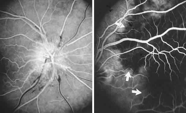
reference to mtDNA mutations. Arch Ophathalmol 103:504, 1996

89. Nikoskelainen E, Hoyt WF, Nummelin K et al: Fundus findings in Leber's hereditary optic neuroretinopathy. III. Fluorescein angiographic studies. Arch Ophthalmol 102:981, 1984

90. Newman NJ: Leber's hereditery optic neuropathy: new genetic considerations. Arch Neurol 50:540, 1993

91. Newman NJ, Lott MT, Wallace DC: The clinical characteristics of pedigrees of Lebers' hereditary optic neuropathy with the 11778 mutation. Am J Ophthalmol 111:750, 1991

92. Riordan-Eva P, Sanders MD, Govan GG et al: The clinical features of Leber's hereditary optic neuropathy defined by the presence of a pathogenic mitochondrial DNA mutation. Brain 118:319, 1995

93. Johns DR, Heher KL, Miller NR et al: Leber's hereditary optic neuropathy: clinical manifestations of the 14484 mutation. Arch Ophthalmol 111:495, 1993

94. Mashima Y, Hiida Y, Saga M et al: Risk of false-positive molecular genetic diagnosis of Leber's hereditary optic neuropathy. Am J Ophthalmol 119:245, 1995

95. Johns DR, Smith KH, Miller NR et al: Identical twins who are discordant for Leber's hereditary optic neuropathy. Arch Ophthalmol 111:1491, 1993

96. Hoyt WF: Charcot-Marie-Tooth disease with primary optic atrophy: report of a case. Arch Ophthalmol 64:925, 1960

97. Bird TD, Griep E: Pattern reversal visual evoked potentials: studies in Charcot-Marie-Tooth hereditary neuropathy. Arch Neurol 38:739, 1981

98. Paradiso G, Micheli F, Taratuto AL et al: Familial bulbospinal neuronopathy with optic atrophy: a distinct entity. J Neurol Neurosurg Psychiatry 61:196, 1996

99. Nicolaides P, Appleton RE, Fryer A: Cerebellar ataxia, areflexia, pes cavus, optic atrophy, and sensorineural hearing loss (CAPOS): a new syndrome. J Med Genet 33:419, 1996

100. Groom M, Kay MD, Corrent GF: Optic neuropathy in familial dysautonomia. J Neuroophthalmol 17:101, 1977

101. Livingston IR, Mastaglia FL, Edis R et al: Visual involvement in Friedreich's ataxia and hereditary spastic ataxia: a clinical and visual evoked response study. Arch Neurol 38:75, 1981

102. Frisen L, Claesson M: Narrowing of the retinal arterioles in descending optic atrophy: a quantitative clinical study. Ophthalmology 94:1020, 1984

103. Papastathopoulos K, Jonas JB: Focal narrowing of retinal arterioles in optic nerve atrophy. Ophthalmology 102: 1706, 1995

104. Ettl A, Kramer J, Daxer A et al: High resolution magnetic resonance imaging of neurovascular orbital anatomy. Ophthalmology 104:869, 1997

105. Mukhenji SK, Tart RP, Fitzsimmons J et al: Fat-suppressed MR of the orbit and cavernous sinus: comparison of fast spin-echo and conventional spin-echo. AJNR Am J Neuroradiol 15:1707, 1994

106. Davis PC, Newman NJ: Advances in neuro-imaging of the visual pathways. Am J Ophthalmol 121:690, 1996

107. Byrne SF, Green RL: Ultrasound of the Eye and Orbit. St. Louis, CV Mosby, 1992

108. Katz B, Hoyt WF: Intrapapillary and peripapillary hemorrhage in young patients with incomplete posterior vitreous detachment. Ophthalmology 102:349, 1995

109. Kokame GT: Intrapapillary, papillary, and vitreous hemorrhage: letters, and reply. Ophthalmology 102:1003, 1995

110. Sher NA, Wirtschafter J, Shapiro SK et al: Unilateral papilledema in “benign” intracranial hypertension (pseudotumor cerebri). JAMA 250:2346, 1983

111. Liu D, Michon J: Measurement of the subarachnoid pressure of the optic nerve in human subjects. Am J Ophthalmol 119:81, 1994

112. Hayreh SS: Pathogenesis of optic disc oedema. In Kennard C, Rose FC (eds): Physiological Aspects of Clinical Neuro-Ophthalmology, p 431. Chicago, Year Book, 1988

113. Liu D, Kahn M: Measurement and relationship of subarachnoid pressure of the optic nerve to intracranial pressure in fresh cadavers. Am J Ophthalmol 116:548, 1993

114. Peterson RA, Rosenthal A: Retinopathy and papilledema in cyanotic congenital heart disease. Pediatrics 49:243, 1972

115. Bucci FA, Krohel GB: Optic nerve swelling secondary to the obstructive sleep apnea syndrome. Am J Ophthalmol 105:428, 1988

116. Hardten DR, Wen DY, Wirtschafter JD et al: Papilledema and intraspinal lumbar paraganglioma. J Clin Neuroophthalmol 12:158, 1992

117. Michowiz SD, Rappaport HZ, Shaked I et al: Thoracic disc herniation associated with papilledema. J Neurosurg 61:1132, 1984

118. Marks SJ, Schick A, Charney JZ et al: The association of papilledema with syringomyelia: case report. Mt Sinai J Med 55:333, 1988

119. Ropper AH, Marmarou A: Mechanism of pseudotumor in Guillain-Barré syndrome. Arch Neurol 41:259, 1984

120. Stern BJ, Gruen R, Koeppel J et al: Recurrent thyrotoxicosis and papilledema in a patient with communicating hydrocephalus. Arch Neurol 41:65, 1984

121. Scharf D: Neurocysticercosis: two hundred thirty-eight cases from a California hospital. Arch Neurol 45:777, 1988

122. Chimowitz MI, Little JR, Awad IA et al: Intracranial hypertension associated with unruptured cerebral arteriovenous malformations. Ann Neurol 27:474, 1990

123. Verm A, Lee AG: Bilateral optic disk with macular exudates as the manifesting sign of a cerebral arteriovenous malformation. Am J Ophthalmol 123:422, 1997

124. Karahalios DG, Rekate HL, Khayata MH et al: Elevated intracranial venous pressure as a universal mechanism in pseudotumor cerebri of varying etiologies. Neurology 46:198, 1996

125. Marr WG, Chambers RG: Pseudotumor cerebri syndrome following unilateral neck dissection. Am J Ophthalmol 51:605, 1961

126. Graus F, Slatkin NE: Papilledema in the metastatic jugular foramen syndrome. Arch Neurol 40:816, 1983

127. Strominger MB, Weiss GB, Mehler MF: Asymptomatic unilateral papilledema in pseudotumor cerebri. J Clin Neuroophthalmol 12:238, 1992

128. Muci-Mendoza R, Arruga J, Hoyt WF: Distensión bilateral del espacio subaracnoideo perioptico en el pseudotumor cerebral con papiledema unilateral: su demonstración a traves de la tomografia computarizada de la orbita. Rev Neurol 39:11, 1981

129. Pagani LF: The rapid appearence of papilledema. J Neurosurg 30:247, 1969

130. Steffen H, Eifert B, Aschoff A et al: The diagnostic value of optic disc elevation in acute elevated intracranial pressure. Ophthalmology 103:1229, 1996

131. Hilton-Jones D, Ponsford JR, Graham N: Transient visual obscurations, without papilledema. J Neurol Neurosurg Psychiatry 45:832, 1982

132. Hedges TR, Baron EM, Hedges TR et al: The retinal venous pulse: its relation to optic disc characteristics and choroidal pulse. Ophthalmology 101:542, 1994

133. Carter SR, Seifrf SR: Macular changes in pseudotumor cerebri before and after optic nerve sheath fenestration. Ophthalmology 102:937, 1995

134. Jacobson DM: Intracranial hypertension and syndrome of acquired hyperopia and choroidal folds. J Neuroophthalmol 15:178, 1995

135. Corbett JJ, Savino PJ, Thompson HS et al: Visual loss in pseudotumor cerebri: follow-up of 57 patients from five to 41 years and a profile of 14 patients with permanent severe visual loss. Arch Neurol 39:461, 1982

136. Baker RS, Buncic JR: Sudden visual loss in pseudotumor cerebri due to central retinal artery occlusion. Arch Neurol 41:1274, 1984

137. Beck RW, Greenberg HS: Post-decompression optic neuropathy. J Neurosurg 63:196, 1985

138. Mashima Y, Oshitari K, Imamura Y et al: High-rsolution magnetic resonance imaging of the intraorbital optic nerve and sub-arachnoid space in patients with papilledema and optic atrophy. Arch Ophthalmol 114:1197, 1996

139. Lessell S: Pediatric pseudotumor cerebri (idiopathic intracranial hypertension). Surv Ophthalmol 37:155, 1992

140. Cinciripini GS, Donahue S, Borchert MS: Idiopathic intracranial hypertension in prepubertal pediatric patients: characteristics, treatment, and outcome. Am J Ophthalmol 127:178, 1999

141. Sorensen PS, Gjerris F, Svenstrup B: Endocrine studies in patients with pseudotumor cerebri: estrogen levels in blood and cerebrospinal fluid. Arch Neurol 43:902, 1986

142. Radhakrishnan K, Ahlskog JE, Garrity JA et al: Idiopathic intracranial hypertension. Mayo Clin Proc 69:169, 1994

143. Digre KB, Corbett JJ: Pseudotumor cerebri in men. Arch Neurol 45:866, 1988

144. Borruat FX, Regli F: Pseudotumor cerebri as a complication of amiodarone therapy. Am J Ophthalmol 116:776, 1993

145. Hamed LM, Glaser JS, Schatz NJ et al: Danazol-induced pseudotumor cerebri. Arch Ophthalmol 100:1000, 1989

146. Sanborn GE, Selhorst JB, Calabrese VP et al: Pseudotumor cerebri and insecticide intoxication. Neurology 29: 1222, 1979

147. Saul RF, Hamburger HA, Selhorst JB: Pseudotumor cerebri secondary to lithium carbonate. JAMA 253: 2869, 1985

148. Green JP, Newman NJ, Stowe ZN et al: “Normal pressure” pseudotumor cerebri. J Neuroophthalmol 16:241, 1996

149. Friedman DI, Forman S, Levi L, Lavin PJ et al: Unusual ocular motility disturbances with increased intracranial pressure. Neurol 50:1893, 1998

150. Moser FG, Hilal SK, Abrams G et al: MR imaging of pseudotumor cerebri. AJNR Am J Neuroradiol 9:39, 1988

151. Wang SJ, Silberstein SD, Patterson S, Young WB: Idiopathic intracranial hypertension without papilledema. A case-control study in a headache center. Neurol 51:245, 1998

151a. Corbett JJ: Problems in the diagnosis and treatment of pseudotumor cerebri. Can J Neurol Sci 10:221, 1983

152. Wall M, George D: Visual loss in pseudotumor cerebri: incidence and defects related to visual field strategy. Arch Neurol 44:170, 1987

153. Hedges TR, Legge RH, Peli E et al: Retinal nerve fiber layer loss in idiopathic intracranial hypertension. Ophthalmology 102:1242, 1995

154. Gu XZ, Tsai JC, Wurdeman A et al: Pattern of axonal loss in longstanding papilledema due to idiopathic intracranial hypertension. Curr Eye Res 14:173, 1995

155. Fan JT, Johnson DH, Burk RR: Transient myopia, angle closure and choroidal detachments after oral acetazolamide. Am J Ophthalmol 115:813, 1993

156. Liu GT, Glaser JS, Schatz NJ: High-dose methylprednisolone and acetazolamide for visual loss in pseudotumor cerebri. Am J Ophthalmol 118:88, 1994

157. Johnson LN, Krohel GB, Madsen RW et al: The role of weight loss and acetazolamide in the treatment of idiopathic intracranial hypertension (pseudotumor cerebri). Ophthalmology 105:2313, 1998

158. Burgett RA, Purvin VA, Kawasaki A: Lumboperitoneal shunting for pseudotumor cerebri. Neurology 49:734, 1997

159. Liu GT, Volpe NJ, Schatz NJ et al: Severe sudden visual loss caused by pseudotumor cerebri and lumboperitoneal shunt failure. Am J Ophthalmol 122:129, 1996

160. Lee AG: Visual loss as the manifesting symptom of ventriculoperitoneal shunt malfunction. Am J Ophthalmol 122:127, 1996

161. Corbett JJ, Nerad JA, Tse DT et al: Results of optic nerve sheath fenestration for pseudotumor cerebri: the lateral orbitotomy approach. Arch Ophthalmol 106:1391, 1988

162. Spoor TC, McHenry JG: Long-term effectiveness of optic nerve sheath decompression for pseudotumor cerebri. Arch Ophthalmol 111:632, 1993

163. Rizzo JF, Lessell S: Choroidal infarction after optic nerve sheath fenestration. Ophthalmology 101:1622, 1994

164. Mauriello JA, Shaderowsky P, Gizzi M et al: Management of visual loss after optic nerve sheath decompression in patients with pseudotumor cerebri. Ophthalmology 102:441, 1995

165. Mathew NT, Ravishankar K, Sanin LC: Coexistence of migraine and idiopathic intracranial hypertension without papilledema. Neurology 46:1226, 1996

166. Magnante DO, Bullock JD: Familial pseudotumor cerebri: occurrence in a mother while pregnant with a subsequently affected daughter. (personal communication)

167. Lam BL, Schatz NJ, Glaser JS et al: Pseudotumor cerebri from cranial venous obstruction. Ophthalmology 99:706, 1992

168. Purvin VA, Trobe JD, Kosmorsky G: Neuro-ophthalmic features of cerebral venous obstruction. Arch Neurol 52:880, 1995

169. Hauser D, Barzilai N, Zalish M et al: Bilateral papilledema with retinal hemorrhages in association with cerebral venous sinus thrombosis and paroxysmal nocturnal hemoglobinuria. Am J Opthalmol 122:592, 1996

170. Ireland B, Corbett JJ, Wallace R: The search for causes of idiopathic intracranial hypertension. Arch Neurol 47:315, 1990

171. FDA Medwatch and Spontaneous Reporting System. Norplant 1991-1993. Obstet Gynecol 85:538, 1995

172. Malozowski S, Tanner LA, Wysowski DK et al: Benign intracranial hypertension in children with growth hormone deficiency treated with growth hormone. J Pediatr 126: 996, 1995

173. Alexandrakis G, Filatov, Walsh T: Pseudotumor cerebri in a 12-year-old boy with Addison's disease. Am J Ophthalmol 116:650, 1993

174. Liu GT, Kay MD, Bienfang DC et al: Pseudotumor cerebri associated with corticosteroid withdrawal in inflammatory bowel disease. Am J Ophthalmol 117:352, 1994

175. Sirdofsky M, Kattah J, Macedo P: Intracranial hypertension in a dieting patient. J Neuro-ophthalmol 14:9, 1994

175a. Chiu AM, Chuenkongkaew WL, Cornblath W, et al: Minocycline treatment and pseudotumor cerebri syndrome. Am J Ophthalmol 126:116, 1998

176. Jennum P, Borgesen SE: Intracranial pressure and obstructive sleep apnea. Chest 95:279, 1989

177. Landau K, Gloor BP: Therapy resistant papilledema in achondroplasia. J Neuroophthalmol 14:24, 1994

178. Stavrou P, Sgouros S, Willshaw HE et al: Visual failure caused by raised intracranial pressure in craniosynostosis. Childs Nerv Syst 13:64, 1997

179. Johnston I, Hawke S, Halmagyi M et al: The pseudotumor syndrome: disorders of cerebrospinal fluid circulation causing intracranial hypertension without ventriculomegaly. Arch Neurol 48:740, 1991

180. Katz B: The dyschromatopsia of optic neuritis. Trans Am Ophthalmol Soc 93:685, 1995

181. Mojon DS, Rösler KM, Oetliker H: A bedside test to determine motion stereopsis using the Pulfrich phenomenon. Ophthalmol 105:1337, 1998

182. Selhorst JB, Saul RF: Uhthoff and his symptom. J Neuroophthalmol 15:63, 1995

183. Keltner JL, Johnson CA, Spurr JO et al: Visual field profile of optic neuritis: one-year follow-up in the optic neuritis treatment trial. Arch Ophthalmol 112:946, 1994

184. Kennedy C, Caroll FD: Optic neuritis in children. Trans Am Acad Ophthalmol Otolaryngol 64:700, 1960

185. Riikonen R: The role of infection and vacination in the genesis of optic neuritis and multiple sclerosis in children. Acta Neurol Scand 80:425, 1989

186. Lucchinetti CF, Kiers L, O'Duffy A et al: Risk factors for developing multiple sclerosis after childhood optic neuritis. Neurology 49:1413, 1997

187. Hamed LM, Silbiger J, Guy J et al: Parainfectious optic neuritis and encephalomyelitis: a report of two cases with thalamic involvement. J Clin Neuroophthalmol 13:18, 1993

188. Purvin VA, Chioran G: Recurrent neuroretinitis. Arch Ophthalmol 112:365, 1994

189. Beck RW, Cleary PA, Trobe JD et al: The effect of corticosteroids for acute optic neuritis on the subsequent development of multiple sclerosis. N Engl J Med 329:1764, 1993

190. Warner JEA, Lessell S, Rizzo JF et al: Does optic disc appearance distinguish ischemic optic neuropathy from optic neuritis. Arch Ophthalmol 115:1408, 1997

191. Guy J, Mao J, Bidgood WD et al: Enhancement and demyelination of the intraorbital optic nerve: fat suppression magnetic resonance imagining. Ophthalmology 99:713, 1992

192. Dunker S, Wiegand W: Prognostic value of magnetic resonance imaging in monosymptomatic optic neuritis. Ophthalmology 103:1768, 1996

193. Beck RW, Kupersmith MJ, Cleary PA et al: Fellow eye abnormalities in acute unilateral optic neuritis. Ophthalmology 100:691, 1993

194. Cleary PA, Beck RW, Bourque LB et al: Visual symptoms after optic neuritis: results from the Optic Neuritis Treatment Trial. J Neuroophthalmol 17:18, 1997

195. Jacobs L, Karpik A, Bozian D et al: Auditory-visual synesthesia. Arch Neurol 38:211, 1981

196. Safran AB, Kine LB, Glaser JS: Positive visual phenomena in optic nerve and chiasm disease: photopsias and photophobia. In Glaser JS (ed): Neuro-ophthalmology, Vol X, p 225. St. Louis, CV Mosby, 1980

197. Kaufman DI, Beck R, ONTT Study Group: The 5-year risk of MS after optic neuritis: experience of the Optic Neuritis Treatment Trial. Neurology 49:1404, 1997

198. Jacobs LD, Kaba SE, Miller CM et al: Correlation of clinical: magnetic resonance imaging, and cerebrospinal fluid findings in optic neuritis. Ann Neurol 41:392, 1997

199. Francis DA, Compston DA, Batchelor JR et al: A reassessment of the risk of multiple sclerosis developing in patients with optic neuritis after extended follow-up. J Neurol Neurosurg Psychiatry 50:6, 1987

200. Soderstom M, Ya-Ping J, Hillert J et al: Optic neuritis. Prognosis for multiple sclerosis from MRI, CSF, and HLA findings. Neurology 50:708, 1998

201. Lightman S, McDonald WI, Bird AC et al: Retinal venous sheathing in optic neuritis: its significance for pathogenesis of multiple sclerosis. Brain 100:405, 1987

202. Engell T: Neurological disease activity in multiple sclerosis patients with periphlebitis retinae. Acta Neurol Scand 73:168, 1986

203. Malinowski SM, Pulido JS, Folk JC: Long-term visual outcome and complications associated with pars planitis. Ophthalmology 100:818, 1993

204. Gass A, Graham E, Moseley IF et al: Cranial MRI in idiopathic retinal vasculitis. J Neurol 242:174, 1995

205. Cole SR, Beck RW, Moke PS et al: The predictive value of CSF oligoclonal banding for MS 5 years after optic neuritis. Optic Neuritis Study Group. Neurol 51:885, 1998

206. Mandler RN, Davis LE, Jeffrey DR et al: Devic's neuromyelitis optica: a clinocopathologic study of 8 patients. Ann Neurol 34:162, 1993

207. O'Riordan JI, Gallagher HL, Thompson AJ et al: Clinical, CSF and MRI findings in Devic's neuromyelitis optica. J Neurol Neurosurg Psychiatry 60:382, 1996

208. Brazis PW, Lee AG: Optic disk edema with a macular star. Mayo Clin Proc 71:1162, 1996

209. Ellis BD, Kosmorsky GS, Cohen BH. Medical and surgical management of acute disseminated encephalomyelitis. J Neuroophthalmol 14:210, 1994

210. Hamed LM, Silbiger J, Guy J et al: Parainfectious optic neuritis and encephalomyelitis. J Neuroophthalmol 13:18, 1993

211. Kawasaki A, Purvin VA, Tang R: Bilateral anterior ischemic optic neuropathy following influenza vaccination. J Neuroophthalmol 18:56, 1998

212. Hull TP, Bates JH: Optic neuritis after influenza vaccination. Am J Ophthalmol 124:703, 1997

213. Pall HS, Williams AC: Subacute polyradiculopathy with optic and auditory nerve involvement. Arch Neurol 44:885, 1987

214. Borruat FX, Schatz NJ, Glaser JS, Forteza A: Central nervous system involvement in Guillain-Barré-like syndrome: clinical and magnetic resonance imaging evidence. Eur Neurol 38:129, 1997

215. Miller DH, Kay R, Schon F et al: Optic neuritis following chickenpox in adults. J Neurol 233:182, 1986

216. Selbst RG, Selhorst JB, Harbison JW et al: Parainfectious optic neuritis. Arch Neurol 40:347, 1983

217. Berrios RR, Serrano LA: Bilateral optic neuritis after a bee sting. Am J Ophthalmol 117:677, 1994

218. Keane J. Neuro-ophthalmologic signs of AIDS: 50 patients. Neurology 41:841, 1991

219. Nichols JW, Goodwin JA: Neuro-ophthalmologic complications of AIDS. Semin Ophthalmol 7:24, 1992

220. Tenhula WN, Xu SZ, Madigan MC et al: Morphometric comparisons of optic nerve axon loss in acquired immunodeficiency syndrome. Am J Ophthalmol 113:14, 1992

221. Patel SS, Rutzen AR, Marx JL et al: Cytomegalovirus papillitis in patients with acquired immune deficiency syndrome. Ophthalmology 103:1476, 1996

222. Cohen DB, Glasgow BJ: Bilateral optic nerve cryptococcosis in sudden blindness in patients with acquired immune deficiency syndrome. Ophthalmology 100:1689, 1993

223. Garrity JA, Herman DC, Imes R et al: Optic nerve sheath decompression for visual loss in patients with acquired immunodeficiency syndrome and cryptococcal meningitis with papilledema. Am J Ophthalmol 116:472, 1993

224. Grossniklaus HE, Specht CS, Allaire G et al: Toxoplasma gondii retinochoroiditis and optic neuritis in acquired immune deficiency syndrome: report of a case. Ophthalmology 97:1342, 1990

225. Shayegani A, Odel JG, Kazim M et al: Varicella-zoster virus optic neuritis in a patient with human immunodeficiency virus. Am J Ophthalmol 122:586, 1996

226. Lee MS, Cooney EL, Stoessel KM et al: Varicella zoster virus retrobulbar optic neuritis in patients with acquired immunodeficiency syndrome. Ophthalmology 105:467, 1998

227. Yau TH, Rivera-Velazquez, Mark AS et al: Unilateral optic neuritis in a patient with the acquired immunodeficiency syndrome. Am J Ophthalmol 121:324, 1996

228. Gabuzda DH, Hirsch MS: Neurologic manifestations of infection with human immunodeficiency virus. Ann Intern Med 107:383, 1987

229. Burton BJL, Leff AP: Plant steroid-responsive HIV optic neuropathy. J Neuroophthalmol 18:25, 1998

230. Margo CE, Hamed LM: Ocular syphilis. Surv Ophthalmol 37:203, 1992

231. Zambrano W, Perez GM, Smith JL: Acute syphilitic blindness in AIDS. J Clin Neuroophthalmol 7:1, 1987

232. Toshniwal P: Optic perineuritis with secondary syphilis. J Clin Neuroophthalmol 7:6, 1987

233. Arruga J, Valentines J, Mauri F et al: Neuroretinitis in acquired syphilis. Ophthalmology 92:262, 1985

234. Balcer LJ, Winterkorn JMS, Galetta SL: Neuro-ophthalmic manifestations of Lyme disease. J Neuroophthalmol 17:108, 1997

235. Schmutzhard E, Pohl P, Stanek G: Involvement of Borrelia burgdorferi in cranial nerve affection. Zentrabl Bakt Hyg A 263:328, 1986

236. Pachner AR, Steere AC: The triad of neurologic manifestations of Lyme disease: meningitis, cranial neuritis, and radiculoneuritis. Neurology 35:47, 1985

237. Wu G, Lincoff H, Ellsworth RM et al: Optic disc edema and Lyme disease. Ann Ophthalmol 18:252, 1986

238. Strominger MB, Slamovits TL, Herskovitz S et al: Transient worsening of optic neuropathy as a sequela of the Jarisch-Herxheimer reaction in the treatment of Lyme disease. J Neuroophthalmol 14:77, 1994

239. Lyme disease: Connecticut. JAMA 259:1147, 1988

240. Wong MT, Dolan MJ, Lattuada CP et al: Neuroretinitis, aseptic meningitis, and lymphadenitis associated with Bartonella henselae infection in immunocompetent patients and patients infected with human immunodeficiency virus type 1. Clin Infect Dis 21:352, 1995

241. Golnik KC, Marotto ME, Fanous MM et al: Ophthalmic manifestations of Rochalimaea species. Am J Ophthalmol 118:145, 1994

242. Reed JB, Scales DK, Wong MT et al: Bartonella henselae neuro-retinitis in cat scratch disease: diagnosis, management, and sequelae. Ophthalmology 105:459, 1998

243. Cox TA, Haskins GE, Gangitano JL et al: Bilateral Toxacara optic neuropathy. J Clin Neuroophthalmol 3:267, 1983

244. Fish RH, Hoskins JC, Kline LB: Toxoplasmosis neuroretinitis. Ophthalmology 100:1177, 1993

245. Lossos A, Eliashiv S, Ben-Chetrit E: Optic neuritis associated with familial Mediterranean fever. J Clin Neuroophthalmol 13:141, 1993

246. Ormerod IEC, McDonald WI: Multiple sclerosis presenting with progressive visual failure. J Neurol Neurosurg Psychiatry 47:943, 1984

247. Eidelberg D, Newton MR, Johnson G et al: Chronic unilateral optic neuropathy: a magnetic resonance study. Ann Neurol 24:3, 1988

248. Fay AM, Kane SA, Kazim M et al: Magnetic resonance imaging of optic perineuritis. J Neuroophthalmol 17: 247, 1997

249. Dutton JJ, Anderson RL: Idiopathic inflammatory perioptic neuritis simulating optic nerve sheath meningioma. Am J Ophthalmol 100:424, 1985

250. Hykin PG, Spalton DJ: Bilateral perineuritis of the optic nerves. J Neurol Neurosurg Psychiatry 54:375, 1991

251. Winterkorn JM, Odel JG, Behrens MM et al: J Neuroophthalmol 14:157, 1994

252. Frohman L, Wolansky L: Magnetic resonance imaginging of syphilitic optic perineuritis. J Neuroophthalmol 17:57, 1997

253. Beardsley TL, Brown SVL, Sydnor CF et al: Eleven cases of sarcoidosis of the optic nerve. Am J Ophthalmol 97:62, 1984

254. Vrabec TR, Augsburger JJ, Fisccher DH et al: Taches de bougie. Ophthalmology 102:1712, 1995

255. Akova YA, Kansu T, Duman S: Pseudotumor cerebri secondary to dural sinus thrombosis in neurosarcoidosis. J Clin Neuroophthalmol 13:188, 1993

256. Beck AD, Newman NJ, Grossniklaus HE et al: Optic nerve enlargement and chronic visual loss. Surv Ophthalmol 38:555, 1994

257. Newman LS, Rose CS, Maier LA: Sarcoidosis. N Engl J Med 336:1224, 1997

258. Belden CJ, Hamed L, Mancuso AA: Bilateral isolated retrobulbar optic neuropathy in limited Wegener's granulomatosis. J Clin Neuroophthalmol 13:119, 1993

259. Slavin M, Glaser JS: Acute severe irreversible visual loss with sphenoethmoiditis: “posterior” orbital cellulitis. Arch Ophthalmol 105:345, 1987

260. Kodsi SR, Younge BR, Leavitt JA et al: Intracranial plasma cell granuloma presenting as an optic neuropathy. Surv Ophthalmol 38:70, 1993

261. Ofner S, Baker RS: Visual loss in cryptococcal meningitis. J Clin Neuropophthalmol 7:45, 1987

262. Lee MS, Cooney EL, Stoessel KM et al: Varicella zoster virus retrobulbar optic neuritis preceding retinitis in patients with acquired immunodeficiency syndrome. Ophthalmology 105:467, 1998

263. Borruat FX, Herbort CP: Herpes zoster ophthalmicus: anterior ischemic optic neuropathy and acyclovir. J Clin Neuroophthalmol 12:37, 1992

264. Lam BL, Barrett DA, Glaser JS et al: Visual loss from idiopathic intracranial pachymeningitis. Neurology 44: 694, 1994

265. Hayreh SS: The optic nerve head circulation in health and disease. Exp Eye Res 61:259, 1995

266. Onda E, Ciofi GA, Bacon DR, van Buskirk EM: Microvasculature of the human optic nerve. Am J Ophthalmol 120:92, 1995

267. Hattenhauer MG, Leavitt JA, Hodge DO et al: Incidence of nonarteritic anterior ischemic optic neuropathy. Am J Ophthalmol 123:103, 1997

268. IONDT Study Group: Characteristics of patients with nonarteritic anterior ischemic neuropathy eligible for the ischemic optic neuropathy decompression trial. Arch Ophthalmol 114:1366, 1996

269. Hayreh SS, Joos KM, Podhajsky PA et al: Systemic diseases associated with nonarteritic anterior ischemic optic neuropathy. Am J Ophthalmol 118:766, 1994

270. Kline LB: Progression of visual field defects in ischemic optic neuropathy. Am J Ophthalmol 106:199, 1988

271. Movsas T, Kelman SE, Elman MJ et al: The natural course of non-arteritic ischemic optic neuropathy. Invest Ophthalmol Vis Sci 42:951, 1991

272. Borchert M, Lessell S: Progressive and recurrent nonarteritic anterior ischemic optic neuropathy. Am J Ophthalmol 106:443, 1988

273. Arnold AC, Hepler RS: Natural history of nonarteritic anterior ischemic optic neuropathy. J Neuroophthalmol 14:66, 1994

274. Gordon RN, Burde RM, Slamovits T: Asymptomatic optic disc edema. J Neuroophthalmol 17:29, 1997

275. Hayreh SS: Acute ischemic disorders of the optic nerve: pathogenesis, clinical manifestations, and management. Ophthalmol Clin North Am 9:407, 1996

276. Olver JM, Spalton DJ, McCartney ACE: Microvascular study of the retrolaminar optic nerve in man: the possible significance in anterior ischemic optic neuropathy. Eye 4:7, 1990

277. Arnold AC, Hepler RS: Fluorescein angiography in acute nonar-teritic anterior ischemic optic neuropathy. Am J Ophthalmol 117:222, 1994

278. Kalenak JW, Kosmorsky GS, Rockwood EJ: Nonarteritic anterior ischemic optic neuropathy and intraocular pressure. Arch Ophthalmol 109:660, 1991

279. Hayreh SS, Podhajsky PA, Zimmerman B: Nonarteritic anterior ischemic optic neuropathy: time of onset of visual loss. Am J Ophthalmol 124:641, 1997

280. Rizzo JF, Lessell S: Optic neuritis and ischemic optic neuropathy: overlapping clinical profiles. Arch Ophthalmol 109:1668, 1991

281. Trobe JD, Glaser JS, Cassady JC et al: Nonglaucomatous excavation of the optic disc. Arch Ophthalmol 98:1046, 1980

282. Trobe JD, Glaser JS, Cassady JC: Optic atrophy: differential diagnosis by fundus observation alone. Arch Ophthalmol 98:1040, 1980

283. Frisen L, Claesson M: Narrowing of the retinal arterioles in descending optic atrophy: a quantitative clinical study. Ophthalmology 91:1342, 1984

284. Beri M, Klugman MR, Kohler JA et al: Anterior ischemic optic neuropathy. VII. Incidence of bilaterality and various influencing factors. Ophthalmology 94:1020, 1987

285. Boone MI, Massry GG, Frankel RA et al: Visual outcome in bilateral nonarteritic anterior ischemic optic neuropathy. Ophthalmology 103:1223, 1996

286. WuDunn D, Zimmerman K, Sadun AA, Feldon SE: Comparison of visual function in fellow eyes after bilateral nonarteritic anterior ischemic optic neutropathy. Ophthalmology 104:104, 1997

287. Lepore FE, Yarian DL: A mimic of the “exact diagnostic sign” of Foster Kennedy. Ann Ophthalmol 17:411, 1985

288. Fry CL, Carter JE, Kanter MC et al: Anterior ischemic optic neuropathy is not associated with carotid artery atherosclerosis. Stroke 24:539, 1993

289. Arnold AC, Hepler RS, Hamilton DR, Lufkin RB: Magnetic resonance imaging of the brain in nonarteritic ischemic optic neuropathy. J Neuroophthalmol 15:158, 1995

290. Giuffre G: Hematologic risk factors for anterior ischemic optic neuropathy. Neuroophthalmology 10:197, 1990

291. Watts MT, Greaves M, Rennie IG et al: Antiphospholipid antibodies in the aetiology of of ischaemic optic neuropathy. Eye 5:75, 1991

292. Worrall BB, Moazami G, Odel JG et al: Anterior ischemic optic neuropathy and activated protein C resistance: a case report and review of the literature. J Neuroophthalmol 17:162, 1997

293. Talks SJ, Chong NH, Gibson JM, Dodson PM: Fibrinogen, cholesterol and smoking as risk factors for non-arteritic anterior ischemic optic neuropathy. Eye 9:85, 1995

294. Beck RW, Servais GE, Hayreh SS: Anterior ischemic optic neuropathy. IX. Cup-to-disc ratio and its role in pathogenesis. Ophthalmology 94:1503, 1987

295. Mansour AM, Shoch D, Logani S: Optic disk size in ischemic optic neuropathy. Am J Ophthalmol 106:587, 1988

296. Katz B, Spencer WH: Hyperopia as a risk factor for nonarteritic anterior ischemic optic neuropathy. Am J Ophthalmol 116:754, 1993

297. Arnold AC, Hepler RS, Lieber M, Alexander JM: Hyperbaric oxygen therapy for nonarteritic anterior ischemic optic neuropathy. Am J Ophthalmol 122:535, 1996

298. Johnson LN, Gould TJ, Krohel GB: Effect of levodopa and carbidopa on recovery of visual function in patients with nonar-teritic anterior ischemic optic neuropathy of longer than six months' duration. Am J Ophthalmol 121:77, 1996

299. Beck RW, Hayreh SS, Podhajsky PA et al: Aspirin in nonarteritic anterior ischemic optic neuropathy. Am J Ophthalmol 123:212, 1997

300. Kupersmith MJ, Frohman L, Sanderson M et al: Aspirin reduces the incidence of second eye NAION: a retrospective study. J Neuroophthalmol 17:250, 1997

301. Ischemic Optic Neuropathy Decompression Trial Research Group: Optic nerve decompression surgery for nonarteritic anterior ischemic optic neuropathy (NAION) is not effective and may be harmful. JAMA 273:625, 1995

302. Glaser JS, Teimory M, Schatz NJ: Optic nerve sheath fenestration for progressive ischemic optic neuropathy. Arch Ophthalmol 112:1047, 1994

303. Yee RD, Selky AK, Purvin VA: Outcomes of surgical and nonsurgical management of nonarteritic ischemic optic neuropathy. Trans Am Ophthmalol Soc 91:227, 1993

304. Johnson LN, Arnold AC: Incidence of nonarteritic and arteritic anterior ischemic optic neuropathy. J Neuroophthalmol 14:38, 1994

305. Chung SM, Gay CA, McCrary JA: Nonarteritic ischemic optic neuropathy: the impact of tobacco use. Ophthalmology 101:779, 1994

306. Hayreh SS, Podhajsky PA, Zimmerman B: Ocular manifestations of cranial arteritis. Am J Ophthalmol 125:509, 1998

307. Tomlinson FH, Lie JT, Nienhuis BJ et al: Juvenile temporal arteritis revisited. Mayo Clin Proc 69:445, 1994

308. Weyand CM, Bartley GB: Giant cell arteritis: new concepts in pathogenesis and implications for management. Am J Ophthalmol 123:392, 1997

309. Wagner AD, Bjornsson J, Bartley GB et al: Interferon gamma-producing T cells in giant cell vasculitis represent a minority of tissue-infiltrating cells and located distant from the site of pathology. Am J Pathol 148:1925, 1996

310. Ghanci FD, Dutton GN: Current concepts in giant cell (temporal) arteritis. Surv Ophthalmol 42:99, 1997

311. Hauser WA, Ferguson RH, Holley KE et al: Temporal arteritis in Rochester, Minnesota, 1951 to 1967. Mayo Clin Proc 46:597, 1971

312. Turnbull J: Temporal arteritis and polymyalgia rheumatica: nosographic and nosologic considerations. Neurology 46:901, 1996

313. Hayreh SS, Podhajsky P: Visual field defects in anterior ischemic optic neuropathy. Doc Ophthalmol Proc Ser 19:53, 1979

314. Beri M, Klugman MR, Kohler JA et al: Anterior ischemic optic neuropathy. VII. Incidence of bilaterality and various influencing factors. Ophthalmology 94:1020, 1987

315. Sebag J, Thomas JV, Epstein EL et al: Optic disc cupping in arteritic anterior ischemic optic neuropathy resembles glaucomatous cupping. Ophthalmology 93:357, 1986

316. Melberg NS, Grand MG, Diekert JP et al: Cotton-wool spots and early diagnosis of giant cell arteritis. Ophthalmology 102:1611, 1995

317. Barricks ME, Traviesa DB, Glaser JS, Levy IS: Ophthalmoplegia in cranial arteritis. Brain 100:209, 1977

318. Bienfang DC: Loss of the ocular pulse in the acute phase of temporal arteritis. Acta Ophthalmol Suppl 191:35, 1989

319. Bosley TM, Savino PJ, Sergott RC et al: Ocular pneumoplethysmography can help in the diagnosis of giant-cell arteritis. Arch Ophthalmol 107:379, 1989

320. Siatkowski RM, Gass JDM, Glaser JS et al: Fluorescein angiography in the diagnosis of giant cell arteritis. Am J Ophthalmol 115:57, 1993

321. Slavin ML, Barondes MJ: Visual loss caused by choroidal ischemia preceding anterior ischemic optic neuropathy in giant cell arteritis. Am J Ophthalmol 117:81, 1994

322. Ho AC, Sergott RC, Regillo CD et al: Color Doppler hemodynamics of giant cell arteritis. Arch Ophthalmol 112:938, 1994

323. Schmidt WA, Kraft HE, Vorpahl K et al: Color duplex ultrasonography in the diagnosis of temporal arteritis. N Engl J Med 337:1336, 1997

324. Andersson R, Malvall BE, Bengtsson BA: Long-term survival in giant cell arteritis including remporal arteritis and polymyalgia rheumatica. Acta Med Scand 220:361, 1986

325. Aiello PD, Trautman JC, McPhee TJ et al: Visual prognosis in giant cell arteritis. Ophthalmology 100:550, 1993

326. Liu GT, Glaser JS, Schatz NJ, Smith JL: Visual morbidity in giant cell arteritis: clinical characteristics and prognosis for vision. Ophthalmology 101:1779, 1994

327. Hayreh SS, Podhajsky PA, Raman R, Zimmerman B: Giant cell arteritis: validity and reliability of various diagnostic criteria. Am J Ophthalmol 123:285, 1997

328. Cullen JF: Ischemic optic neuropathy. Trans Ophthalmol Soc UK 87:759, 1967

329. Miller A, Green M: Simple rule for calculating normal erythrocyte sedimentation rate. BMJ 286:266, 1983

330. Gruener G, Merchut MP: Renal causes of elevated sedimentation rate in suspected temporal arteritis. J Clin Neuroophthalmol 12:272, 1992

331. Klein RG, Campbell RJ, Hunder GG, Carney JA: Skip lesions in temporal arteritis. Mayo Clin Proc 51:504, 1976

332. Nishino H, DeRemee RA, Rubino FA et al: Wegener's granulomatosis associated with vasculitis of the temporal artery: report of five cases. Mayo Clin Proc 68:115, 1993

333. Levy MH, Margo CE: Temporal artery biopsy and sarcoidosis. Am J Ophthalmol 117:409, 1994

334. Goodman BW: Temporal arteritis. Am J Med 67:839, 1979

335. Wilkinson IMS, Russell RWR: Arteries of the head and neck in giant cell arteritis: a pathological study to show the pattern of arterial involvement. Arch Neurol 27:378, 1972

336. Cornblath WT, Eggenberger ER: Progressive visual loss from giant cell arteritis despite high-dose intravenous methylprednisolone. Ophthalmology 104:854, 1997

337. Gardiner PVG, Griffiths ID: Sudden death after treatment with pulsed methylprednisolone. BMJ 300:125, 1990

338. Matteson EL, Gold KN, Bloch DA et al: Long-term survival of patients with giant cell arteritis in the American College of Rheumatology giant cell arteritis criteria cohort. Am J Med 100:193, 1996

339. van der Veen MJ, Dinant HJ, Vam Booma-Frankfort C et al: Can methotrexate be used as a steroid sparing agent in the treatment of polymyalgia rheumatica and giant cell arteritis? Ann Rheum Dis 55:218. 1996

340. Demaziere A: Dapsone in the long-term treatment of temporal arteritis. Am J Med 87:3, 1989

341. Skillern PG, Lockhart G: Optic neuritis and uncontrolled diabetes mellitus in 14 patients. Ann Intern Med 51:468, 1959

342. Lubow M, Makley TA: Pseudopapilledema of juvenile diabetes mellitus. Arch Ophthalmol 85:417, 1971

343. Barr CC, Glaser JS, Blankenship G: Acute disc swelling in juvenile diabetes: clinical profile and natural history of 12 cases. Arch Ophthalmol 98:2185, 1980

344. Regillo CD, Brown GC, Savino PJ et al: Diabetic papillopathy: patient characteristics and fundus findings. Arch Ophthalmol 113:889, 1995

345. Carroll FD: Optic nerve complications of cataract extraction. Trans Am Acad Ophthalmol Otolaryngol 77:623, 1973

346. Hayreh SS: Anterior ischemic optic neuropathy. IV. Occurrence after cataract extraction. Arch Ophthalmol 98:1410, 1980

347. Serrano LA, Behrens MM, Carroll FD: Postcataract extraction ischemic optic neuropathy. Arch Ophthalmol 100:1177, 1982

348. Hamed LM, Purvin V, Rosenberg M: Recurrent anterior ischemic optic neuropathy in young adults. J Clin Neuroophthalmol 8:239, 1988

349. O'Hara M, O'Connor PS: Migrainous optic neuropathy. J Clin Neuro Ophthalmol 4:85, 1984

350. Katz B: Bilateral sequential migrainous ischemic optic neuropathy. Am J Ophthalmol 99:489, 1985

351. Toshniwal P: Anterior ischemic optic neuropathy secondary to cluster headache. Acta Neurol Scand 73:213, 1986

352. Hayreh SS: Anterior ischemic optic neuropathy. VIII. Clinical features and pathogenesis of post-hemorrhagic amaurosis. Ophthalmology 94:1488, 1987

353. Williams EL, Hart WM, Tempelhoff R: Postoperative ischemic optic neuropathy. Anesth Analg 80:1018, 1995

354. Hollenhorst RW, Svein HJ, Benoit CF: Unilateral blindness occurring during anesthesia for neurosurgical operations. Arch Ophthalmol 52:819, 1954

355. Katz DM, Trobe JD, Cornblath WT, Kline LB: Ischemic optic neuropathy after lumbar spine surgery. Arch Ophthalmol 112:925, 1994

356. Jaben SL, Glaser JS, Daily M: Ischemic optic neuropathy following general surgical procedures. J Clin Neuroophthalmol 3:239, 1983

357. Johnson MW, Kincaid MC, Trobe JD: Bilateral retrobulbar optic nerve infarctions after blood loss and hypotension: a clinicopathologic case study. Ophthalmology 94:1577, 1987

358. Connolly SE, Gordon KB, Horton JC: Salvage of vision after hypotension-induced ischemic optic neuropathy. Am J Ophthalmol 117:235, 1994

359. Shapira OM, Kimmel WA, Lindsey PS et al: Anterior ischemic optic neuropathy after open heart operations. Ann Thorac Surg 61:660, 1996

360. Slavin ML: Ischemic optic neuropathy after cardiac arrest. Am J Ophthalmol 104:435, 1987

361. Knox DL, Hanneken AM, Hollows FC et al: Uremic optic neuropathy. Arch Ophthalmol 106:50, 1988

362. Hamed LM, Winward KE, Glaser JS et al: Optic neuropathy in uremia. Am J Ophthalmol 108:30, 1989

363. Michaelson C, Behrens M, Odel J: Bilateral anterior ischemic optic neuropathy associated with optic disc drusen and systemic hypotension. Br J Ophthalmol 73:767, 1989

364. Waybright EA, Selhorst JB, Combs J: Anterior ischemic optic neuropathy with internal carotid artery occlusion. Am J Ophthalmol 93:42, 1982

365. Brown GC: Anterior ischemic optic neuropathy occurring in association with carotid artery obstruction. J Clin Neuroophthalmol 6:39, 1986

366. Bogousslavsky J, Regli F, Zografos L et al: Optico-cerebral syndrome: simultaneous hemodynamic infarction of optic nerve and brain. Neurology 37:263, 1987

367. Leonard TJK, Sanders MD: Ischaemic optic neuropathy in pulseless disease. Br J Ophthalmol 67:389, 1983

368. Hall S, Barr W, Lie JT et al: Takayasu arteritis: a study of 32 North American patients. Medicine 64:89, 1995

369. Liebermann MF, Shahi A, Grenn WR: Embolic ischemic optic neuropathy. Am J Ophthalmol 86:206, 1978

370. Tomsak RL: Ischemic optic neuropathy associated with retinal embolism. Am J Ophthalmol 99:590, 1985

371. Beck RW, Gamel JW, Willcourt RJ et al: Acute ischemic optic neuropathy in severe preeclampsia. Am J Ophthalmol 90:342, 1980

372. DeFrancisco M, Savino PJ, Schatz NJ: Optic atrophy in acute intermittent porphyria. Am J Ophthalmol 87:221, 1979

373. Manor RS, Axer-Siegal R, Cohenn S et al: Bilateral anterior ischemic optic neuropathy, pseudoxanthoma elasticum, and platelet hyperaggregability. Neuroophthalmology 6:173, 1986

374. Slavin ML, Barondes MJ: Ischemic optic neuropathy in sickle cell disease. Am J Ophthalmol 105:212, 1988

375. Perlman JI, Forman S, Gonzalez ER. Retrobulbar ischemic optic neuropathy associated with sickle cell disease. J Neuroophthalmol 14:45, 1994

376. Sklar EML, Schatz NJ, Glaser JS et al: MR of vasculitis-induced optic neuropathy. AJNR Am J Neuroradiol 17: 121, 1996

377. Ahmadieh H, Roodpeyma S, Azarmina M et al: Bilateral simultaneous optic neuritis in chidhood systemic lupus erythematosus. J Neuroophthalmol 14:84, 1994

378. Frohman LP, Lama P: Annual review of systemic diseases: 1991996. I. J Neuroophthalmol 18:67, 1998

379. Tesar JT, McMillan V, Molina R et al: Optic neuropathy and central nervous system disease assoiciated with Sjögren's syndrome. Am J Med 92:686, 1992

380. Hutchinson CH: Polyarteritis nodosa presenting as posterior ischemic optic neuropathy. J R Soc Med 77:1043, 1984

381. Massry GG, Chung SM, Selhorst JB: Optic neuropathy, headache, and diplopia with MRI suggestive of cerebral arteritis in relapsing polychondritis. J Neuroophthalmol 15:171, 1995

382. Schmidt MH, Fox AJ, Nicolle DA: Bilateral anterior ischemic optic neuropathy as a presentation of Takayasu's disease. J Neuroophthalmol 17:156, 1997

383. Isayama Y, Takahashi T, Inoue M et al: Posterior ischemic optic neuropathy. III. Clinical diagnosis. Ophthalmologica 187:141, 1983

384. Sawle GV, Sarkies NJC: Posterior ischaemic optic neuropathy due to internal artery occlusion. Neuroophthalmology 7:349, 1987

385. Sommer A, Tielsch JM, Katz J et al: Relationship between intra-ocular pressure and primary open-angle glaucoma among white and black Americans. Arch Ophthalmol 109:10980, 1991

386. Caprioli J: Recognizing structural damage to the optic nerve head and nerve fiber layer in glaucoma. Am J Ophthalmol 124:516, 1997

387. Kalenak JW, Kosmorsky GS, Hassenbusch SJ: Compression of the intracranial optic nerve mimicking unilateral normal-pressure glaucoma. J Clin Neuroophthalmol 12: 230, 1992

388. Greenfield DS, Siatkowski RM, Glaser JG et al: The cupped disc: who needs neuroimaging? Ophthalmology 105:1866, 1998

389. Siegner SW, Netland PA: Optic disc hemorrhages and progression of glaucoma. Ophthalmology 103:1014, 1996

390. Brown GC, Shields JA: Tumors of the optic nerve head. Surv Ophthalmol 29:239, 1985

391. Dutton JJ: Gliomas of the anterior visual pathway. Surv Ophthalmol 38:427, 1994

392. Listernick R, Louis DN, Packer RJ et al: Optic pathway gliomas in children with neurofibromatosis 1: consensus statement from NF1 Optic Pathway Glioma Task Force. Ann Neurol 41:143, 1997

393. Anderson DR, Spencer WH: Ultrastructural and histochemical observations of optic nerve gliomas. Arch Ophthalmol 83:324, 1970

394. Grimson BS, Perry DD: Enlargement of the optic disk in childhood optic nerve tumors. Am J Ophthalmol 97: 627, 1984

395. McDonnell P, Miller NR: Chiasmatic and hypothalamic extension of optic nerve glioma. Arch Ophthalmol 101:1412, 1983

396. Imes RK, Hoyt WF: Magnetic resonance imaging signs of optic nerve gliomas in neurofibromatosis 1. Am J Ophthalmol 111:729, 1991

397. Brodsky MC: The “pseudo-CSF” signal of orbital optic glioma on magnetic resonance imaging: a signature of neurofibromatosis. Surv Ophthalmol 38:213, 1993

398. Gans MS, Frazier Byrne S, Glaser JS: Standardized a-scan echography in optic nerve disease. Arch Ophthalmol 105:1232, 1987

399. Sadun F, Hinton DR, Sadun AA: Rapid growth of an optic nerve ganglioglioma in a patient with neurofibromatosis 1. Ophthalmology 103:794, 1996

400. Jenkin D, Angyalfi S, Becker L et al: Optic glioma in children: surveillance, resection or irradiation. Int J Radiat Oncol Biolm Phys 25:215, 1993

401. Dutton JJ: Optic nerve sheath meningiomas. Surv Ophthalmol 37:167, 1992

402. Karp LA, Zimmerman LE, Borit A et al: Primary intraorbital meningiomas. Arch Ophthalmol 91:24, 1974

403. Ing EB, Garrity JA, Cross SA et al: Sarcoid masquerading as optic nerve sheath meningioma. Mayo Clin Proc 72:38, 1997

404. Zimmerman CF, Schatz NJ, Glaser JS: Magnetic resonance imaging of optic nerve meningiomas. Ophthalmology 97:585, 1990

405. Lindblom B, Truwit CL, Hoyt WF: Optic nerve sheath meningioma: definition of intraorbital, intracanalicular, and intracranial components with magnetic resonance imaging. Ophthalmology 99:560, 1992

406. Eng TY, Albright NW, Kuwahara G et al: Precision radiation therapy for optic nerve sheath meningiomas. Int J Rad Oncol Biol Phys 22:1093, 1992

407. Lee AG, Woo SY, Miller NR et al: Improvement in visual function in an eye with presumed optic nerve sheath meningioma after treatment with three-dimensional conformal radiation therapy. J Neuroophthalmol 16:247, 1996

408. Hsu DW, Efird JT, Hedley-White ET: Progesterone and estrogen receptors in meningiomas: prognostic considerations. J Neurosurg 86:113, 1997

409. Olsen ME, Chernik NL, Posner JB: Infiltration of the leptomeninges by systemic cancer: a clinical and pathologic study. Arch Neurol 30:122, 1974

410. Little JR, Dale AJD, Okazaki H: Meningeal carcinomatosis. Arch Neurol 30:138, 1984

411. Christmas NJ, Mead MD, Richardson EP, Albert DM: Secondary optic nerve tumors. Surv Ophthalmol 36:196, 1991

412. McFadzean R, Brosnahan D, Doyle D et al: A diagnostic quartet in leptomeningeal infiltration of the optic nerve sheath. J Neuroophthalmol 14:175, 1994

413. Krol G, Sze G, Malkin M et al: MR of cranial and spinal meningeal carcinomatosis: comparison with CT and myelography. AJNR Am J Neuroradiol 9:709, 1988

413a. de la Sayette V, Bertran F, Honnorat J et al: Paraneoplastic cerebellar syndrome and optic neuritis with anti-CV2 antibodies: clinical response to excision of the primary tumor. Arch Neurol 55:405, 1998

414. Siatkowski RM, Lam BL, Schatz NJ et al: Optic neuropathy in Hodgkin's disease. Am J Ophthalmol 114:625, 1992

415. Strominger MB, Schatz NJ, Glaser JS: Lymphomatous optic neuropathy. Am J Ophthalmol 116:774, 1993

416. Kashani AA, Kerman BM: Central nervous system malignant B-cell lymphoma identified with standardized echography of the optic nerve. J Neuroophthalmol 17:243, 1997

417. Kodsi SR, Younge BR, Leavitt JA et al: Intracranial plasma cell granuloma presenting as an optic neuropathy. Surv Ophthalmol 38:70, 1993

418. Nikaido H, Mishima H, Ono H et al: Leukemic involvement of the optic nerve. Am J Ophthalmol 105:294, 1988

419. Kaikov Y: Optic nerve head infiltration in acute leukemia in children: an indication for emergency optic nerve radiation therapy. Med Pediatr Oncol 826:101, 1996

420. Cramer SC, Glaspy JA, Efird JT et al: Chronic lymphocytic leukemia and the central nervous system: a clinical and pathologic study. Neurology 46:19, 1996

421. Purvin V, vanDyk HJL: Primary reticulum cell sarcoma of the brain presenting as steroid-responsive optic neuropathy. J Clin Neuroophthalmol 4:15, 1984

422. Wagoner MD, Gonder JR, Albert DM: Intraocular reticulum cell sarcoma. Ophthalmology 87:724, 1980

423. Khawly JA, Rubin P, Petros W et al: Retinopathy and optic neuropathy in bone marrow transplantation for breast cancer. Ophthalmology 103:87, 1996

423a. Purvin, V: Anterior ischemic optic neuropathy secondary to interferon-alpha. Arch Ophthalmol 113:1041, 1995

424. Caraceni A, Martini C, Spatti G et al: Recovering optic neuritis during systemic cisplatin and carboplatin chemotherapy. Acta Neurol Scand 96:260, 1997

425. DeLano MC, Fun FY, Zinreich SJ: Relationship of the optic nerves to the posterior paranasal sinuses: a CT anatomic study. AJNR Am J Neuroradiol 17:669, 1996

426. Buss DR, Tse Dt, Farris BK: Ophthalmic complications of sinus surgery. Ophthalmology 97:612, 1990

427. Hayman, Carter K, Schiffman JS et al: A sellar misadventure: imaging considerations. Surv Ophthalmol 41:252, 1996

428. Goodwin JA, Glaser JS. Chiasmal syndrome in sphenoid sinus mucocele. Ann Neurol 4:440, 1978

429. Johnson LN, Hepler RS, Yee RD et al: Sphenoid sinus mucocele (anterior clinoid variant) mimicking diabetic ophthalmoplegia and retrobulbar neuritis. Am J Ophthalmol 102:111, 1986

430. Dooley DP, Hollsten DA, Grimes SR et al: Indolent orbital apex syndrome caused by occult mucormycosis. J Clin Neuroophthalmol 12:245, 1992

431. Hutnik, Nicolle DA, Munoz DG: Orbital aspergillosis: a fatal masquerader. J Neuroophthalmol 17:257, 1997

432. Frohman LP, Lama P: Annual review of systemic diseases: 1991996. J Neuroophthalmol 18:67, 1998

433. Newman NJ, Slamovits TL, Friedland S et al: Neuro-ophthalmic manifestations of meningocerebral inflammation from the limited form of Wegener's granulomatosis. Am J Ophthalmol 120:613, 1995

434. Goldberg RA, Weisman JS, McFarland JE et al: Orbital inflammation and optic neuropathies associated with chronic sinusitis of intranasal cocaine abuse: possible role of contiguous inflammation. Arch Ophthalmol 107:831, 1989

435. Harbison JW, Lessell S, Selhorst JB: Neuro-ophthalmology of sphenoid sinus carcinoma. Brain 107:855, 1984

436. Berman EL, Chu A, Wirtschafter JD et al: Esthesioneuroblastoma presenting as sudden unilateral visual loss. J Clin Neuroophthalmol 12:31, 1992

437. Neigel JM, Rootman J, Belkin RI et al: Dysthyroid optic neuropathy: the crowded orbital apex syndrome. Ophthalmology 95:1515, 1988

438. Glatt HJ: Optic nerve dysfunction in thyroid eye disease: a clinician's perspective. Radiology 200:26, 1996

439. Birchall D, Goodall KL, Noble JL et al: Graves' ophthalmopathy: intracranial prolapse on CT as an indicator of optic nerve compression. Radiology 200:123, 1996

440. Hufnagel TJ, Hickey WF, Cobbs WH et al: Immunohistochemical and ultrastructural studies on the exenterated orbital tissues of a patient with Graves' disease. Ophthalmology 91:1411, 1984

441. Bartley GB, Fatourechi V, Kadrmas EF et al: Clinical features of Graves' ophthalmopathy in an incidence cohort. Am J Ophthalmol 121:284, 1996

442. Trobe JD, Glaser JS, LaFlamme P: Dysthyroid optic neuropathy: clinical profile and rationale for management. Arch Ophthalmol 179:285, 1978

443. Kao SCS, Kendler DL, Nugent RA et al: Radiotherapy in the management of thyroid orbitopathy: computed tomography and clinical outcomes. Arch Ophthalmol 111:819, 1993

444. Jacobson DM, Warner JJ, Broste SK: Optic nerve contact and compression by the carotid artery in asymptomatic patients. Am J Ophthalmol 123:677, 1997

445. Lindenberg R, Walsh FB, Sacks JG: Neuropathology of Vision: An Atlas. Philadelphia, Lea & Febiger, 1973

446. Golnik KC, Hund PW, Stroman GA et al: Magnetic resonance imaging in patients with unexplained optic neuropathy. Ophthalmology 103:515, 1996

447. Colapinto EV, Cabeen MA, Johnson LN: Optic nerve compression by a dolichoectatic internal carotid artery: case report. Neurosurgery 39:604, 1996

448. Miller NR, Savino PJ, Schneider T: Rapid growth of an intracranial aneurysm causing apparent retrobulbar optic neuritis. J Neuroophthalmol 15:212, 1995

449. Chan JW, Hoyt WF, Ellis WG et al: Pathogenesis of acute monocular blindness from leaking anterior communicating artery aneurysms: report of six cases. Neurology 48:680, 1997

450. Samples JR, Younge BR: Tobacco-alcohol amblyopia. J Clin Neuroophthalmol 1:213, 1981

451. Frisen L: Fundus changes in acute malnutritional optic neuropathy. Arch Ophthalmol 101:577, 1983

452. Kupersmith MJ, Weiss PA, Carr RE: The visual-evoked potential in tobacco-alcohol and nutritional amblyopia. Am J Ophthalmol 95:307, 1983

453. van Noort BAA, Bos PJM, Klopping C et al: Optic neuropathy from thiamine deficiency in a patient with ulcerative colitis. Doc Ophthalmol 67:45, 1987

454. Stambolian D, Behrens MM: Optic neuropathy associated with B12 deficiency. Am J Ophthalmol 83:465, 1977

455. Troncoso J, Mancall EL, Schatz NJ: Visual evoked responses in pernicious anemia. Arch Neurol 36:168, 1979

456. Rizzo JF, Lessell S: Tobacco amblyopia. Am J Ophthalmol 116:84, 1993

457. Cuba Neuropathy Field Investigation Team: Epidemic optic neuropathy in Cuba: clinical characterization and risk factors. N Engl J Med 333:1176, 1955

458. Freeman AG: Optic neuropathy and chronic cyanide toxicity. Lancet 1:441, 1986

459. McKellar MJ, Hidajat RR, Elder MJ: Acute ocular methanol toxicity: clinical and electrophysiologic features. Aust N Z J Ophthalmol 3:225, 1997

460. Sharpe JA, Hostovsky M, Bilbao JM et al: Methanol optic neuropathy: a histopathological study. Neurology 32: 1093, 1982

461. Fasler JJ, Rose FC: West Indian amblyopia. Postgrad Med J 56:494, 1980

462. Grant WM: Toxicology of the Eye. Springfield, IL, Charles C Thomas, 1986

463. Kumar A, Sandramouli S, Verma L et al: Ocular ethambutol toxicity: is it reversible? J Clin Neuroophthalmol 13:15, 1993

464. Helm G, Holland G: Ocular tuberculosis. Surv Ophthalmol 38:230, 1993

465. Salmon JF, Carmichael TR, Welsh NH: Use of contrast sensitivity measurement in the detection of subclinical ethambutol toxic optic neuropathy. Br J Ophthalmol 71: 192, 1987

466. Ricoy JR, Ortega A, Cabello A: Subacute myelo-optic neuropathy (SMON). J Neurol Sci 53:241, 1982

467. Pittman FE, Westphal MC: Optic atrophy following treatment with diiodohydroxyquin. Pediatrics 53:81, 1974

468. Godel V, Nemet P, Lazar M: Chloramphenicol optic neuropathy. Arch Ophthalmol 98:1417, 1980

469. Klingele TG, Burde RM: Optic neuropathy associated with penicillamine therapy in a patient with rheumatoid arthritis. J Clin Neuroophthalmol 4:75, 1984

470. Ehyai A, Freemon FR: Progressive optic neuropathy and sensorineural hearing loss due to chronic glue sniffing. J Neurol Neurosurg Psychiatry 46:349, 1983

471. Adams JW, Bofenkamp TM, Kobrin J et al: Recurrent acute toxic optic neuropathy secondary to 5-FU. Cancer Treat Rep 68:565, 1984

472. Pickrell L, Purvin V: Ischemic optic neuropathy secondary to intracarotid infusion of BCNU. J Clin Neuroophthalmol 7:87, 1987

473. Slamovits TL, Burde RM, Klingele TG: Bilateral optic atrophy caused by chronic oral ingestion and topical application of hexachlorophene. Am J Ophthalmol 89:676, 1980

474. Coskucan NM, Jabs DA, Dunn JP et al: The eye in bone marrow transplantation. VI. Retinal complications. Arch Ophthalmol 112:372, 1994

475. Gittinger JW, Asdourian GK: Papillopathy caused by amiodarone. Arch Ophthalmol 105:349, 1987

476. Garrett SN, Kennedy JJ, Schiffman JS: Amiodarone optic neuropathy. J Clin Neuroophthalmol 8:105, 1988

477. Matthews GP, Sandberg MA, Berson EL: Foveal cone electro-retinograms in patients with central visual loss of unexplained etiology. Arch Ophthalmol 110:1568, 1992

478. Steinsapir KD, Goldberg RA: Traumatic optic neuropathy. Surv Ophthalmol 38:487, 1994

479. Kline LB, McCluskey MM, Skalka HW: Imaging techniques in optic nerve evulsion. J Clin Neuroophthalmol 8:281, 1988

480. Brodsky MC, Wald KJ, Chen S et al: Protracted posttraumatic optic disc swelling. Ophthalmology 102:1628, 1995

481. Quigley HA, Davis EB, Anderson DR: Descending optic nerve degeneration in primates. Invest Ophthalmol Vis Sci 16:861, 1977

482. Cheney ML, Blair PA: Blindness as a complication of rhinoplasty. Arch Otolaryngol Head Neck Surg 113:768, 1987

483. Callahan MA: Prevention of blindness after blepharoplasty. Ophthalmology 90:1047, 1983

484. Horton JC, Hoyt WF, Foreman DS et al: Confirmation by magnetic resonance imaging of optic nerve injury after retrobulbar anesthesia. Arch Ophthalmol 114:351, 1996

485. Devoto MH, Kersten RC, Zalta AH et al: Optic nerve injury after retrobulbar anesthesia. Arch Ophthalmol 115:687, 1997

486. Liu C, Youl B, Moseley I: Magnetic resonance imaging of the optic nerve in extremes of gaze: implications for the positioning of the globe for retrobulbar anaesthesia. Br J Ophthalmol 76:728, 1992

487. Hollenhorst RW, Svien HJ, Benold CF: Unilateral blindness occurring during anesthesia for neurosurgical operations. Arch Ophthalmol 52:819, 1954

488. Jampol LM, Goldbaum M, Rosenberg M, Bahr R: Ischemia of ciliary arterial circulation from ocular compression. Arch Ophthalmol 93:1311, 1975

489. Chou P-I, Sadun AA, Lee H: Vasculature and morphometry of the optic canal and intracanalicular optic nerve. J Neuroophthalmol 15:186, 1995

490. Lessell S: Indirect optic nerve trauma. Arch Ophthalmol 107:382, 1989

491. Cook MW, Levin LA, Joseph MP et al: Traumatic optic neuropathy: a meta-analysis. Arch Otolaryngol Head Neck Surg 122:389, 1996

492. Ross HS, Rosenberg S, Friedman AH: Delayed radiation necrosis of the optic nerve. Am J Ophthalmol 76:683, 1973

493. Shukovsky LJ, Fletcher GH: Retinal and optic nerve complications in a high dose irradiation technique of ethmoid sinus and nasal cavity. Radiology 104:629, 1972

494. Salz JJ, Donin JF: Blindness after burns. Can J Ophthalmol 7:243, 1972

Back to Top