|
|
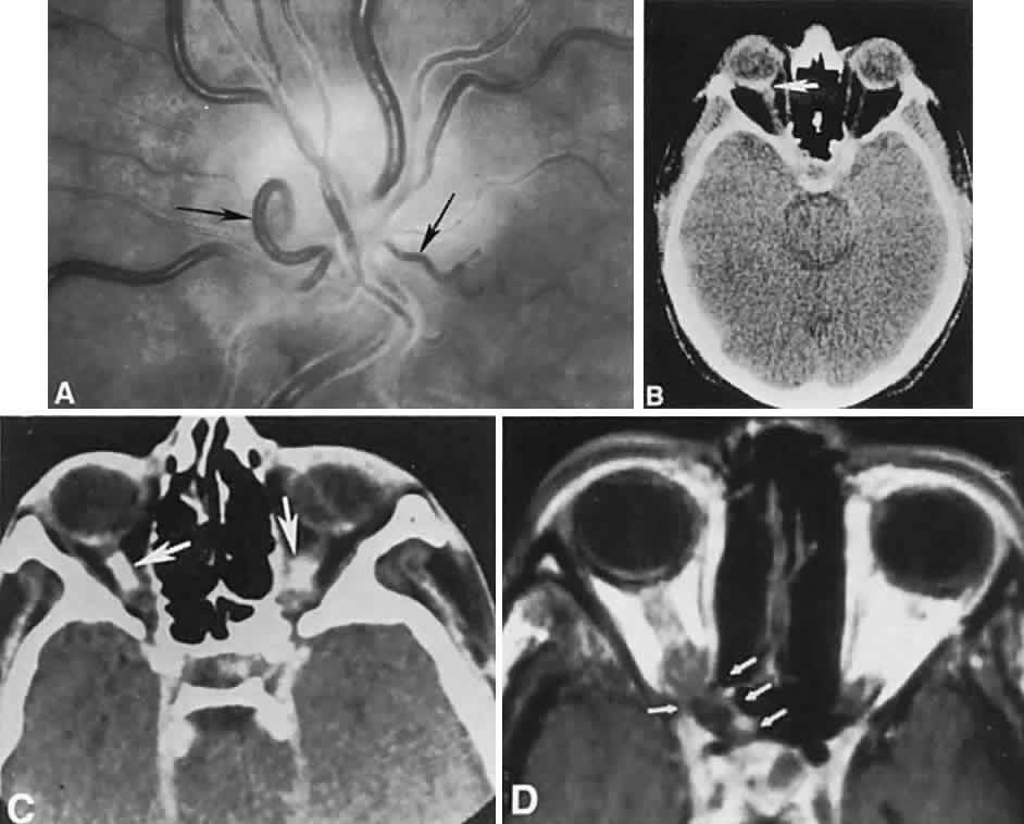
| Fig. 43. Optic nerve sheath (perioptic) meningioma. A. Chronic disc edema with progressive atrophy and retinociliary venous shunts (arrows). B. Computed tomography (CT) axial section of nerve (from A) shows irregular thickening of the sheath (arrow). C. CT axial section disclosed bilateral bone-density calcification (arrows). D. Magnetic resonance axial section (TR, 900 msec; TE, 20 msec, gadolinium-enhanced). Note evidence of meningioma spread in the canal area (arrows). |