|
|
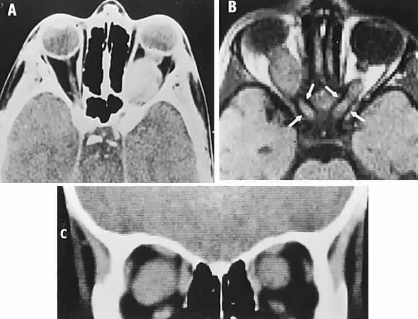
| Fig. 42. Optic nerve glioma. A. Computed tomography (CT) axial section shows smoothly fusiform enlargement of one optic nerve; chronic mass effect has expanded the orbit diameter. B. Magnetic resonance T1-weighted axial section; note moderate enlargement of both optic canals (arrows). C. CT coronal section of bilateral optic nerve gliomas. |