|
|
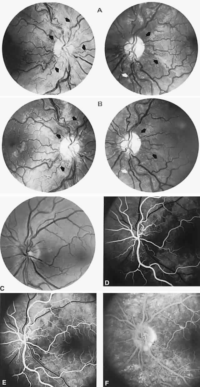
| Fig. 24. (continued) C. Left optic disc of a 17-year-old boy with Leber's optic neuropathy. D through F. Fluorescein angiography demonstrates peripapillary microangiopathy, with mildly dilated retinal arterioles (D), capillary shunts in arteriovenous phase (E). The late phase (F) shows no fluorescein leakage from disc tissue. |